Management of Localized Renal Cell Carcinoma


Management of Localized Renal Cell Carcinoma


▪ 44A Open Radical Nephrectomy for Localized Renal Cell Carcinoma

Amit R. Patel

Matthew N. Simmons

Steven C. Campbell


INTRODUCTION

Advances in molecular biology, histopathology, epidemiology, radiographic imaging, and surgical techniques have revolutionized how we approach the treatment of renal cell carcinoma (RCC) in the 21st century. Historically, the surgical standard for treatment of all renal masses was open radical nephrectomy (ORN). Introduction of CT imaging in the 1980s resulted in a trend of decreasing tumor size at initial diagnosis, with more than 50% of tumors measuring <4 cm currently. Data pertaining to the risks of chronic kidney disease (CKD), cardiovascular disease, and overall survival have led to the increased use of nephron-sparing surgery for treatment of localized RCC. Furthermore, laparoscopic radical nephrectomy was introduced in the 1990s, and this technique has largely superceded the open approach. While the role of ORN has become more limited, the fundamental aspects of the surgery remain the foundation for all current treatments. This chapter discusses the historical and current indications for ORN, surgical techniques, and outcomes related to ORN for localized RCC.


HISTORICAL PERSPECTIVE OF THE RADICAL NEPHRECTOMY

Langenbuch performed the first nephrectomy for malignant disease in 1875 through a lumbar incision (1). From this point through the mid-1900s, studies debated the surgical approach including transperitoneal versus lumbar incision, methods of ligating the renal pedicle (en bloc vs. individual vessel clamping), and early ureter ligation for greater mobilization of the kidney. In 1948, Chute et al. advocated anterior removal of the kidney due to easier access and preliminary ligation of the renal hilum (2). Similarly, Mortensen reported a similar technique to excise large renal tumors along with removal of perirenal fat, adrenal gland, and adjacent lymph nodes via a thoracoabdominal incision (3). Charles Robson et al. described their thoracoabdominal approach, and the modern ORN was popularized after their description of the technique and outcomes in 1963 (4). In 1969, Robson published updated outcomes and proposed a modification of the staging system based on the correlation between stage/histology and survival in their study (5).

Diagnosis of kidney tumors in the midtwentieth century was largely based on symptoms characterized by the classic triad of gross hematuria, flank pain, and a palpable abdominal mass. The varied signs and symptoms due to paraneoplastic effects led to RCC being termed the “internist’s tumor.” Given that most patients presented with constitutional symptoms and locally advanced tumors this era, treatments options were limited to ORN. The 10-year mortality following nephrectomy for RCC in the early 1900s ranged from 17% to 22% (6). Surgical mortality was as high as 37% in 1920 and was attributed to shock from toxins released during tumor manipulation, hemorrhage, and pulmonary embolism from tumor emboli released from tumor manipulation (1). In the 1970s, overall 10-year survival increased to 44% to 49%, and only one third of patients had localized disease (5,6). Incidentally, detected renal masses accounted for only 7% of renal masses detected in the 1970s primarily due to poor detection rates of intravenous pyelograms and arteriograms (6).

In the modern era, CT imaging has enabled early detection of tumors and also differentiation of certain tumor types based on their radiologic appearance (7). Because of this, RCC has shifted more toward becoming the “radiologist’s tumor.” Incidentally detected tumors are usually localized and of lower stage than symptomatic renal masses. Mean tumor sizes steadily decreased at diagnosis from 7.8 to 5.3 cm from 1989 to 1998, respectively (8). The largest contributor to the increase in incidence from 1983 to 2002 was T1a renal masses (9). While the largest increased has been observed for smaller localized tumors, the incidence of all stages of RCC is also rising. Comparison of RCC incidence rates between 1973-1985 and 1986-1998 shows that localized disease rose by an annual rate of 3.7%, regional disease by 1.9%, and distant metastatic disease by 0.68% (10). Thus, urologists today manage two main categories of patients presenting with RCC: patients with large, symptomatic, locally advanced disease with possible regional adenopathy, adrenal invasion, or renal vein/inferior vena cava invasion (≥T2) and patients who have small localized renal masses (T1a) with good prognostic features, low rates of recurrence, and higher survival.


CONTEMPORARY INDICATIONS FOR OPEN RADICAL NEPHRECTOMY

Contemporary surgical management of kidney tumors can be conducted using partial or radical nephrectomy, and this can be approached using either open or laparoscopic techniques. Partial nephrectomy (PN) was initially considered only for imperative indications including solitary kidney, bilateral renal tumors, or in patients with CKD. The role of PN has expanded to include any sized kidney tumor that is amenable to this approach based on its anatomic location (11,12). The basis of this shift is multifactorial and due to mounting evidence that PN is associated with equivalent cancer control, and decreased risk of postoperative CKD and associated morbid cardiovascular disease (13). Primary indications for ORN include tumor location in a central position that prohibits PN or presence of extrarenal extension. Other indications include presence of T3b disease requiring renal vein or IVC thrombectomy and disease extension into the collecting
system. ORN is also preferentially conducted for patients with bulky tumors, or in patients with nodal and metastatic involvement (i.e., “cytoreductive” ORN). In terms of indications for open versus laparoscopic technique selection, this is primarily guided by patient and surgeon preference and expertise as well as the expected level of technical difficulty. In many patients with large tumors, prior abdominal surgery, complex or anomalous vasculature, or vascular involvement, the open approach is often preferable to limit OR time and to maximize safety of the operation.


EVALUATION AND MANAGEMENT OF PATIENTS UNDERGOING OPEN RADICAL NEPHRECTOMY

Initial evaluation of patients with a suspected malignant kidney tumor should focus on tumor staging and functional evaluation. Cross-sectional imaging of the kidney using CT or MRI provides detailed information regarding tumor size and morphology, renal vascular anatomy, and staging information including retroperitoneal lymph node assessment and visceral organ metastasis status. Plain film chest radiography is often sufficient for metastatic evaluation in patients with small renal masses, but a CT of the chest is justifiable in any context to rule out lung metastasis. In patients with neurological complaints or focal neurological deficits a CT or MRI of the head should be conducted. Bone scan should be reserved for patients with bone pain, elevated alkaline phosphatase, locally advanced disease or declining performance status (14).

Functional studies should include measurement of serum electrolytes and creatinine, complete blood count, and coagulation studies. Calculation of preoperative glomerular filtration rate should be conducted using one of several available formulas to better assess how intervention will impact postoperative function. A MAG-3 scan to assess differential kidney function should be considered selectively, such as when the contralateral kidney appears atrophic on radiologic imaging. This will allow for identification of patients who will develop severe CKD or those who may require postoperative hemodialysis. The MAG-3 scan can also be helpful to make treatment decisions in patients requiring reconstructive procedures or those who are being considered for PN in atrophic kidneys. If the total functional contribution of the kidney is <15% to 20%, it is likely that the kidney will provide negligible function postoperatively, and ORN may be elected in lieu of a more complicated procedure. Accurate assessment of kidney function is essential for counseling patients regarding the possible increased risk of CKD, CAD, and hemodialysis after surgery.


RADICAL NEPHRECTOMY: SURGICAL ANATOMY AND SURGICAL APPROACHES

Understanding of kidney anatomy and surgical planes is key to safe kidney removal and preservation of oncologic boundaries. The kidneys are retroperitoneal organs that lie along the 12th thoracic vertebral body and the third lumbar vertebral body. They are surrounded by numerous muscle groups, including the diaphragm at the posterior upper one third of each kidney, the psoas muscle at the medial aspect posteriorly, and the quadratus lumborum and transversalis posterolaterally. Perinephric fat and Gerota’s fascia envelop the kidney laterally, superiorly, and medially. The right kidney is bordered by the liver and adrenal gland at the upper pole, medially by the duodenum, and anteriorly by the ascending colon and hepatic flexure. The left kidney is bordered by the spleen superiorly, the adrenal gland, pancreas, and splenic vessels superomedially, and the descending colon and splenic flexure anteriorly.

Classic ORN was originally described by Robson as removal of the kidney including all tissues within the confines of Gerota’s fascia, in combination with adrenalectomy and regional lymph node dissection. Contemporary ORN entails removal of the kidney and the tissues within the Gerota’s fascia with or without adrenalectomy, and often does not involve formal lymphadenectomy. Depending on tumor stage, location, size, and surgeon preference access to the kidney during ORN can be achieved through a flank, transabdominal, or thoracoabdominal approach. The advantages of the flank incision are rapid access to the kidney and limitation of the surgical field to the retroperitoneum. A disadvantage of the flank approach is that the renal hilum is the deepest structure in the surgical field, and exposure or access to the hilum may be limited by larger renal masses. In cases of bulky tumors, caval thrombus extension or extrarenal invasion, a transabdominal approach provides optimal exposure to the great vessels and adjacent organs. At our institution, the most commonly used anterior approach is via a modified chevron incision. Either Thompson or Bookwalter-type retractors allow for excellent retraction using this approach. A thoracoabdominal approach allows for excellent exposure for right-sided tumor excision; however, this incision may convey significant morbidity and requires placement of a chest tube.

Patients are placed in the lateral decubitus position for flank ORN. The patient should be positioned with their iliac crest at the break of the table. A double arm board is used for arm support, and the hips, knees, and ankles are padded with foam. The bottom thigh should be flexed and the knee bent while the upper leg is kept straight. Pillows should be placed between the legs for support. The kidney rest is raised, and the table is flexed to place the flank muscles under tension. In patients with a prior contralateral PN use of the kidney rest should be omitted to prevent contralateral renal compression. A towel roll or intravenous fluid bag is placed under the axilla to protect against brachial nerve palsy.

A classic flank incision is typically made between the 10th and 11th ribs parallel to the axis of the 11th rib extending 12 to 15 cm from the midaxillary line toward the umbilicus (Fig. 44A.1A). This incision affords improved exposure to the kidney compared to the rib-sparing approach. The intercostal neurovascular bundles should be identified between the internal oblique and transversalis muscle layers, and mobilized along the length of the incision (Fig. 44A.1B,C). Transection or entrapment of the intercostal nerves can result in paresthesias and pseudoherniation. The retroperitoneum is entered bluntly just proximal to the tip of the 11th rib. Blunt dissection is conducted to sweep the peritoneum and pleura off of the anterior abdominal wall along the length of the intended incision, prior to division of the abdominal wall muscle and fascia (Fig. 44A.1D). An incision is made in the investing fascia underlying the perirenal fat running parallel to the psoas muscle on the posterolateral aspect of the kidney (Fig. 44A.1E). A plane is then developed on the anterior kidney surface progressing medially toward the renal hilum between the peritoneum and Gerota’s fascia. Development of this plane allows for exposure of duodenum and inferior vena cava for the right kidney or the aorta for left-sided kidneys. For right nephrectomy, the duodenum is mobilized medially using a Kocher maneuver to expose the underlying IVC. For left nephrectomy, development of this plane allows for safe mobilization of the spleen, splenic vessels, and pancreas from the superomedial aspect of the kidney. Once this plane is developed, a Bookwalter retractor with a bent oval ring will allow for optimal exposure (Fig. 44A.1F). Care should be taken to apply adequate padding under retractor blades to prevent injury to the pancreas, duodenum, spleen, and liver.


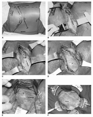
FIGURE 44A.1. (A) Layout of external anatomy for classic flank incision. A “mini-incision” or rib-sparing approach is now often utilized in this era. (B) Exposure of external oblique muscle layer and mobilization of 11th rib from intercostal muscles. The rib is then excised as lateral as possible. (C) Division of external oblique muscle layer exposing the internal oblique muscle. The 11th rib has been excised to expose the diaphragm (D), tranversalis muscle (T), and prerenal fat (asterisk). (D) Transversalis muscle (T) divided exposing the peritoneum (P) and prerenal fat (asterisk). (E) Exposure of the lateral flank plane with the prerenal fat (asterisk) and peritoneum (P) retracted medially. Pleural edge is seen at the anterior apical edge of the incision. Diaphragmatic muscle fibers (D) are seen as they merge with the fascia of quadratus lumborum. (F) Bookwalter retractor in place. Peritoneum (P) is retracted medially to expose the kidney. (G) Kidney is retracted laterally to exposure renal hilum. Vessel loops are placed around the renal artery (RA) and renal vein (RV) proximal to the gonadal vein (GV) draining into the renal vein.


FIGURE 44A.1. Continued.

A key initial step in the hilar dissection is early identification of the ureter and gonadal vein near the lower pole of the kidney. The gonadal vein can be used as a guide for dissection to the renal vein (Fig. 44A.1G). A plane can be dissected along the lateral surface of the IVC for right nephrectomy or the lateral surface of the aorta for left nephrectomy in order to maximize the medial resection margin. These areas are rich in lymphatics and small blood vessels, and it is advisable to place clips during this dissection to prevent lymphocele or chylous ascites. Dissection along these planes automatically results in a limited paracaval or paraaortic lymphadenectomy. Additionally, ligating the ureter and gonadal vessels early during the dissection allows for lateral retraction on the kidney and hilum to assist in safely mobilizing the renal vessels for both the flank and abdominal approaches.

Preoperative cross-sectional imaging will allow for determination of the number and location of renal arteries and veins. The main renal vein should be skeletonized, and a vessel loop applied to allow for easy identification and retraction during exploration for the arteries. Arteries should be approached as proximally as possible to allow for ligation of a common trunk. Redundant ligation of the proximal arterial stump should consist of a suture ligature and an additional suture or clip. Ligation of the arterial supply prior to ligation of venous outflow allows for a decrease in the size of the kidney and a decrease in venous outflow via the main renal veins as well as though parasitic veins. “Deflation” of the renal vein is a reliable indicator that all arterial inflow has been controlled, and failure to deflate should prompt reexploration for an unidentified artery. Ligation of the renal veins can be problematic due to their fragility and large diameter. Suture ligatures allow for secure control of the cut vessel with minimal risk of ligature slippage, and all attempts should be made to leave a sizable cuff of vein distal to the suture ligature to prevent slippage. The use of a vascular stapler is also an excellent option for securing the renal vein. After the hilum is controlled, the kidney is circumferentially mobilized taking care to remain outside of the Gerota’s fascia. To spare the adrenal, a bloodless plane can be found just under the inferior surface of the adrenal. Electrocautery dissection can be used to free the adrenal from the perinephric fat. The kidney is then circumferentially mobilized and the specimen is extracted.

For the bilateral subcostal transabdominal approach the patient is positioned supine with the umbilicus positioned at the break of the table. A rolled blanket is placed under the patient at the level of the lumbar spine. The table is flexed approximately 15 degrees to expand the abdomen. A subcostal incision is made 2 to 3 cm below the costal margin extending from the midaxillary line on the side of nephrectomy to the lateral rectus margin on the opposite side (Fig. 44A.2A). A vertical midline extension from the chevron incision to the xiphisternum can be made to increase exposure. A Thompson or Bookwalter retractor is used to expose the abdomen by placing retractors on the abdominal wall. During a right ORN, the liver is mobilized if necessary by dividing the ligamentum teres, the falciform ligament, and the lateral peritoneal attachments. This allows for placement of a Harrington retractor under the liver to retract it away from the upper pole. The ipsilateral colon is mobilized along the line of Toldt to expose the retroperitoneum. For a right ORN, a Kocher maneuver is conducted and dissection is carried out to expose both the aorta and IVC. For left nephrectomy, dissection is carried medially to allow for clear identification of the aorta, the origin of the superior mesenteric artery, and the left renal artery and vein. The superior mesenteric artery should be identified coursing anterior to the left renal vein, and clearly differentiated from the left renal artery to prevent inadvertent ligation. The small bowel is packed medially with moist sponges. Care should be taken to adequately pad the bowel, mesentery, and pancreas during exposure with a self-retaining retractor (Fig. 44A.2B). The kidney is retracted laterally to increase exposure to the renal hilum. The ureter and gonadal vein can be ligated early to assist in mobilizing the lower pole laterally. This maneuver will facilitate dissection of the renal hilum from caudal to cranial direction. The artery and vein are tied, suture ligated, and divided as described previously (Fig. 44A.2C). To avoid injury to the spleen or splenic hilum, dissection in this area is minimized until the very end of the procedure. Sharp release of the remaining attachments allows the spleen to fall away undisturbed. If splenic injury is encountered earlier in the procedure, the L upper quadrant can be packed until the nephrectomy is completed, and the spleen can be more readily addressed and removed if necessary. In cases where IVC thrombectomy is required, the IVC should be circumferentially mobilized above and below the renal hilum and vessels loops should be applied. Similar circumferential mobilization should also be conducted for both renal veins and any associated lumbar veins. Clamping of these vessels will allow for cessation of venous flow to the IVC to facilitate venotomy and thrombectomy.


SURGICAL COMPLICATIONS

Regardless of the approach used, care must be taken to maintain the integrity of Gerota’s fascia and avoid direct tumor entry. Tumor spillage or positive margins result in an increased rate of recurrence and cancer-specific mortality (15). Splenic injury can occur during left nephrectomy especially during the preliminary dissection of the splenic flexure, particularly if this is done too exuberantly. In cases of small splenic tears, hemostasis can often be obtained using plasma beam electrocautery and application of hemostatic agents (cellulose sheets or proprietary biologic preparations). Splenectomy should be considered in cases of extensive injury or in patients with mild to moderate injury who will be initiated on a postoperative anticoagulation regimen for preexisting conditions such as DVT or atrial fibrillation. Postoperative hemorrhage from splenic injury can occur days to weeks after surgery and can result in problematic bleeding.

Vascular injury is the most substantive intraoperative complication of ORN. In cases of large tumors, there are often
multiple large-caliber parasitic vessels. By remaining in the correct plane outside of Gerota’s fascia, most of these vessels can be avoided. Lumbar veins drain into the vena cava at each vertebral level and can be avulsed when too much traction is placed on the vena cava. In addition, there are typically large lumbar veins that can drain directly into the renal vein or vena cava at the level of the renal veins, particularly on the left side. The renal veins should be mobilized sufficiently prior to ligation to allow for identification of these lumbar vessels posteriorly. Other sources of possible hemorrhage are the gonadal and adrenal veins. The gonadal veins on either side can be ligated early in the procedure to prevent avulsion during kidney retraction and hilar dissection. Care should be taken on the superior aspect of the left renal vein to avoid bleeding from the adrenal vein. Likewise, care should be taken near the posterior infrahepatic IVC to avoid the right adrenal vein. Pinhole or subcentimeter venotomies can often be controlled by placement of figure-of-eight 5-0 vascular sutures. Larger lacerations can be controlled with a series of Allis clamps followed by sewing of the caval wall with a running 5-0 vascular suture. In cases of large venotomies, compression should be applied on the area of hemorrhage, and the IVC and other venous tributaries should be controlled with atraumatic vascular clamps so that formal venous reconstruction can be conducted.


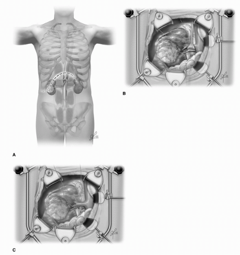
FIGURE 44A.2. (A) Location of incision for right-sided bilateral subcostal incision. (B) Thompson retractor is in place. The liver is retracted off of the upper pole. Careful medial retraction on the duodenum will help to expose the inferior vena cava. The body wall and colon are retracted to expose the lower pole of the kidney. (C) The kidney can be retracted laterally to facilitate hilar dissection. A single tie with a suture ligature is recommended for both the renal vein and artery.

Complications following ORN can also include renal insufficiency, pancreatitis, ileus and wound infection. Stephenson et al. reported a comprehensive analysis of complication rates for contemporary ORN in a cohort of 688 patients (16). Of the patients in this cohort, 51% had T1 disease, 13% T2, 22% T3a, and 14% T3b-4. Overall, 16% (112) of patients had a perioperative complication. Less than 1% of patients had acute renal failure, retroperitoneal hemorrhage, adjacent organ
injury, bowel obstruction, or pneumothorax. Three patients required splenectomy for splenic injuries. Median hospital stay duration was 6 days. Length of stay and complication rates were similar in other studies; however, reporting of complications was not standardized (17,18). A recent meta-analysis for the management of clinical T1 renal masses found that ORN had a urological complication rate of 1.3%, which was significantly lower than all other treatment modalities including laparoscopic RN, open or laparoscopic PN, and ablative therapies (11). Patients with T2 disease or greater, high operative blood loss, and increased length of stay had an increased risk of perioperative DVT and PE (19). The role of heparin DVT prophylaxis in preventing venous thromboembolic events remains to be studied in patients undergoing RN or PN.


RADICAL NEPHRECTOMY: CONTEMPORARY OUTCOMES

The incidence of RCC in 2009 in the United States is estimated at 57,760 new cases with an associated 12,980 deaths (20). Notably, the incidence of kidney cancer has risen 62% over the past 5 years with an increase in the death rate of 4% (21). The cause of this rise in incidence is multifactorial, and only partly explained by the increased rate of detection of incidental renal masses (7). Contemporary outcomes following surgical excision of localized RCC have improved significantly since the original outcome report by Robson in 1969, where 10-year overall survival was 62% (5). The improvement in survival after surgery is also multifactorial and largely due to the wide availability and improvement in cross-sectional imaging, improvement in perioperative surgical care, and an enhanced understanding of RCC and the histologic subtypes. Table 44A.1 lists contemporary outcomes in the literature for ORN for localized RCC published since 1999. Overall, for T1 masses, 5- and 10-year cancer-specific survival rates range from 84% to 98%. Meta-analysis of outcomes studies for T1 renal masses found that local recurrence-free survival was 98% and metastatic recurrence-free survival was 89% at a mean follow-up duration of 5 years, representing a gold standard for oncologic outcomes in this disease (11).


TABLE 44A.1 CONTEMPORARY RESULTS FOR CANCER-SPECIFIC AND OVERALL SURVIVAL FOR RADICAL NEPHRECTOMY FOR LOCALIZED RCC


Cancer-specific
Survival (%)


Overall
Survival (%)


Author


Year


N


Stage


5-yr


10-yr


5-yr


10-yr


Kinouchi et al. (22)


157


T1a-T2


96


89


1999


65


T3a


67


49


Ljungberg et al. (23)


187


T1


95


T2


87


1999


T3


37


Lau et al. (24)


2000


164


T1*


97


96


88


74


Russo (25)


2000


183


T1a


95


85


Ficarra et al. (26)


326


T1


91


89


133


T2


85


73


2002


66


T3a


57


36


Stephenson et al. (27)


T1


93


T2


82


T3a


67


89


2004


T3b


57


Mitchell et al. (28)


2005


30


> T1b


98


82


Hemal et al. (29)


2007


71


T2


94


84


68


Antonelli et al. (30)


137


T1a


98


2008


277


T1b


93


Thompson et al. (31)


2008


290


T1a


82


Zini (32)


2008


112


T1


99


92


Crepel et al. (33)


2009


4866


T1b


95


Zini et al. (34)


2009


5616


T1a


84


68


Thompson et al. (35)


2009


704


T1b


95


86


79


57


Epidemiologic studies have identified factors that influence survival after ORN. Table 44A.2 summarizes various prognostic models that have been developed to predict outcomes in patients preoperatively and postoperatively (41). The University of California, Los Angeles Integrated Stage System (UISS) calculates risk of postnephrectomy disease recurrence based on ECOG performance status, nuclear grade, and tumor stage (36). The SSIGN scoring system predicts 1-, 5-, and 10-year cancer-specific survival in patients with conventional RCC following ORN based on tumor stage, size, grade, and pathologic features (37). All prediction models have limitations including the number of prognostic features they utilize, the type of prognostic variables (molecular markers, histologic features, clinical symptoms) utilized, and the use of targeted or adjuvant therapies. Future algorithms are likely to be more robust to provide more accurate predictions with respect to treatment outcomes and survival.


FOLLOW-UP STRATEGIES FOLLOWING RADICAL NEPHRECTOMY

Follow-up strategies have been proposed by investigators based on long-term oncologic outcomes in patients who underwent ORN for RCC. Despite numerous studies, there is no consensus regarding surveillance protocols. Stage and grade have been
mainstays in prediction of RCC recurrence after ORN. Compared to patients with T1 tumors, those with T2, T3, and T4 tumors have a relative risk of disease progression of 2.3, 4.5, and 11.4, respectively. Compared to patients with Fuhrman grade 1 tumors, those with grade 3 and 4 histology have a relative risk of disease progression of 2.7 and 4.0, respectively (42). The greatest risk for recurrence for RCC occurs within the first 5 years following nephrectomy, although this remains one of the least predictable malignancies. For patients who recur after radical nephrectomy, 43% occur in the first year, 70% within 2 years, 80% within 3 years, and 93% within 5 years (23). Recurrence for T1 disease is reported to be 7% at a median time from diagnosis of 38 months, 26% for T2 disease at a median time of 32 months, and 39% for T3 disease at a median time of 17 months (43). Stage-based surveillance protocols stratify the intensity of surveillance based on the pathologic stage of the tumor (23,27,43,44,45), and reduce the costs of surveillance substantially by rationally reducing the frequency of follow-up CT scans. Radiologic surveillance for T1 and T2 tumors includes yearly history and physical examination for up to 5 years, with chest radiography and biochemical profile every 6 months to 1 year for up to 5 years. Abdominal imaging is not usually recommended for T1 tumors, but some authors recommend abdominal CT scans at 2 and 5 years. Surveillance for T3 and above is more rigorous, and involves clinical assessment and physical examination with biochemical profile and chest radiography every 6 months for up to 3 years, and yearly thereafter. Abdominal CT scan should be performed starting 6 to 12 months postoperatively, yearly for up to 3 years, and then every 2 years thereafter.


TABLE 44A.2 SELECTION OF VALIDATED POSTOPERATIVE PROGNOSTIC MODELS AND ALGORITHMS FOR RCC


Author


Name


Type


Year


Predicts


CI


Zisman et al. (36)


Modified UCLA interated staging system (UISS)


Algorithm


2002


Risk group assessment based on stage, grade, and performance status for clinical outcomes


na


Frank et al. (37)


SSIGN score


Algorithm


2002


Prediction model based on tumor stage, size, grade, and necrosis


na


Sorbellini et al. (38)


Postoperative prognostic nomogram


Nomogram


2005


Recurrence following surgery for conventional RCC using pathologic features, tumor size, and presenting symptoms


0.82


Sorbellini et al. (39)


Postoperative renal insufficiency prognostic nomogram


Nomogram


2006


Risk of renal insufficiency following surgery for RCC using preoperative serum creatinine, stage, % change in kidney volume after surgery, age, and sex


0.84


Karakiewicz et al. (40)


Condition survival nomogram


Nomogram


2009


Prediction of freedom from RCC-specific mortality at 1, 2, 5, and 10 yr following nephrectomy using stage, nodal status, tumor size, grade, and symptoms


0.87-0.91


CONCLUSIONS

Considerable progress has been made in the treatment of patients with localized RCC over the last decade. Radiologic and surgical innovations have transformed the management of the disease. While ORN historically was the surgical gold standard for all renal cortical tumors, today it is an alternate standard of care for small renal masses. This is due in part to the stage and size migration of tumors given the increased percentage of incidentally detected renal masses. Concerns about diminution in cardiovascular health and overall survival postnephrectomy have also led to expanded indications for nephron-sparing surgery. Nevertheless, ORN remains the gold standard for patients with larger tumors not amenable to PN. ORN per historical definition was defined as resection of kidney within all Gerota’s fascia, adrenalectomy, and regional lymph node dissection; however, the latter two maneuvers are now performed selectively. Survival remains high for patients with T2 or T1 tumors, and complication and mortality rates remain low. There remains no consensus on surveillance strategies; however, these should adapt to each patient’s individual risk factors for recurrence using established prediction models.


References

1. Herr HW. Surgical management of renal tumors: a historical perspective. Urol Clin North Am 2008;35:543-549; v.

2. Chute R., Soutter L, Kerr WS Jr. The value of the thoracoabdominal incision in the removal of kidney tumors. N Engl J Med 1949;241:951-660 (illust).

3. Mortensen H. Transthoracic nephrectomy. J Urol 1948;60:855-858.

4. Robson CJ. Radical nephrectomy for renal cell carcinoma. J Urol 1963;89:37-42.

5. Robson CJ, Churchill BM, Anderson W. The results of radical nephrectomy for renal cell carcinoma. J Urol 1969;101:297-301.

6. Skinner DG, Colvin RB, Vermillion CD, et al. Diagnosis and management of renal cell carcinoma. A clinical and pathologic study of 309 cases. Cancer 1971;28:1165-1177.

7. Chow WH, Devesa SS, Warren JL, et al. Rising incidence of renal cell cancer in the United States. JAMA 1999;281:1628-1631.

8. Lee CT, Katz J, Shi W, et al. Surgical management of renal tumors 4 cm or less in a contemporary cohort. J Urol 2000;163:730-736.

9. Hollingsworth JM, Miller DC, Daignault S, et al. Rising incidence of small renal masses: a need to reassess treatment effect. J Natl Cancer Inst 2006;98:1331-1334.

10. Hock LM, Lynch J, Balaji KC. Increasing incidence of all stages of kidney cancer in the last 2 decades in the United States: an analysis of surveillance, epidemiology and end results program data. J Urol 2002;167:57-60.

11. Campbell SC, Derweesh IH, Faraday MM, et al. Guideline for management of the clinical T1 renal mass. J Urol 2009;182:1271-1279.

12. Simmons MN, Weight CJ, Gill IS. Laparoscopic radical versus partial nephrectomy for tumors > 4 cm: intermediate-term oncologic and functional outcomes. Urology 2009;73:1077-1082.

13. Weiner DE, Tabatabai S, Tighiouart H, et al. Cardiovascular outcomes and all-cause mortality: exploring the interaction between CKD and cardiovascular disease. Am J Kidney Dis 2006;48:392-401.

14. Shvarts O, Lam JS, Kim HL, et al. Eastern Cooperative Oncology Group Performance status predicts bone metastasis in patients presenting with renal cell carcinoma: implication for preoperative bone scans. J Urol 2004;172:867-870.

15. Itano NB, Blute ML, Spotts B, et al. Outcome of isolated renal cell carcinoma fossa recurrence after nephrectomy. J Urol 2000;164:322-325.


16. Stephenson AJ, Hakimi AA, Snyder ME, et al. Complications of radical and partial nephrectomy in a large contemporary cohort. J Urol 2004;171:130-134.

17. Corman JM, Penson DF, Hur K, et al. Comparison of complications after radical and partial nephrectomy: results from the National Veterans Administration Surgical Quality Improvement Program. BJU Int 2000;86:782-789.

18. Shekarriz B, Upadhyay J, Shekarriz H, et al. Comparison of costs and complications of radical and partial nephrectomy for treatment of localized renal cell carcinoma. Urology 2002;59:211-215.

19. Pettus JA, Eggener SE, Shabsigh A, et al. Perioperative clinical thromboembolic events after radical or partial nephrectomy. Urology 2006;68:988-992.

20. Jemal A, Seigel R, Ward E, et al. Cancer statistics, 2009. CA Cancer J Clin 2009;59:225-249.

21. Jemal A, Tiwari RC, Murray T, et al. Cancer statistics, 2004. CA Cancer J Clin 2004;54:8-29.

22. Kinouchi T, Saiki S, Meguro N, et al. Impact of tumor size on the clinical outcomes of patients with Robson State I renal cell carcinoma. Cancer 1999;85:689-695.

23. Ljungberg B, Alamdari FI, Rasmuson T, et al. Follow-up guidelines for nonmetastatic renal cell carcinoma based on the occurrence of metastases after radical nephrectomy. BJU Int 1999;84:405-411.

24. Lau WK, Blute ML, Weaver AL, et al. Matched comparison of radical nephrectomy vs nephron-sparing surgery in patients with unilateral renal cell carcinoma and a normal contralateral kidney. Mayo Clin Proc 2000;75:1236-1242.

25. Russo P. Renal cell carcinoma: presentation, staging, and surgical treatment. Semin Oncol 2000;27:160-176.

26. Ficarra V, Galfano A, Verhoest G, et al. Prognostic factors in patients with renal cell carcinoma: retrospective analysis of 675 cases. Eur Urol 2002;41:190-198.

27. Stephenson AJ, Chetner MP, Rourke K, et al. Guidelines for the surveillance of localized renal cell carcinoma based on the patterns of relapse after nephrectomy. J Urol 2004;172:58-62.

28. Mitchell RE, Gilbert SM, Murphy AM, et al. Partial nephrectomy and radical nephrectomy offer similar cancer outcomes in renal cortical tumors 4 cm or larger. Urology 2006;67:260-264.

29. Hemal AK, Kumar A, Kumar R, et al. Laparoscopic versus open radical nephrectomy for large renal tumors: a long-term prospective comparison. J Urol 2007;177:862-866.

30. Antonelli A, Cozzoli A, Nicolai M, et al. Nephron-sparing surgery versus radical nephrectomy in the treatment of intracapsular renal cell carcinoma up to 7 cm. Eur Urol 2008;53:803-809.

31. Thompson RH, Boorjian SA, Lohse CM, et al. Radical nephrectomy for pT1a renal masses may be associated with decreased overall survival compared with partial nephrectomy. J Urol 2008;179:468-471; discussion 472-473.

32. Zini L, Patard JJ, Capitano U, et al. Cancerspecific and non-cancer-related mortality rates in European pateints with T1a and T1b renal cell carcinoma. BJU Int. 2009 103: 894-898.

33. Crepel M, Jeldres C, Perrotte P, et al. Nephron-sparing surgery is equally effective to radical nephrectomy for T1BN0M0 renal cell carcinoma: a population-based assessment. Urology 2010;75:271-275.

34. Zini L, Perrotte P, Capitanio U, et al. Radical versus partial nephrectomy: effect on overall and noncancer mortality. Cancer 2009;115:1465-1471.

35. Thompson RH, Siddiqui S, Lohse CM, et al. Partial versus radical nephrectomy for 4 to 7 cm renal cortical tumors. J Urol 2009;182:2601-2606.

36. Zisman A, Pantuck AJ, Dorey F, et al. Mathematical model to predict individual survival for patients with renal cell carcinoma. J Clin Oncol 2002;20:1368-1374.

37. Frank I, Blute ML, Cheville JC, et al. An outcome prediction model for patients with clear cell renal cell carcinoma treated with radical nephrectomy based on tumor stage, size, grade and necrosis: the SSIGN score. J Urol 2002;168:2395-2400.

38. Sorbellini M, Kattan MW, Snyder MW, et al. A postoperative prognostic nomogram predicting recurrence for patients with conventional clear cell renal cell carcinoma. J Urol 2005;173:48-51.

39. Sorbellini M, Kattan MW, Snyder MW, et al. Prognostic nomogram for renal insufficiency after radical or partial nephrectomy. J Urol 2006;176:472-476; discussion 476.

40. Karakiewicz PI, Suardi N, Capitanio U, et al. Conditional survival predictions after nephrectomy for renal cell carcinoma. J Urol 2009;182:2607-2612.

41. Lane BR, Kattan MW. Prognostic models and algorithms in renal cell carcinoma. Urol Clin North Am 2008;35:613-625; vii.

42. Russo P, Jang TL, Pettus JA, et al. Survival rates after resection for localized kidney cancer: 1989 to 2004. Cancer 2008;113:84-96.

43. Levy DA, Slaton JW, Swanson DA, et al. Stage specific guidelines for surveillance after radical nephrectomy for local renal cell carcinoma. J Urol 1998;159:1163-1167.

44. Sandock DS, Seftel AD, Resnick MI. A new protocol for the followup of renal cell carcinoma based on pathological stage. J Urol 1995;154:28-31.

45. Lam JS, Klatte T, Kim HL, et al. Postoperative surveillance protocol for patients with localized and locally advanced renal cell carcinoma based on a validated prognostic nomogram and risk group stratification system. J Urol 2005;174:466-472; discussion 472; quiz 801.


▪ 44B Laparoscopic Radical Nephrectomy

Manish A. Vira

Louis R. Kavoussi


INTRODUCTION

Surgical excision remains as the standard of care in the treatment of loco-regional renal cell carcinoma (RCC). Although recent trends have shifted the treatment of small renal masses toward nephron-sparing surgery, radical nephrectomy remains as the gold standard for the surgical management of clinical stage ≥T1b renal tumors. Since the first published report of laparoscopic nephrectomy in 1991, the technique has now become adopted internationally and in many academic centers is the preferred approach for most tumors treated by radical nephrectomy (1,2). However, despite its increasing use in advanced cases, recent reports indicate that the majority of radical nephrectomies continue to be performed through open flank approaches. Miller et al. analyzed a cohort of 63,000 patients from the Nationwide Inpatient Sample undergoing treatment for RCC between 1991 and 2003. While the incidence of laparoscopic treatment increased over time, by 2003, only 16.1% of all surgeries (radical and partial) were performed laparoscopically (3). Similarly, an analysis of the SEER database from 1997 to 2002 revealed that in 2002, tumors ≥ 4 cm were five times more likely to be treated with open radical nephrectomy (ORN) as compared to laparoscopic radical nephrectomy (LRN) (˜80% vs. 16%) (4). Further analysis from the latter report and from Yabroff et al. indicates that surgeon- and hospital-attributable factors, more so than patient and tumor factors, seem to influence the surgical approach (4,5). Unfortunately, recent surgical trends from the last 5 years are not available. Although the barriers to the diffusion of laparoscopic approaches are multifactorial, education and training of the benefits and proper surgical technique should continue to push laparoscopy forward and expand its use beyond high-volume academic centers. In the current chapter, we discuss the indications for, preoperative evaluation and preparation of, surgical approaches, operative and oncologic results, complications, and future directions of LRN.


INDICATIONS

While initially laparoscopic nephrectomy was reserved for benign conditions and small renal masses, the indications have now expanded to extremely large tumors (>20 cm) and tumors with inferior vena cava (IVC) thrombus (6). Steinberg et al. compared LRN for the treatment of T1 (mean 4.5 cm) versus T2 tumors (mean 9.2 cm) and reported no difference in operative times, complication rate, hospital stay, and convalescence (7). Furthermore, the same group compared LRN and ORN for the treatment of T2 tumors (mean 9.2 and 9.9 cm, respectively) and reported decreased operative time, blood loss, and hospital stay and similar complication rate. Similarly, multiple single-institution series have confirmed the feasibility and safety
of LRN for large tumors (8,9,10,11). The initial case series of LRN for tumors with renal vein involvement was reported in 2003, and with larger series now reported in the literature, renal vein involvement is no longer a contraindication for LRN (12,13,14). In our own experience, LRN for T3b tumors was associated with similar operative times, complication rate, and hospital stay when compared to tumors ≤T2 (unpublished data). Patients with more advanced disease, node positive or metastatic, are also candidates for LRN. While the role of lymphadenectomy in the surgical treatment of RCC can be debated (discussed elsewhere in the kidney cancer section), extensive lymphadenectomy can be performed laparoscopically with good outcomes and excellent node counts (15,16). Cytoreductive nephrectomy in patients with distant metastases is indicated in selected patients prior to systemic therapy. LRN in these patients may be preferable to minimize the time to convalescence and potentially the time to initiation of systemic therapy (17,18,19).

Overall, the tumor-related contraindications to LRN are limited to tumors with extensive inferior vena caval involvement and with bulky hilar lymphadenectomy compromising safe vascular control of the renal pedicle. Although adjacent organ involvement can be troublesome, with laparoscopic experience, concomitant resection can be feasible of the spleen, tail of the pancreas, and bowel segments. Patient-related factors that may favor partial or open nephrectomy include existing or future risk of renal insufficiency, extensive prior abdominal surgery, and inability to tolerate pneumoperitoneum. Renal insufficiency is always a risk following tumor excision for RCC. In a landmark study, Go et al. reported a 43% increased risk of cardiovascular events and 17% increased risk of mortality after 3 years among patients with estimated glomerular filtration rate (GFR)of 45 to 59 mL/min per 1.73 m2 compared to patients with GFR > 60 mL/min per 1.73 m2 (20). In order to determine the contribution of radical versus partial nephrectomy to risk of renal insufficiency, Huang et al. analyzed 662 patients with unilateral, solitary renal tumors <4 cm and with normal serum creatinine (21). Patients who underwent radical nephrectomy were 3.8 times more likely to develop chronic kidney disease when controlling for age, presence of hypertension, and comorbidity index. Therefore, our approach has been to recommend nephron-sparing surgery whenever possible via a laparoscopic or open technique in order to minimize the risk of future renal insufficiency. Given the availability of both transperitoneal and retroperitoneal approaches, prior abdominal surgery is not a relative contraindication to LRN. Once safe intra-abdominal access is obtained, even the most extensive of adhesions can be lysed with careful dissection. Parsons et al. reported that prior abdominal surgery in the same anatomical region was associated with longer operative time and hospital stay but not with increased complication or conversion to open surgery (22). Finally, in our experience, inability to tolerate pneumoperitoneum is a relatively infrequent barrier to LRN. These patients typically with severe chronic obstructive pulmonary disease or significant cardiac dysfunction are at high risk regardless of laparoscopic or open approach. Although abdominal insufflation can adversely affect cardiac and pulmonary dynamics, several reports have documented the decreased risk of laparoscopic versus open surgery in high-risk populations (23,24,25).


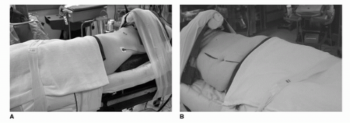
FIGURE 44B.1. Patient position. (A) The patient is secured to the operating table at a 45-degree angle. (B) The ipsilateral arm is padded and flexed over a gel chest roll.


PREOPERATIVE EVALUATION AND PREPARATION

The preoperative evaluation for patients undergoing LRN is generally similar to the workup for any patient undergoing renal surgery. Most patients should undergo routine presurgical testing including complete blood count, metabolic panel, urine culture, chest x-ray, and electrocardiogram. With regard to the renal tumor, the urologic surgeon should ensure that adequate cross-sectional imaging (with computed tomography or magnetic resonance imaging) has been performed. Although not necessary, several centers advocate three-dimensional reconstructions to assess the vasculature and the relationship of the primary tumor with adjacent organs. Imaging is especially important to rule out or to define the proximal extent of vein thrombus. Depending on the clinical stage of the primary tumor, selected metastatic evaluation should be performed prior to surgery. In most patients, a bone scan and brain imaging need to be performed only in the presence of signs or symptoms, that is, bone pain, increased alkaline phosphatase, or abnormal neurological exam. In preparation for LRN, we recommend patients to undergo 1-day bowel prep with magnesium citrate and clear liquid diet. Although there is no significant literature to support the assertions, most laparoscopic surgeons believe that bowel preparation improves visualization during surgery and decreases time to resumption of bowel function following surgery. Finally, as per recent guidelines from the American Urological Association, antibiotic prophylaxis is indicated in all patients undergoing LRN.

In the operating room, a relatively common and almost always avoidable complication results from improper patient positioning. For all laparoscopic renal procedures, we recommend placing the patient in a modified lateral decubitus position (Fig. 44B.1). For transperitoneal procedures, we employ a modified position in which the patient is bumped 30 to
45 degrees. For retroperitoneal procedures, we place patients in the full flank position at 90 degrees. Proper padding of the patient along the entire length of the table is important along with adequate anterior and posterior support to maintain the decubitus position. In the full flank position, the table should be flexed 30 to 45 degrees to maximize the space between inferior costal margin and the superior iliac spine (very important in retroneoscopic procedures). The most vulnerable point involves positioning of the arms and the potential for brachial plexus injury. In the full 90-degree flank position, an axillary roll should be placed just caudal to the axilla (this is generally not necessary in the 45-degree modified flank position). The ipsilateral arm should be placed on a gel pad over the chest or an elevated padded arm rest depending on the degree of flank position. Care should be taken to avoid anterior flexion of the shoulder beyond 90 degrees. The contralateral arm should be placed on an arm board with proper ulnar padding. After securing the patient with 3 inch wide tape, the operating room table must be fully rotated prior to draping to ensure that the patient is stable in all positions. Finally, orogastric drainage, lower extremity sequential compression devices, and Foley catheter drainage are recommended.


TABLE 44B.1 RANDOMIZED CONTROLLED TRIALS TRANSPERITONEAL VERSUS RETROPERITONEAL NEPHRECTOMY


N


Tumor Size


OR Time


Estimated Blood Loss


Complications


Length of Stay


Nambirajan et al. (49)


p = N.S.


p = N.S.


p = N.S.


p = N.S.


p = N.S.


Transperitoneal


20


4.6 cm


181 min


179 mL


No difference


7.2 d


Retroperitoneal


20


4.3 cm


213 min


208 mL


No difference


7.6 d


Desai et al. (50)


p = N.S.


p = 0.001


p = N.S.


p = N.S.


p = N.S.


Transperitoneal


50


5.3 cm


207 min


180 mL


10%


1.8 d


Retroperitoneal


52


5.0 cm


150 min


242 mL


7.7%


1.9 d


Nadler et al. (51)


p = N.S.


p = N.S.


p = N.S.


p = N.S.


p = N.S.


Transperitoneal


11


No difference


196 min


127 mL


No difference


2.1 d


Retroperitoneal


11


No difference


185 min


107 mL


No difference


3.6 d


N.S., not significant (p > 0.05).


SURGICAL APPROACHES

Soon after the first transperitoneal laparoscopic nephrectomy was reported, Kerbl et al. described their experience with the retroperitoneal approach (26). Although authors describe some difficulty with the reduced operating space of the retroperitoneum, with more experience, the retroneoscopic approach has become a standard alternative. We prefer the transperitoneal approach for most LRN because of the more familiar anatomic landmarks and wider working space; however, for patients with extensive prior transabdominal surgery and for morbidly obese patients, the retroperitoneal approach offers distinct advantages. Therefore, ability with both approaches is necessary. Tables 44B.1 and 44B.2 list reports in the literature comparing intraoperative and postoperative parameters for transperitoneal, retroperitoneal, and hand-assisted LRN. Overall, the differences between the approaches are relatively minor and likely clinically insignificant. The retroperitoneal approach allows more direct access to the renal vasculature although only one of the trials demonstrated an operative time benefit.


TABLE 44B.2 TRIALS COMPARING CONVENTIONAL LAPAROSCOPIC VERSUS HAND-ASSISTED LAPAROSCOPIC NEPHRECTOMY


N


Tumor Size


OR Time


Estimated Blood Loss


Complications


Length of Stay


Nadler et al. (51)


p = N.S.


p = 0.01


p = N.S.


p = N.S.


p = 0.05


Laparoscopic


11


No difference


196 min


127 mL


No difference


2.1 d


Hand assisted


11


No difference


139 min


133 mL


No difference


3.4 d


Venkatesh et al. (52)


p = N.S.


p = N.S.


p = N.S.


p = N.S.


p = N.S.


Laparoscopic


12


5.8 cm


171 min


No difference


16%


2.7 d


Hand assisted


9


5.6 cm


142 min


No difference


22%


3.0 d


Matin et al. (53)a


p = N.S.


p = 0.001


p = N.S.


p = N.S.


p = 0.001


Laparoscopic


113


6.0 cm


180 min


100 mL


8%


2 d


Hand assisted


158


6.0 cm


120 min


100 mL


10%


4 d


Gabr et al. (54)a


p = 0.0003


p = N.S.


p = 0.0001


p = N.S.


p = N.S.


Laparoscopic


147


4.9 cm


209 min


283 mL


38.1%


2.4 d


Hand assisted


108


6.9 cm


220 min


406 mL


56.5%


2.8 d


a Nonrandomized, retrospective.


N.S., not significant (p >0.05).


In an effort to simplify laparoscopic nephrectomy, Nakada et al. in 1997 introduced the concept of hand-assisted transperitoneal LRN arguing that this approach would decrease the number of ports needed, decrease the operative time, and decrease the overall learning curve (27). While the hand-assisted approach may decrease the overall operative time, this benefit is counterbalanced by the increased estimated blood loss and slightly increased length of stay.


Transperitoneal Laparoscopic Nephrectomy

After the patient is placed in the modified flank position, the procedure begins with transabdominal peritoneal insufflation. While we prefer closed Veress needle insertion to
begin insufflation, two recent comprehensive reviews of the literature have failed to show a difference between the closed and direct (Hassan open) techniques with regard to prevention of major complications (28,29,30). In patients without prior umbilical incisions, we recommend transumbilical insertion of the Veress needle. After insertion, low-flow insufflation is begun to ensure initial pressures of <10 mm Hg. Once safe entry has been established, high-flow insufflation is begun to peak intra-abdominal pressure of 20 mm Hg followed by initial trocar placement. Stabilization of the abdominal wall may or may not be used during insertion of the Veress needle although routine elevation does not seem to prevent needle bowel injuries. In patients with prior abdominal surgery, the Veress needle can be inserted into any of the four quadrants furthest away from the site of prior surgery. In both the left and right upper quadrant, the needle should be inserted at least two finger breadths inferior to the costal margin in the midclavicular line. In the lower quadrants, the needle should be inserted three to four finger breadths medial to the anterior superior iliac spine. Although multiple maneuvers have been described to confirm safe intraperitoneal placement, such as, injection/aspiration of saline and the drop test, we do not employ these maneuvers routinely. Furthermore, none of these techniques have demonstrated efficacy in the literature more so than ensuring that intra-abdominal pressure on entry is <10 mm Hg. If after several attempts, safe intraperitoneal needle placement is not successful, the direct technique as described by Hasson can be performed (31). Once insufflation to 20 mm Hg is achieved, a blunt tip trocar (with or without the laparoscope) can be advanced into the abdomen. The remaining trocars are then placed under direct laparoscopic vision. For the vast majority of LRN, a three port (with additional 3-mm instrument for liver retraction on the right side) can be used. Fig. 44B.2A and B shows our typical port placements for left-and right-sided approach; however, multiple different configurations have been described in the literature. Additional trocars can be placed in the lower midline or in the subcostal space at the anterior axillary line as necessary for assistants. Once the trocars are in position, the table is fully rotated toward the contralateral side to maximize bowel displacement.


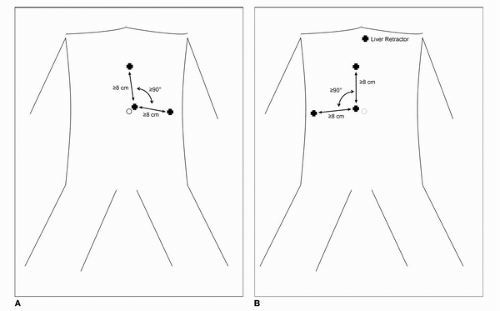
FIGURE 44B.2. (A,B) Trocar position for left and right transperitoneal laparoscopic approach. Trocars should be at least 8 cm apart. On the right side an additional trocar can be used for liver retraction.

On the left side, the line of Toldt is incised sharply from the iliac vessels inferiorly to the lateral border of the spleen superiorly. At the superior extent of this incision, careful dissection is done to avoid injury to the diaphragm or the greater curvature of the stomach. The left colon and colonic mesentery is then bluntly dissected from the anterior surface of Gerota’s fascia. Care should be taken to avoid division of the lateral attachments of Gerota’s as this complicates the exposure of the renal hilum. Complete mobilization of the mesentery results in medial displacement of the colon and exposure of the left gonadal vein and paraaortic lymphatic tissue. At the superior extent of the mesenteric mobilization, the tail of the pancreas should be recognized and bluntly mobilized medially. This plane of dissection can be followed around the superior pole of the kidney resulting in division of the lino-renal ligaments. At this point, the spleen and tail of the pancreas generally fall superior medially and out of the operative field. On the left side, we recommend elevating the gonadal vein at the level of the inferior pole of the kidney to expose the ureter. The entire kidney and posterior aspect of Gerota’s fascia can then be elevated off the psoas fascia. By following the gonadal vein superiorly, the left renal vein is rapidly identified. The junction of the left gonadal vein and the renal vein generally marks the junction of the left adrenal vein and often the junction of a lumbar vein. The thin veil of tissue over the anterior renal vein is then carefully incised allowing dissection superior and posterior to the
renal vein. It is during this dissection that the renal artery is encountered. We have found that the laparoscopic DeBakey instrument is especially useful during this dissection.

On the right side, the line of Toldt is incised from the iliac vessels inferiorly to the lower pole of the kidney superiorly. The line of dissection is then carried over the medial border of the kidney toward the junction of the IVC and the posterior edge of the liver. Again the colon and mesentery are bluntly dissected medially. Once the hepatic flexure is mobilized, the second portion of the duodenum is encountered and should be mobilized medially (Kocher maneuver) to expose the right renal hilum and IVC. Thermal injury to the duodenum can be devastating and, therefore, we recommend athermal sharp dissection. As on the left side, the right gonadal vein is elevated to expose the ureter at the level of the lower pole of the kidney. Again, the ureter and lower pole of the kidney is elevated off the anterior surface of the psoas fascia. However, since the gonadal vein inserts into the IVC, we recommend dropping the gonadal vein medially. The ureter can be followed superiorly to the right renal vein. Dissection of the superior edge of the right renal vein should be performed cautiously to avoid inadvertent injury to the right adrenal vein as it enters the posterior-lateral aspect of the IVC. Dissection posterior to the right renal vein generally exposes the right renal artery.

On both the right and left side, safe dissection of the renal vasculature is predicated on adequate anterior displacement of the lower pole of the kidney and resultant “stretch” on the renal vessels. Often in the case of renal vein thrombus or very large tumors, aberrant or collateral vessels arising from the lumbar vasculature can be encountered. These vessels can generally be controlled with titanium or locking clips. However, careful attention should be given to avoid excessive clips around the renal pedicle as the clips may interfere with placement and proper deployment of the endovascular stapler. Once the artery is identified, the endovascular stapler with 2.0 to 2.5 mm staples is used to ligate and divide the vessel. Similarly, the renal vein is ligated and divided. Note that complete isolation is not mandated. Under routine circumstances, we advocate individual ligation of the artery and vein; however, in situations of uncontrolled bleeding or extensive adhesions in the hilum, en bloc ligation and division can be considered. Buse et al. recently reported a prospective analysis of elective en bloc ligation during laparoscopic nephrectomy and found minimal bleeding complications and no development of clinically evident arteriovenous fistula at 12 months follow-up (32).

In the cases of renal vein or inferior vena caval thrombus, several additional points should be made. First of all, during the hilar dissection, an effort should be made to avoid unnecessary vein manipulation to minimize risk of embolism. Once the artery is divided, often the thrombus will decrease in size and proximal extent allowing for safe placement of the vascular stapler across the vein. We recommend use of laparoscopic ultrasound to define the proximal extent and to confirm complete isolation of the thrombus following placement of the stapler. In situation in which there is no adequate vein length or in which the thrombus extends just into the IVC, several maneuvers can be applied to safely excise the thrombus. The DeBakey clamp can be used to “milk” the thrombus distally into the vein. This can also be done with a vessel loop around the vein. Alternatively, a laparoscopic Satinsky clamp can be used to “side bite” the IVC and encompass the tumor thrombus with the renal vein. After excision the IVC can be repaired with nonabsorbable polypropylene suture. As with any laparoscopic procedure, the urologist should be ready to convert to an open approach in cases of adherence to the vein wall or more proximal IVC thrombus.

Following division of the vascular pedicle the upper pole of the kidney can be mobilized. On the left side, the medial border of the adrenal gland is dissected and divided carefully given the arterial blood supply from the aorta. The remaining lino-renal ligaments are divided, and finally, the lateral attachments of Gerota’s fascia are divided. On the right side, the lateral border of the IVC is mobilized and the right adrenal vein is ligated and divided. The posterior coronary hepatic ligament is divided sharply allowing dissection of the superior aspect of Gerota’s fascia. The ureter is ligated with titanium clips and divided. For mid or lower pole tumors, adrenal-sparing approaches can be performed, in which case, Gerota’s fascia is entered at the superior border of the renal vein. The wedge shaped cone of Gerota’s encompassing the adrenal gland can then be dissected and left behind.

If intact specimen extraction is to be performed (recommended), then a 10-mm or more often a 15-mm Endocatch bag (US Surgical Norwalk, Connecticut) is inserted through either the lateral trocar site or through separate stab incision in the lower midline (depending on extraction site). The specimen is then safely placed into the bag for extraction. For larger tumors, it is especially important that the tumor is inserted first; otherwise, placement of the entire specimen into the bag can be difficult. The bag can be extracted via lower midline, Pfannestiel, or muscle-splitting lower quadrant incision (Gibson incision).


Hand-Assisted Laparoscopic Nephrectomy

The hand-assisted technique is almost always performed via a transperitoneal approach. The patient is placed in a modified flank position and a 7 to 8 cm incision is made at the side of the hand port. There are several hand ports commercially available, but we prefer the Gelport (Applied Medical, Rancho Santa Margarita, California) since it allows removal of the hand without loss of the pneumoperitoneum. Most surgeons prefer to use the nondominant hand inside the abdomen. Figure 44B.3 shows hand port and trocar position for a right hand-dominant surgeon for both the left and right sides. Multiple different configurations have been described in the literature. Once the trocars are in position, the dissection is carried out in similar technique as described above. Generally, the hand inside the abdomen is used for retraction and exposure of dissection planes. In the hilum, palpation of the renal artery can facilitate early identification. Furthermore, with the vessels on stretch, the hand can guide the tip of the instrument into the proper plane between the vein and the artery. We always recommend placing the specimen in the Endocatch bag as hand port site metastasis has been reported following removal of the specimen via unprotected wound edges (33). A note of caution should be taken regarding risk of incisional hernia from the hand port site which seems to be higher than with conventional laparoscopic approaches (34).


Retroperitoneal Laparoscopic Nephrectomy

As mentioned above, the retroperitoneal approach is useful in patients with extensive prior abdominal surgical history to avoid potential adhesions to the intra-abdominal space. In addition, for morbidly obese patients, this approach may allow more direct and easy access to the kidney. For all retroneoscopic procedures, the patient should be placed in the full flank position (90 degrees) with the flank positioned above the kidney rest at the break of the table. After proper positioning and securing of the patient, the tip of the 12th rib is palpated and a 1.5 cm incision is made just off the tip. The incision is deepened under direct vision with direct opening of the fascia. A finger is then inserted beyond the fascia and a small space is bluntly dissected in the retroperitoneum. Once dissected, the surgeon’s finger should feel the psoas fascia and the lower pole of the kidney covered by Gerota’s fascia. A Balloon Dissector (Covidien, Mansfield, Massachusetts) is then inserted via the incision into the space and inflated with approximately 800 mL of air
(30-40 pumps). The advantage of the commercially available device is that a laparoscope can be inserted via the dissector for direct visualization of the balloon dissection. This process is repeated in the cephalad direction to ensure adequate development of the retroperitoneal space. A blunt tip trocar (Covidien, Mansfield, Massachusetts) is then inserted to seal the incision site. The first working port is placed directly posterior to the middle port just lateral to the paraspinous muscles. Through this port, a blunt instrument is used to mobilize the peritoneum medially for placement of the second working trocar. Figure 44B.4 shows patient with trocars in place.


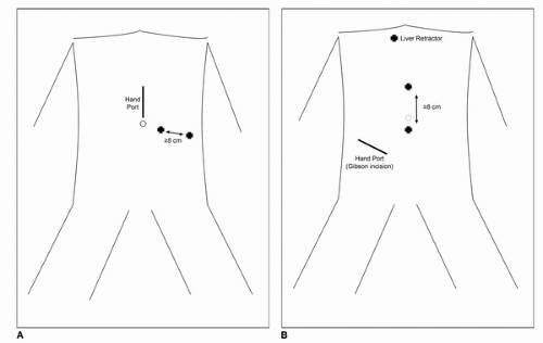
FIGURE 44B.3. A: Hand port and trocar position for left hand-assisted laparoscopic approach for a right handed surgeon with nondominant hand in the abdomen. B: Hand port and trocar position for right hand-assisted laparoscopic approach for a right handed surgeon with nondominant hand in the abdomen.


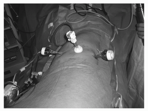
FIGURE 44B.4. Patient with trocars in place for retroneoscopic approach. Camera trocar is placed just off the tip of the 12th rib.

Once the trocars are in place, the initial dissection is aimed at adequately exposing the psoas muscle and tendon. This is the key to successful retroneoscopic surgery as the proper spatial orientation of the psoas maintains the orientation of the kidney. Unlike the transperitoneal approach, the technique is virtually the same on the right and left side. Anterior medial to the psoas the ureter is identified coursing through the retroperitoneal fat. The lower pole of the kidney is then identified and elevated anteriorly placing stretch on the renal hilum. At this point, the pulsations of the renal artery are evident in the hilum. With the posterior approach, the veil of adventitial and lymphatic tissue overlying the renal artery can be easily dissected. The artery is mobilized and divided using the endovascular stapler. With the artery divided, the renal vein comes into view and similarly it is ligated and divided. The ureter and gonadal vessels are divided. The kidney is then mobilized in the retroperitoneum from the posterior peritoneal surface and the psoas muscle. Again, the decision for adrenal sparing is made based on the tumor size and location. Once the kidney is freed, the specimen is placed into the Endocatch bag and extracted through a Gibson or Pfannestiel (extraperitoneal) incision.


ONCOLOGIC AND OPERATIVE RESULTS

Although the technique was first reported in 1991, LRN did not become more widely accepted until the late 1990s when the first multi-institutional large series with intermediate-term follow-up was published. Cadeddu et al. reported on
157 patients from five institutions undergoing LRN with mean follow-up of 19.2 months (with 51 patients with at least 24 months follow-up) (35). Overall, the authors reported at 91% 5-year actuarial disease-free survival for the entire cohort, 89% for clinical stage T2, and 86.9% for high-grade clinical stage T2 patients. With a modest complication rate of 9.6%, this report established the safety and short-term oncologic efficacy of LRN in the treatment of RCC. Since then, multiple single institution series have established similar long-term oncologic outcomes for LRN when compared to ORN (Table 44B.3). As such, in many academic centers, LRN has become the standard of care for surgical extirpation of RCC that is not amenable to nephron-sparing surgery. Contemporary series of LRN have focused on outcomes associated with high-risk tumors (Table 44B.4). Again, long-term follow-up demonstrates similar cancer-specific survival compared to historical controls of ORN. Recently, Berger et al. reported a multi-institutional series of LRN with follow-up of 11.2 years, by far the longest follow-up in the literature (36). With a mean age of 60 years and mean tumor size 5 cm, the authors reported an actual 10-year recurrence-free, cancer-specific, and overall survival of 86%, 92%, and 65%, respectively. These figures are comparable to established ORN series.


TABLE 44B.3 LONG-TERM OUTCOMES LAPAROSCOPIC VERSUS ORN


N
LRN Vs. ORN


Eligibility
LRN Vs. ORN


Tumor Size
LRN Vs. ORN


Follow-up
LRN Vs. ORN


Outcome


Luo et al. (55)


142 vs. 194


T1-T3


a


44 mo


7 yr CSS
92.5% vs. 91.4%


Bensalah et al. (56)


44 vs. 135


T3a/b


5.0 vs. 5.3 cm


28 vs. 55 mo


No difference in CSS


Colombo et al. (57)


63 vs. 53


T1-T3


5.4 vs. 6.4 cm


65 vs. 78 mo


7 yr CSS
91% vs. 93%


Chung et al. (58)


54 vs. 70


T1-T2


5.2 vs. 5.3 cm


44 vs. 68 mo


5 yr CSS
93.8% vs. 94%


Hemal et al. (11)


41 vs. 71


≥T2


9.9 vs. 10.1 cm


51 vs. 57 mo


5 yr CSS
95.1% vs. 94.4%


Kawauchi et al. (59)


123 vs. 70


T1-T2


4.4 vs. 4.4 cm


41 vs. 75 mo


5 yr CSS
92% vs. 94%


Harano et al. (60)


96 vs. 86


T1-T2


4.3 vs. 4.9 cm


25 vs. 86 mo


4 yr CSS
88% vs. 93%


Portis et al. (61)


64 vs. 69


T1-T3


4.3 vs. 6.2 cm


54 vs. 69 mo


5 yr CSS
98% vs. 92%


a Tumor size not available.


CSS, cancer-specific survival.


With regard to perioperative and quality-of-life outcomes, most historical series reported longer operative times and decreased blood loss with LRN. There has been one randomized trial evaluating open versus laparoscopic nephrectomy for pathological disease. Burgess et al. randomized 45 patients presenting for nephrectomy for both benign and malignant conditions (37). Their results revealed no difference in operative parameters (time, blood loss, and complications) and significantly decreased postoperative pain scores and earlier return to normal activities in the laparoscopic group. Harryman et al. prospectively analyzed a nonrandomized series of 100 patients undergoing open and laparoscopic nephrectomy (38). The laparoscopic group despite a higher comorbidity symptom score had a significantly shorter length of stay (4 vs. 6 days, p < 0.001) and significantly lower pain visual analog scale scores 1 month postoperatively (p = 0.007). Although certainly a different cohort of patients, some conclusions can be drawn from the laparoscopic donor nephrectomy literature. A recent meta-analysis and randomized control trial confirmed that patients undergoing laparoscopic donor nephrectomy had significantly less pain scores and analgesic requirements (39,40). Perhaps more importantly, in a recent randomized trial, the laparoscopic cohort had significantly decreased rate of complications per donor (0.3 vs. 0.6, p = 0.033) and earlier actual return to work (42 vs. 67 days, p = 0.004) (40).


TABLE 44B.4 EFFICACY OF LRN FOR HIGH-RISK TUMORS (≥PT2)


N


Stage


Follow-up


Outcome


Luo et al. (55)


75


≥T2


44 mo


Mean CSS 69 mo


Rosoff et al. (62)


30


≥T2


30 mo


CSS at 30 mo 97%


Gabr et al. (54)


64


Intermediate riska


35 mo


5 yr CSS 88.3%


Bensalah et al. (56)


44


T3a/T3b


28 mo


5 yr CSS >80%


Hemal et al. (11)


41


≥T2


51 mo


5 yr CSS 95.1%


CSS, cancer-specific survival.


The first port site recurrence was reported by Fentie et al. in a patient with a large, high-grade renal cell carcinoma with sarcomatoid features (41). In that particular case, the tumor was morcellated without leakage of contents. There are now a total of 10 cases of port site recurrences with renal cell carcinoma including three cases following laparoscopic partial nephrectomy (42,43,44). Although the exact mechanisms of port site recurrence are not known, it is believed that deviation from basic oncologic surgical principles is the common factor. Similar care in tumor manipulation as is taken with
open surgery should be followed to minimize the risks. These principles include minimizing direct tumor handling, avoidance of tumor spillage in the operative field, placement of the tumor in an impermeable sac prior to extraction, and removal of all contaminated instrumentation including the surgeon’s gloves following extraction.


COMPLICATIONS

As with any laparoscopic surgery, the risk of elective or emergent conversion to laparotomy exists and as such, laparotomy instruments including vascular clamps should be readily available. Richstone et al. recently performed an analysis of the indications and risk factors for conversion (3.3%) in a series of more than 2,000 cases (45). As would be expected, vascular injury was the most frequent cause accounting for 38.5% of conversions, followed by concern with margins in 13.5%, bowel injury in 13.5%, failure to progress in 11.5%, adhesions in 9.6%, diaphragmatic injury in 1.9%, and other in 11.5%. Intraoperative vascular injuries can occur in up to 2% of transperitoneal and retroperitoneal LRN (46,47). The immediate action for any significant vascular injury is control of the bleeding either by manual compression or by laparoscopic clamp. Depending on the injury and the skill of the surgeon, the injury can be repaired laparoscopically or via an open approach after conversion to laparotomy. The second most common significant intraoperative complication is adjacent organ injury, primarily bowel injury. Bowel injuries are most often secondary to thermal injury from excessive cautery during dissection. Early recognition and appropriate management are the keys to avoiding potentially disastrous complication. Unrecognized bowel injuries often present insidiously, and as described by Bishoff et al., typically are associated with severe single trocar site pain (48). In their review, Bishoff et al. reported an overall 0.8% incidence of bowel injuries during laparoscopic renal surgery. Other organs at risk during LRN include the spleen, liver, and pancreas. The incidence of these injuries is low (<0.5%), but again, early recognition is the key to successful management. Pareek et al. published a comprehensive review of complication rates of laparoscopic renal surgery and reported an overall 10.7% major and 3.3% minor complication rate for LRN.


FUTURE DIRECTIONS AND CONCLUSIONS

While the early experience with LRN involved the management of early stage, localized RCC, advanced laparoscopic skill sets have pushed LRN for treatment of extremely large tumors (up to 25 cm), tumors with renal and minimal IVC vein involvement, and tumors with extensive retroperitoneal lymphadenopathy. While the indications for LRN may not change, the future directions lie in access. There are several reports of laparo-endoscopic single site nephrectomy and even natural orifice transluminal endoscopic surgery transvaginal nephrectomy. At the present time, LRN is established as the standard of care and in experienced hands, the preferred method of extirpation in the treatment of renal cell carcinoma.


References

1. Clayman RV, Kavoussi LR, Soper NJ, et al. Laparoscopic nephrectomy. N Engl J Med 1991;324(19):1370-1371.

2. Clayman RV, Kavoussi LR, Soper NJ, et al. Laparoscopic nephrectomy: initial case report. J Urol 1991;146(2):278-282.

3. Miller DC, Taub DA, Dunn RL, et al. Laparoscopy for renal cell carcinoma: diffusion versus regionalization? J Urol 2006;176(3):1102-1106; discussion 1106-1107.

4. Miller DC, Saigal CS, Banerjee M, et al. Diffusion of surgical innovation among patients with kidney cancer. Cancer 2008;112(8):1708-1717.

5. Yabroff KR, Harlan L, Stevens J, et al. Patterns of care in adults with renal cell carcinoma: findings in a population based sample. J Urol 2009;181(6):2462-2468; discussion 2469.

6. Varkarakis IM, Bhayani SB, Allaf ME, et al. Laparoscopic-assisted nephrectomy with inferior vena cava tumor thrombectomy: preliminary results. Urology 2004;64(5):925-929.

7. Steinberg AP, Finelli A, Desai MM, et al. Laparoscopic radical nephrectomy for large (greater than 7 cm, T2) renal tumors. J Urol 2004;172(6 Pt 1):2172-2176.

8. Bird VG, Shields JM, Aziz M, et al. Laparoscopic radical nephrectomy for patients with T2 and T3 renal-cell carcinoma: evaluation of perioperative outcomes. J Endourol 2009;23(9):1527-1533.

9. Conley SP, Humphreys MR, Desai PJ, et al. Laparoscopic radical nephrectomy for very large renal tumors (> or =10 cm): is there a size limit? J Endourol 2009;23(1):57-61.

10. Gong EM, Lyon MB, Orvieto MA, et al. Laparoscopic radical nephrectomy: comparison of clinical Stage T1 and T2 renal tumors. Urology 2006;68(6):1183-1187.

11. Hemal AK, Kumar A, Kumar R, et al. Laparoscopic versus open radical nephrectomy for large renal tumors: a long-term prospective comparison. J Urol 2007;177(3):862-866.

12. Desai MM, Gill IS, Ramani AP, et al. Laparoscopic radical nephrectomy for cancer with level I renal vein involvement. J Urol 2003;169(2):487-491.

13. Guzzo TJ, Schaeffer EM, McNeil BK, et al. Laparoscopic radical nephrectomy for patients with pathologic T3b renal-cell carcinoma: the Johns Hopkins experience. J Endourol 2009;23(1):63-67.

14. Martin GL, Castle EP, Martin AD, et al. Outcomes of laparoscopic radical nephrectomy in the setting of vena caval and renal vein thrombus: seven-year experience. J Endourol 2008;22(8):1681-1685.

15. Chapman TN, Sharma S, Zhang S, et al. Laparoscopic lymph node dissection in clinically node-negative patients undergoing laparoscopic nephrectomy for renal carcinoma. Urology 2008;71(2):287-291.

16. Simmons MN, Kaouk J, Gill IS, et al. Laparoscopic radical nephrectomy with hilar lymph node dissection in patients with advanced renal cell carcinoma. Urology 2007;70(1):43-46.

17. Eisenberg MS, Meng MV, Master VA, et al. Laparoscopic versus open cytoreductive nephrectomy in advanced renal-cell carcinoma. J Endourol 2006;20(7):504-508.

18. Rabets JC, Kaouk J, Fergany A, et al. Laparoscopic versus open cytoreductive nephrectomy for metastatic renal cell carcinoma. Urology 2004;64(5):930-934.

19. Walther MM, Lyne JC, Libutti SK, et al. Laparoscopic cytoreductive nephrectomy as preparation for administration of systemic interleukin-2 in the treatment of metastatic renal cell carcinoma: a pilot study. Urology 1999;53(3):496-501.

20. Go AS, Chertow GM, Fan D, et al. Chronic kidney disease and the risks of death, cardiovascular events, and hospitalization. N Engl J Med 2004;351(13):1296-1305.

21. Huang WC, Levey AS, Serio AM, et al. Chronic kidney disease after nephrectomy in patients with renal cortical tumours: a retrospective cohort study. Lancet Oncol 2006;7(9):735-740.

22. Parsons JK, Jarrett TJ, Chow GK, et al. The effect of previous abdominal surgery on urological laparoscopy. J Urol 2002;168(6):2387-2390.

23. Fazeli-Matin S, Gill IS, Hsu TH, et al. Laparoscopic renal and adrenal surgery in obese patients: comparison to open surgery. J Urol 1999;162(3 Pt 1):665-669.

24. Schwenk W, Bohm B, Witt C, et al. Pulmonary function following laparoscopic or conventional colorectal resection: a randomized controlled evaluation. Arch Surg 1999;134(1):6-12; discussion 13.

25. Grabowski JE, Talamini MA. Physiological effects of pneumoperitoneum. J Gastrointest Surg 2009;13(5):1009-1016.

26. Kerbl K, Figenshau RS, Clayman RV, et al. Retroperitoneal laparoscopic nephrectomy: laboratory and clinical experience. J Endourol 1993;7(1):23-26.

27. Nakada SY, Moon TD, Gist M, et al. Use of the pneumo sleeve as an adjunct in laparoscopic nephrectomy. Urology 1997;49(4):612-613.

28. Neudecker J, Sauerland S, Neugebauer E, et al. The European Association for Endoscopic Surgery clinical practice guideline on the pneumoperitoneum for laparoscopic surgery. Surg Endosc 2002;16(7):1121-1143.

29. Ahmad G, Duffy JM, Phillips K, et al. Laparoscopic entry techniques. Cochrane Database Syst Rev 2008(2):CD006583.

30. Vilos GA, Ternamian A, Dempster J, et al. Laparoscopic entry: a review of techniques, technologies, and complications. J Obstet Gynaecol Can 2007;29(5):433-465.


31. Hasson HM. A modified instrument and method for laparoscopy. Am J Obstet Gynecol 1971;110(6):886-887.

32. Buse S, Gilfrich C, Pfitzenmaier J, et al. En bloc stapler ligation of the renal vascular pedicle during laparoscopic nephrectomy. BJU Int 2008;101(7):878-882.

33. Chen YT, Yang SS, Hsieh CH, et al. Hand port-site metastasis of renal-cell carcinoma following hand-assisted laparoscopic radical nephrectomy: case report. J Endourol 2003;17(9):771-775.

34. Montgomery JS, Johnston WK 3rd, Wolf JS Jr. Wound complications after hand assisted laparoscopic surgery. J Urol 2005;174(6):2226-2230.

35. Cadeddu JA, Ono Y, Clayman RV, et al. Laparoscopic nephrectomy for renal cell cancer: evaluation of efficacy and safety: a multicenter experience. Urology 1998;52(5):773-777.

36. Berger A, Brandina R, Atalla MA, et al. Laparoscopic radical nephrectomy for renal cell carcinoma: oncological outcomes at 10 years or more. J Urol 2009;182(5):2172-2176.

37. Burgess NA, Koo BC, Calvert RC, et al. Randomized trial of laparoscopic v open nephrectomy. J Endourol 2007;21(6):610-613.

38. Harryman OA, Davenport K, Keoghane S, et al. A comparative study of quality of life issues relating to open versus laparoscopic nephrectomy: a prospective pragmatic study. J Urol 2009;181(3):998-1003; discussion 1003.

39. Antcliffe D, Nanidis TG, Darzi AW, et al. A meta-analysis of mini-open versus standard open and laparoscopic living donor nephrectomy. Transpl Int 2009;22(4):463-474.

40. Nicholson ML, Kaushik M, Lewis GR, et al. Randomized clinical trial of laparoscopic versus open donor nephrectomy. Br J Surg;97(1):21-28.

41. Fentie DD, Barrett PH, Taranger LA. Metastatic renal cell cancer after laparoscopic radical nephrectomy: long-term follow-up. J Endourol 2000;14(5):407-411.

42. Castillo OA, Vitagliano G, Diaz M, et al. Port-site metastasis after laparoscopic partial nephrectomy: case report and literature review. J Endourol 2007;21(4):404-407.

43. Greco F, Wagner S, Reichelt O, et al. Huge isolated port-site recurrence after laparoscopic partial nephrectomy: a case report. Eur Urol 2009;56(4):737-739.

44. Masterson TA, Russo P. A case of port-site recurrence and locoregional metastasis after laparoscopic partial nephrectomy. Nat Clin Pract Urol 2008;5(6):345-349.

45. Richstone L, Seideman C, Baldinger L, et al. Conversion during laparoscopic surgery: frequency, indications and risk factors. J Urol 2008;180(3):855-859.

46. Breda A, Finelli A, Janetschek G, et al. Complications of laparoscopic surgery for renal masses: prevention, management, and comparison with the open experience. Eur Urol 2009;55(4):836-850.

47. Meraney AM, Samee AA, Gill IS. Vascular and bowel complications during retroperitoneal laparoscopic surgery. J Urol 2002;168(5):1941-1944.

48. Bishoff JT, Allaf ME, Kirkels W, et al. Laparoscopic bowel injury: incidence and clinical presentation. J Urol 1999;161(3): 887-890.

49. Nambirajan T, Jeschke S, Al-Zahrani H, et al. Prospective, randomized controlled study: transperitoneal laparoscopic versus retroperitoneoscopic radical nephrectomy. Urology 2004; 64(5):919-924.

50. Desai MM, Strzempkowski B, Matin SF, et al. Prospective randomized comparison of transperitoneal versus retroperitoneal laparoscopic radical nephrectomy. J Urol 2005;173(1):38-41.

51. Nadler RB, Loeb S, Clemens JQ, et al. A prospective study of laparoscopic radical nephrectomy for T1 tumors—is transperitoneal, retroperitoneal or hand assisted the best approach? J Urol 2006;175(4):1230-1233; discussion 1234.

52. Venkatesh R, Belani JS, Chen C, et al. Prospective randomized comparison of laparoscopic and hand-assisted laparoscopic radical nephrectomy. Urology 2007;70(5):873-877.

53. Matin SF, Dhanani N, Acosta M, et al. Conventional and hand-assisted laparoscopic radical nephrectomy: comparative analysis of 271 cases. J Endourol 2006;20(11):891-894.

54. Gabr AH, Gdor Y, Strope SA, et al. Patient and pathologic correlates with perioperative and long-term outcomes of laparoscopic radical nephrectomy. Urology 2009;74(3):635-640.

55. Luo JH, Zhou FJ, Xie D, et al. Analysis of long-term survival in patients with localized renal cell carcinoma: laparoscopic versus open radical nephrectomy. World J Urol 2010;28(3):289-93.

56. Bensalah K, Salomon L, Lang H, et al. Survival of patients with nonmetastatic pT3 renal tumours: a matched comparison of laparoscopic vs open radical nephrectomy. BJU Int 2009;104(11):1714-7.

57. Colombo JR Jr, Haber GP, Jelovsek JE, et al. Seven years after laparoscopic radical nephrectomy: oncologic and renal functional outcomes. Urology 2008;71(6):1149-1154.

58. Chung SD, Huang KH, Lai MK, et al. Long-term follow-up of hand-assisted laparoscopic radical nephrectomy for organ-confined renal cell carcinoma. Urology 2007;69(4):652-655.

59. Kawauchi A, YonedalK, Fujito A, et al. Oncologic outcome of hand-assisted laparoscopic radical nephrectomy. Urology 2007;69(1):53-56.

60. Harano M, Eto M, Omoto K, et al. Long-term outcome of hand-assisted laparoscopic radical nephrectomy for localized stage T1/T2 renal-cell carcinoma. J Endourol. Sep 2005;19(7):803-807.

61. Portis AJ, Yan Y, Landman J, et al. Long-term followup after laparoscopic radical nephrectomy. J Urol 2002;167(3):1257-1262.

62. Rosoff JS, Raman JD, Sosa RE, et al. Laparoscopic radical nephrectomy for renal masses 7 centimeters or larger. JSLS 2009;13(2):148-153.


▪ 44C Open Partial Nephrectomy

Paul Russo


INTRODUCTION

Historically, partial nephrectomy (PN) was performed under restricted, essential conditions for patients with a tumor in a solitary kidney, bilateral kidney tumors, or for patients with chronic renal insufficiency due to intrinsic renal dysfunction or calculus disease. Due to the increased use of cross-sectional imaging for nonspecific musculoskeletal or abdominal complaints or during unrelated cancer care, approximately 70% of renal tumors are now detected incidentally at a small size (<4 cm). Although the traditional radical nephrectomy (RN) was liberally used to resect these small tumors in patients with a normal contra lateral kidney, the realization that at least 20% of these tumors were benign and 25% were indolent coupled with equivalent oncological outcomes whether RN or PN was performed leads to the current era of kidney or nephron sparing surgery. Recent data associating RN with the development of chronic kidney disease (CKD), cardiovascular morbidity, and worse overall survival when compared to PN have led to the recommendation by the 2009 AUA Guidelines committee that PN should be performed whenever technically feasible for the management of the T1 renal mass. In this chapter, a discussion of the pathological, oncological, and medical rationale for PN is ensued. In addition, a full discussion of intraoperative and perioperative management of patients undergoing PN is presented.


THE OPEN PARTIAL NEPHRECTOMY: HISTORICAL EVOLUTION

PN was first performed in 1887 when Czerny resected an angiosarcoma from the kidney of a 30-year-old man. Subsequent animal experiments demonstrated that gentle pressure could control bleeding and that suture approximation of the kidney could lead to primary kidney healing often without urinary fistula. Important observations concerning compensatory hypertrophy and the minimal amount of overall renal tissue required to support life were also described. In the 1930s, pathological studies indicated that only 7% of renal cancers <5 cm metastasized compared to 83% >10 cm, and it was observed that the local growth pattern of renal tumors was expansile from the renal cortex with rare adjacent organ invasion. In 1950, Vermooten proposed a 1-cm margin as adequate to achieve local tumor control when he performed a 10-cm PN. As more surgeons attempted PN, enthusiasm for the operation waned because of bleeding and urinary fistula
complications, while RN enjoyed increasing success especially when performed by urologists (1,2).

Progress in open stone surgery in the 1960s and 1970s, an enhanced understanding of renal vascular and collecting system anatomy, utilization of techniques such as the Gil Vernet extended pyelolithotomy, anatrophic nephrolithotomy, and PN for urinary calculus led to a resurgence of interest in PN for renal tumors. Elaborate collecting system repairs of the kidney, kidney “splits” using ice slush renoprotection, and methods to drain and stent the kidney were also described (3,4,5). In the 1980s, kidney trauma surgeons described techniques to control major bleeding from penetrating injuries including early vascular control of the renal hilum, complete exposure of the kidney, partial polar nephrectomy, collecting system repair, omental pedicle flap to augment repairs, absorbable gelatin sponge bolsters to prevent tearing of the renal capsule during renorrhaphy, and direct vascular repair of injuries to the renal vein and artery (6). These techniques are fully integrated into contemporary PN, particularly in the resection of endophytic and perihilar renal tumors.

Modern radiological imaging techniques of CT, MRI, and renal ultrasound, often performed to evaluate abdominal and musculoskeletal complaints, created a new class of small, incidentally detected renal masses far different from the massive, symptomatic, and often metastatic tumors common earlier in the 20th century (7). Also, a shift in the principles of surgical oncology was occurring away from the radical Halstedian view toward one of organ preservation in the treatment of such malignancies as breast cancer and extremity sarcoma. Small, incidentally discovered kidney tumors were perfectly suitable for an organ-sparing operation. Concerns for local tumor recurrence and the observation of small satellite tumors seen in RN specimens were voiced by skeptics as major objections to elective PN.

The phrase “nephron sparing” was introduced by Licht and Novick in 1993 in a report of 241 patients who underwent PN despite a normal contra lateral kidney from 1967 to 1991. With a median tumor size of 3.5 cm and a median follow-up of 3 years, only two local recurrences were reported and survival was 95% (8). Similar results were reported by Herr (9), and long-term follow-up from the Cleveland Clinic group indicated that elective PN was safe and effective (10). The use of hemostatic agents, argon beam coagulation for the cut renal surface, and intraoperative ultrasound gave renal surgeons more tools to safely approach PN. Ice slush renoprotection allowed for resection of endophytic tumors, renal sinus tumors, and tumors in a solitary kidney. In the last decade, an enhanced understanding of the diversity of renal cortical tumors (RCTs) histological subtypes and their variable metastatic threats coupled with concerns that RN could cause CKD made the case for PN even stronger.


SMALL RENAL TUMORS: PATHOLOGICAL AND ONCOLOGICAL RATIONALE FOR PARTIAL NEPHRECTOMY

RCTs are members of a complex family with unique histologies, cytogenetic defects, and variable metastatic potentials ranging from the benign oncocytoma and metanephric adenoma, to the indolent papillary and chromophobe carcinomas, to the more potentially malignant conventional clear cell carcinoma (11,12). Of 1,863 surgically treated patients with malignant RCTs (excluding benign tumors such as oncocytoma) at MSKCC from 1989 to 2006, 72% were conventional clear cell carcinoma, 17% were papillary carcinoma, and 12% were chromophobe carcinoma. Of these, 187 patients developed metastasis, 161 (86%) had the conventional clear cell carcinoma, 17 (9.1%) had papillary renal cell carcinoma, and 9 (4.8%) had chromophobe carcinoma. On a multivariate analysis, chromophobe (HR 0.40) and papillary carcinomas (HR 0.62) were significantly associated with a better prognosis than clear cell carcinoma (13).

An active surveillance study of small renal tumors pooled 234 patients from nine centers, followed for a mean of 34 months, indicated a mean growth rate of 0.28 cm/year (14). In another study, 36% of tumors under active surveillance had zero growth, but this feature alone did not predict for benign tumor histology in cases where tissue was obtained (15). Despite modern imaging techniques, none could predict tumor histology and between 16.4% and 23% of resected tumors were ultimately benign (16,17,18). Even a renal mass without evidence of macroscopic fat on noncontrast CT, despite a highly suspicious appearance, can still be benign angiomyolipoma (19). Of 2,675 tumors resected at MSKCC, the incidence of benign tumors decreased from 38% for tumors of 1 cm or less to 7% for tumors of 7 cm or greater, and the incidence of high-grade clear cell carcinoma increased from 0% for tumors of 1 cm or less to 59% for tumors of 7 cm or greater (20). Only 1 patient in 781 with a tumor <3 cm presented to the MSKCC Urology Service with metastatic disease (21). When taken together, the bulk of the clinical evidence now suggests that for small renal tumors referred to urologists without evidence of metastases, particularly those of <3 cm, the likelihood of a low grade, benign or indolent tumor with limited metastatic potential is extremely high, creating the perfect conditions to utilize PN as means of achieving the dual goals of local tumor control and maximum organ preservation. Alternatively, these above-described data linking small renal masses with a generally nonaggressive natural history provide an excellent basis for active surveillance strategies in elderly or comorbidly ill patients (22,23). The historical dual use of RN to achieve a diagnosis and treat the renal tumor must now be considered surgical overkill and potentially deleterious to the patient with the small renal mass.

Reports indicated that PN was not compromising the local tumor control or metastasis-free survival when compared to RN for patients with T1a renal tumors (4 cm or less) across all histological subtypes (24,25,26,27) (Fig. 44C.1). The rationale for expanding PN to larger RCTs of 4 to 7 cm was articulated (28) and initial reports were met with similarly favorable results (29,30,31). Combining the Mayo Clinic and MSKCC databases, investigators evaluated 1,159 patients with renal tumor between 4 to 7 cm treated with RN (N = 873, 75%) and PN (N = 286, 25%) and demonstrated no significant difference in survival between the groups (32) (Fig. 44C.2). A series of even larger PN for T2 disease was performed in 34 patients in which 6 patients (16.2%) had benign masses and 12 patients (35%) had indolent (papillary or chromophobe) pathology. After 17 months of follow-up, 71% of patients with a malignant diagnosis are alive without evidence of disease. Although the resection of massive renal tumors (in this series up to 18 cm in diameter) is largely a function of favorable tumor location and careful case selection, it is clear that local tumor control can be effectively achieved with results similar to those found in series of tumors of 7 cm or less (33). Mayo Clinic investigators reported similar results in 276 patients with T2 or greater tumors treated with either PN (N = 69) or RN (N = 207) (34). These data indicate clearly that the ultimate oncological threat of a given tumor depends upon biological factors of the tumor including histological subtype, the presence or absence of symptoms, tumor grade, and tumor size with prognostic nomograms and algorithms available to predict outcomes with reasonable accuracy (35,36).


FIGURE 44C.1. MSKCC: DFS partial and radical nephrectomy: tumors 4 cm or less across all histologies. (Adapted from Lee CT, Katz J, Shi W, et al. Surgical management of renal tumors of 4 cm or less in a contemporary cohort. J Urol 2000;163:730-736.)


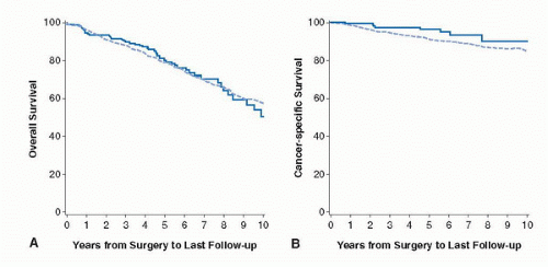
FIGURE 44C.2. PN versus RN for clear cell RCC 4 to 7 cm. (A) Overall survival in 873 patients treated with RN (dotted curve) and 286 treated with PN (solid curve) (p = 0.8). (B) Cancerspecific survival in 704 patients treated with RN and 239 treated with PN (p = 0.039.). (Adapted from Thompson HR, Siddiqui S, Lohse CM, et al. Evaluation of partial versus radical nephrectomy for renal cortical tumors 4-7 cm. J Urol 2009; 182:2601-2606.)


RENAL MEDICAL RATIONALE FOR PARTIAL NEPHRECTOMY

A historical misconception exists that RN can cause a permanent rise in serum creatinine due to the sacrifice of normal renal parenchyma not involved by tumor but will not cause serious long-term side effects as long as the patient has a normal contralateral kidney. The renal transplant literature is cited as the clinical evidence to support this view since patients undergoing donor nephrectomy have not been reported to have higher rates of kidney failure requiring dialysis or death (37). However, distinct differences between kidney donors and kidney tumor patients exist. Donors tend to be carefully screened for medical comorbidities and are generally young (age 45 or less) (38,39). In contrast, renal tumor patients are not screened, are older (mean age 61 years), and many have significant comorbidities affecting baseline kidney function including metabolic syndrome, hypertension, coronary artery disease, obesity, vascular disease, and diabetes. In addition, as patients age, particularly beyond the age of 60, nephrons atrophy and glomerular filtration rate progressively decreases (40). A study of 110 nephrectomy specimens in which the non-tumor-bearing kidney was examined demonstrated unsuspected underlying renal disease including vascular sclerosis, diabetic nephropathy, glomerular hypertrophy, mesangial expansion, and diffuse glomerulosclerosis (41). Only 10% of patients had completely normal renal tissue adjacent to the tumor.

Evidence that RN could cause a significant rise in the serum creatinine when compared to PN in patients with RCTs of 4 cm or less was published by investigators from Mayo Clinic and MSKCC in 2000 and 2002, respectively. RN patients were more likely to have elevated serum creatinine levels to >2.0 ng/mL and proteinuria (Mayo Study) (42), a persistent finding even when study patients were carefully matched for associated risk factors (MSKCC study) including diabetes, smoking history, preoperative serum creatinine, and ASA score (43). In both studies, oncological outcomes were highly favorable (>90% survival rates) whether PN or RN was done. CKD, defined as an estimated glomerular filtration rate (eGFR) of <60 mL/min/1.73 m2, is increasingly viewed as
a major public health problem in the United States, and since 2003, it is considered an independent cardiovascular risk factor (44,45,46,47,48). An estimated 19 million adults in the United States have CKD and by the year 2030, 2 million will be in need of chronic dialysis or renal transplantation (49). Traditional risk factors for CKD include age >60, hypertension, diabetes, cardiovascular disease, and family history of renal disease, factors also common in the population of patients who develop RCTs.

A study involving 1,120,295 patients demonstrated a direct correlation between CKD and rates of hospitalization, cardiovascular events, and death, which occurred before overt renal failure requiring dialysis or renal transplantation (50). As kidney function deteriorated, the percentage of patients with two associated cardiovascular risk factors increased from 34.7% (stage 1 and 2 CKD) to 83.6% (for stage 3) to 100% for stage 4 and 5 subjects. Patients with CKD are more likely to require medical interventions to treat cardiovascular disease than those with normal renal function. The low prevalence of patients with stage 4 or 5 CKD is attributable to their 5-year survival rates of only 30% (51).

A concern that the overzealous use of RN, particularly in patients with small renal masses and common comorbidities that can affect renal function, could be causing or worsening preexisting CKD became a focus of intense research. MSKCC investigators used a widely available formula, the Modification in Diet and Renal Disease (MDRD) equation (52) (http://www.nephron.com/MDRD_GFR.cgi), to estimate the glomerular filtration rate in a retrospective cohort study of 662 patients with a normal serum creatinine and two healthy kidneys who underwent either elective PN or RN for a RCT 4 cm or less in diameter. To their surprise, 171 patients (26%) had preexisting CKD (GFR < 60) prior to operation. Data were analyzed using two threshold definitions of CKD, a GFR < 60 mL/min/1.73 m2 or a GFR < 45 mL/min/1.73 m2. After surgery, the 3-year probability of freedom from new onset of GFR < 60 was 80% after PN but only 35% after RN. Corresponding values for 3-year probability of freedom from a GFR < 45, a more severe level of CKD, was 95% for PN and 64% for RN. Multivariable analysis indicated that RN was an independent risk factor for the development of new-onset CKD (53) (Fig. 44C.3).

Mayo Clinic investigators identified 648 patients from 1989 to 2003 treated with RN or PN for a solitary renal tumor ≤4 cm with a normal contra lateral kidney. In 327 patients younger than 65, it was found that RN was significantly associated with an increased risk of death, which persisted after adjusting for a year of surgery, diabetes, Charlson-Romano index, and tumor histology (32). Using the Surveillance, Epidemiology and End Results cancer registry data linked with Medicare claims, MSKCC investigators studied 2,991 patients older than 65 years for resected renal tumors of 4 cm or less from 1995 to 2002. A total of 2547 patients (81%) underwent RN and 556 patients underwent PN. During a median follow-up of 4 years, 609 patients experienced a cardiovascular event and 892 patients died. After adjusting for preoperative demographic and comorbidity variables, RN was associated with a 1.38 times increased risk of overall mortality and a 1.4 times greater number of cardiovascular events (54) (Fig. 44C.4). Similar results were reported in patients undergoing laparoscopic RN and PN (55).


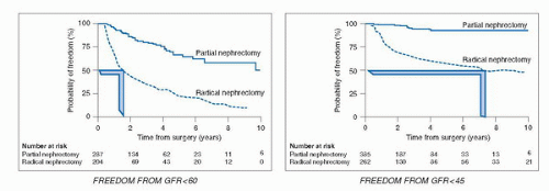
FIGURE 44C.3. Adverse impact on eGFR of radical nephrectomy versus partial nephrectomy leading to CKD status. (Adapted from Huang WC, Levey AS, Serio A, et al. Chronic kidney disease following nephrectomy for small renal cortical tumors. Lancet Oncol 2006;7:735-740.)

Because of these reports, urologists are now increasingly aware that CKD status can be created or preexisting CKD significantly worsened by the liberal use of RN for the treatment of the small renal mass (56). Short-term end points, including length of hospital stay, analgesic requirements, and cosmetic elements viewed by many as the reason to elect laparoscopic RN, must now be tempered by concerns that RN causes CKD and decreases overall patient survival. The most recent AUA guidelines for the management of the small renal tumor emphasize these points and strongly support the use of PN whenever technically feasible (57).


PARTIAL NEPHRECTOMY: PREOPERATIVE ASSESSMENT AND SURGICAL PLANNING

A careful history with a special emphasis on medical comorbidities, which can affect the cardiovascular system and kidney, including heavy cigarette smoking, hypertension, diabetes, and coronary artery disease, and a physical examination are essential elements of the initial office visit. These factors can contribute to perioperative complications and may also be etiological factors in the development of kidney cancer (58,59). Patients with cardiac disease need an evaluation including echocardiogram, stress test, and carotid duplex studies. Appropriate medical or surgical therapy of these conditions should be initiated as indicated prior to
elective kidney surgery. In addition, calculation of eGFR should be done using the following web link for the MDRD equation (http://www.nephron.com/MDRD_GFR.cgi) and in approximately 26% of patients, many of whom were previously unaware, CKD will be diagnosed (53) because of an eGFR < 60 mL/min/1.73 m2. If so, the PN would be classified as essential rather than elective. A risk stratification system, SCORED (screening for occult renal disease), was created to predict patients likely to develop CKD after kidney surgery. Patients with high SCORED values were most in need of kidney-sparing surgical approaches (60). A careful evaluation of outside imaging needs to be done. A surgeon should not operate on a renal mass without a noncontrast CT image to rule out the possibility of detecting macroscopic fat indicative of angiomyolipoma (7) and a contrast-enhanced study (MRI, CT, or renal perfusion/excretion scan) indicating bilateral renal function. Renal protocol CT imaging, which consists of three imaging sequences including precontrast, corticomedullary phase, and late nephrogenic excretory phase, provides a high degree of diagnostic accuracy in diagnosing RCTs (100% specificity, 95% sensitivity) (61) but cannot determine if the mass is benign or malignant. The degree to which a renal mass enhances is dependent on the CT scanner being utilized. For a single detector scanner, 10 HU was considered suspicious for a RCT, but with more modern multidetector scanners, “pseudo enhancement” of renal cysts secondary to volume averaging and beam hardening effects, particularly in smaller lesions, could lead to operation for a benign cyst. Enhancement of 10 to 20 HU in this setting may lead the radiologist to ask for further imaging often in the form of renal ultrasound with Doppler imaging to assess for vascular flow within the mass (62). Ultrasound can more fully characterize cystic lesions and serves as an effective template for intraoperative ultrasound, which can be highly effective in locating small subcortical renal tumors.


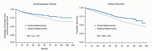
FIGURE 44C.4. Adverse impact of RN versus PN on subsequent cardiovascular events and overall survival. (Adapted from Huang W, Elkin E, Jang T, et al. Partial nephrectomy vs. radical nephrectomy in patients with small renal tumors: is there a difference in mortality and cardiovascular outcomes? J Urol 2009;181:55-62.)


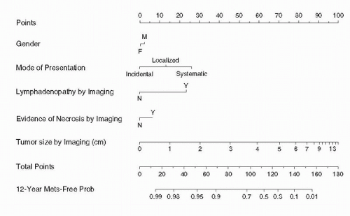
FIGURE 44C.5. Preoperative nomogram predicting 12-year metastasis-free survival based on clinical features combining data on 2,517 patients from MSKCC and the Mayo Clinic. (Adapted from Raj GV, Thompson RH, Leibovich BC, et al. Preoperative nomogram predicting 12-year probability of metastatic renal cancer. J Urol 2008;179:2146-2151.)

The surgeon should address the likelihood of executing a PN and its degree of difficulty, estimate postoperative renal functional status, carefully review the major complications related to PN (bleeding, infection, urinary fistula, and the need for a prolonged urinary drain), and discuss the likelihood of conversion to RN if, for technical reasons, a PN cannot be executed. A preoperative nomogram, derived from data on 2,517 patients, is highly effective in reassuring patients with small renal masses that a favorable long-term prognosis is achievable with an effective resection (63) (Fig. 44C.5). A scoring system (R.E.N.A.L. for Radius of tumor, Exophytic/Endophytic,
Nearness to collecting system or sinus, Anterior or posterior, and Location relative to the polar line) was developed as a means to document and describe surgical difficulty for a planned PN. Examples of scoring in the RENAL system are as follows; tumors that were 4 cm or less are given 1 point, 2 points for 4 to 7 cm, 3 points for >7 cm. Tumors that are 50% or more exophytic are assigned 1 point, tumors <50% exophytic are assigned 2 points, and tumors that are endophytic are assigned 3 points. As more points are accumulated, the degree of difficulty to perform PN increases. The authors used their system to score 50 consecutive PN and RN performed by open, laparoscopic, or robotic-assisted techniques and correlated the operative experience as the masses were deemed low (score 4-6), moderate (score 7-9), and high complexity (score 10-12). Few RN were performed by any technique for the low scored patients (64).

The likelihood of a subsequent ipsilateral (<5%) or contralateral (<5%) tumor recurrence in a patient’s lifetime is also discussed. In the face of a small renal mass, if medical comorbidities seem great, renal functional reserve minimal, and or the patient is elderly, a proposal for active surveillance can readily be made (15,23,65).


TECHNICAL FEATURES OF OPEN PARTIAL NEPHRECTOMY: SUPRA-11TH RIB MINI-FLANK SURGICAL INCISION

The traditional open PN utilized a large flank incision with resection of the distal third of the 11th rib. Although this approach provided wide exposure to the kidney and retroperitoneum, patients complained of significant postoperative pain, a prolonged recovery, and for many an uncomfortable and unsightly flank bulge usually from muscle atony from nerve damage as opposed to fascial hernia. Canadian investigators described 70 patients who underwent formal flank or thoracoabdominal incision for RN (69%) or PN (31%). Fifty percent of patients experienced a flank bulge (66). Besides the associated discomfort and poor cosmetic appearance, associated parathesias and neuralgic pain around the incision were also reported. For the surgeon, resection of the rib and closure of the large incision also added significant operating time. These significant wound difficulties were major drivers for the development of laparoscopic RN for small renal tumors and a normal contra lateral kidney in the late 1990s.


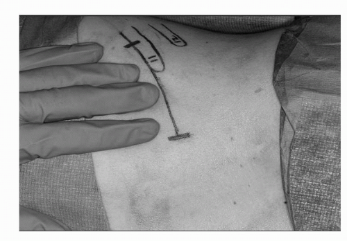
FIGURE 44C.6. Mini-flank surgical incision, approximately 8 to 10 cm, with incision above the 11th rib and in the space between the 10th rib. (Adapted from Diblasio CJ, Snyder ME, Russo P. Mini flank supra-eleventh incision for open partial or radical nephrectomy. BJU Int 2006;97:149-156.) (See color insert.)

An effective alternative to the classic flank incision or laparoscopic RN for small renal tumors is the supra-11th rib “mini-flank” retroperitoneal approach, which provides rapid and excellent exposure to the kidney without the need for a rib resection (67). With the patient in the standard flank position, an 8 to 10 cm extraperitoneal incision is performed between the bed of the 10th and 11th ribs (Fig. 44C.6). The latissimus dorsi and external oblique and internal oblique muscles are transected, and the transversus abdominus is divided in the direction of its fibers while preserving the intercostal neurovascular bundle. Using blunt dissection, the peritoneal cavity is mobilized medially, the perinephric soft tissues laterally, and the diaphragmatic fibers and pleural superiorly. A small incision in the plane between the soft tissues overlying the psoas muscle and Gerota fascia is then bluntly developed exposing the kidney, ureter, and ipsilateral great vessel (vena cava, aorta). The Bookwalter retractor (Codman and Shurtleff, Inc., Raynham, Massachusetts) is placed using the bladder blade attachment to retract the 10th rib and rib cage superiorly, while the short right-angle blade retracts the 11th rib inferiorly. A malleable blade retracts the peritoneal cavity and its intestinal contents medially. Careful dissection is conducted to isolate the ureter, renal artery, and renal vein, each of which is surrounded by a different colored vessel loop (Fig. 44C.7). On the left side, division of the gonadal vein and adrenal vein liberates the renal vein more fully, facilitates identification and isolation of the renal artery, and allows for substantial upward mobilization of the renal hilum and easy access to the entire kidney. Dense lymphatic vessels that commonly surround the renal artery are ligated and divided further facilitating upward mobilization of the kidney, which allows a decrease in venous bleeding during the tumor resection and allows easy identification and repairs of rents in renal sinus veins. After the renal hilum has been fully dissected, the upper pole of the kidney is separated from the adrenal using blunt dissection between the two and perforating vessels and soft tissues are ligated and divided.

With the kidney completely mobilized, careful palpation and inspection of its entire surface is performed in order to confirm the presence of the tumor and seek any satellite lesions. All preoperative imaging is available in the operating room and intraoperative ultrasound is routinely utilized to confirm the presence of the tumor, seek satellite lesions, and assess the proximity of the tumor to intrarenal veins and to determine if
there is an intrarenal vein thrombus or tumor encroachment upon the renal collecting system (67) (Fig. 44C.8). Occasionally, a polar segmental artery may feed the exact tumor bearing area of the kidney. Ligation and division of this artery allows for a “regional ischemia” and precise resection with little damage to non-tumor-bearing kidney. When there is a completely intrarenal tumor without any evidence on the renal cortical surface, measurements from each pole of the involved kidney in millimeters are made using the preoperative CT scan with subsequent corresponding marks made on the renal cortex, the position of the endophytic tumor is then confirmed precisely with the intraoperative ultrasound. For a purely exophytic tumor or in a patient with significant underlying CKD, resection of the tumor without renal artery occlusion is carried out. For other patients with large, endophytic, or perihilar tumors who require renal artery occlusion, renoprotective measures including mannitol infusion (12.5 g/200 mL of normal saline) and ice slush are routinely used. It is no longer necessary to place the kidney in a plastic bag prior to ice slush placement since the small surgical mini-flank incision does not lead to patient hypothermia (Fig. 44C.9). In no cases of open PN is renal artery occlusion used without such renoprotective measures recommended (warm ischemia).


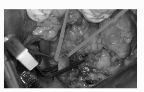
FIGURE 44C.7. The renal vein and renal artery (arteries) are carefully dissected from surrounding lymphatic soft tissues and identified by red (artery) and blue (vein) vessel loops. During cold ischemia, a bulldog vascular clamp is applied to the renal artery. (See color insert.)


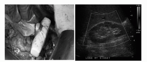
FIGURE 44C.8. Intraoperative ultrasound is routinely performed to locate endophytic tumors, assess kidney for satellite tumors, collecting system invasion, or branched renal vein thrombus invasion. (See color insert.)

Once the tumor is isolated with its surrounding perinephric fat, the renal cortex is scored with a 1-cm margin using the electrocautery (Fig. 44C.10). Sharp scissor dissection is utilized with a careful eye to keep the plain of surgical dissection within the renal cortex (pink kidney tissue) and not get too close to the renal tumor and its pseudocapsule. If dissection is too close, readjustment to a deeper plane of dissection is made. Once the renal sinus is entered beneath the tumor, 3-0 absorbable sutures are used to close any open small veins and arteries, or breaches in the collecting system both to secure these structures (Fig. 44C.11). A later search for venous bleeding can be accomplished by simply dropping the kidney into the wound and then raising it again. Once the specimen is delivered, it is
carefully inspected to be certain that the lesion is intact and that there is a complete covering layer of kidney and soft tissue. The deep tumor surgical margin is marked with a silk suture to orient the specimen, which is then delivered to the pathology department fresh and in sterile condition. Frozen section of the deep margin and specimen can provide immediate reassurance to the surgeon and the family, but a final pathological diagnosis of the renal cortical tumor may not be available by frozen section due to the need to perform immunohistochemical or cytogenetic analysis. With the aide of 2.5× loupe magnification, defects in collecting system and small vessels are easily identified and closed/ligated using 3-0 or 4-0 absorbable suture with special care taken to separately repair veins and arteries and avoid large bulky deep bites into the renal sinus, which could cause an iatrogenic a-v fistula or pseudoaneurysm.


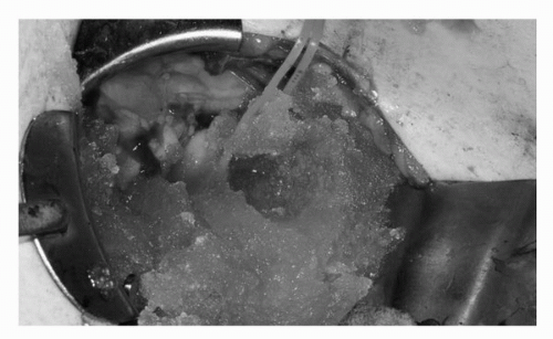
FIGURE 44C.9. Renoprotective ice slush is applied to the kidney with careful time kept to accurately record the cold ischemia time. A protective bag is no longer employed since the mini-flank incision is small and patients do not become hypothermic during the resection. (See color insert.)


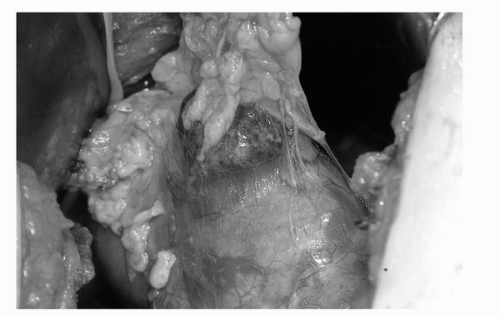
FIGURE 44C.10. Exophytic tumor with surrounding fat intact. A 1-cm margin of normal renal cortex is scored using electrocautery prior to commencing the PN. (See color insert.)


FIGURE 44C.11. Repairs are made in the collecting system and rents in renal sinus arteries and veins are repaired with absorbable 3-0 and 4-0 sutures. (See color insert.)

Endophytic tumors can emanate from elements of the renal cortex facing the renal sinus and be impalpable. Intraoperative ultrasound is essential to precisely locate the tumor and plan the nephrotomy incision. Cold ischemia with ice slush is utilized. Access to the renal sinus is achieved by going through the cortex preferably in avascular plane (Brodel’s line) whereupon the renal tumor is palpated and carefully resected with care being taken not to get too close to the tumor or enter its pseudocapsule. Following ligation of all renal sinus vessels and collecting system repair, the argon beam coagulator is used on the parenchymal surface and perinephric fat and hemostatic agents such as FloSeal (Baxter, Deerfield, Illinois) and Surgicel packing (Johnson and Johnson, New Brunswick, New Jersey) are then placed in the resection cavity. 0 chromic blunt-tipped liver sutures are then placed between pledgets of Surgicel to reapproximate the edges of renal cortex and close the resection cavity. The renal artery is unclamped and gentle pressure over the entire kidney is utilized for 3 to 5 minutes. If no bleeding is observed and the collecting system was entered, a closed suction Jackson-Pratt drain (Allegiance Healthcare, McGaw Park, Illinois) is placed through a separate stab wound in the retroperitoneal space in a dependent position posterior to the kidney. For exophytic tumors excised completely for entry into collecting system, the drain can safely be omitted. If oozing from the resection bed persists, another 5-minute period of gentle compression is applied. If brisk arterial bleeding is observed, inspection of the surgical bed and ligation of the bleeder are performed. Reclamping of the renal artery is avoided to prevent reperfusion injury to the kidney. The surgical incision is closed in two layers using No. 1 PDS, and the skin incision is reapproximated using 4-0 absorbable sutures in a subcuticular fashion.

In the first published series of 167 consecutive patients undergoing open PN (N = 133) or RN (N = 34) from 2000 to 2003 using the supra-11th mini-flank incision, excellent kidney exposure without rib exposure with decreased intraoperative EBL and length of stay (LOS) as well as better cosmetic results compared to traditional open techniques were obtained. In the original open PN group, the median LOS was 4.5 days (range 2-8) and the median EBL was 375 mL (range 50-2,000). At the median follow-up of 18 months, 3.6% of patients reported a bulge (no hernia but muscular atony) at the incision site, and one patient was diagnosed with an incisional hernia requiring surgical intervention. There were no intraoperative complications, although one patient had a prolonged hospitalization due to a concomitant urinary fistula with a urinary tract infection, which also resulted in delayed removal of the drain (67). In an update of 280 additional cases of open PNs (4/03-1/07) using the supra-11th mini-flank incision, the median LOS decreased further to 4 days (range 2-12) with a median EBL of 300 mL (range 50-3,000). There was one reported major intraoperative complication (bleeding), but it did not result in loss of the kidney. At a median follow-up of 8 months for this cohort, 1.8% of patients reported a flank bulge (68). Muscle atony/bulge at the incision site without hernia can be a disconcerting finding ameliorated or improved completely by exercises that passively twist the upper torso (using an exercise bar, broom, or golf club), which thereby strengthens collateral muscle groups leading to resolution of the bulge. For the rare flank hernia, complex repair with synthetic mesh is more effective than attempted primary repair, which is much more prone to recurrence. Today, with the added benefit of clinical pathways, LOS has been further reduced to 2.6 days.

The management of the ipsilateral adrenal gland during PN is controversial. Cleveland Clinic investigators performed ipsilateral adrenalectomy only if there was a suspicious adrenal mass on preoperative imaging or if intraoperative findings suggested a direct tumor extension from an upper pole renal mass. Ipsilateral adrenalectomy was performed only in 48/2065 PN (2.3%) with direct extension of a renal cancer found in only one patient, noncontiguous metastatic disease in two patients, and other adrenal pathology in three patients. In 42 patients (87%), the adrenal gland was benign despite an abnormal preoperative imaging appearance. During long term follow up, 15 patients underwent subsequent adrenalectomy (0.74%) revealing metastatic disease in 11 patients, 2 of which were bilateral and two of which were contralateral. The authors concluded that in the absence of abnormal preoperative imaging or obvious intraoperative findings, ipsilateral adrenalectomy is not necessary during PN (69).


RENAL ISCHEMIA DURING OPEN PARTIAL NEPHRECTOMY

With increasing PN experience, surgeons have increasingly pursued more complex PN (large, endophytic, perihilar) tumors, which usually require clamping of the renal artery to limit blood loss and facilitate necessary vascular and collecting system repairs. Renal metabolism is predominantly aerobic, and hence the kidney is highly sensitive to warm ischemic damage. Historical investigations using canine models suggested that warm ischemia could be safely tolerated for 30 to 90 minutes (70), but the extrapolation of these studies to humans, the vast majority of whom have intrinsic renal abnormalities related to comorbid diseases and the aging process (41), is speculative at best. Renal ischemia and reperfusion injury have been extensively studied in the transplant donor setting with extrapolation to the PN setting. Following renal artery occlusion, immediate complex vascular, inflammatory and sublethal injury repair responses lead to arteriolar vasoconstriction, disruption of the counter current mechanisms, and decreases in GFR and urinary production (71). Reactive oxygen radicals that result can further damage the glomerular components. Renoprotective mannitol infusion may ameliorate these effects (72). Classic studies indicating that renoprotective measures including ice slush could provide surface cooling, decrease renal energy expenditure, and ameliorate the adverse impact of warm ischemia made cold ischemia an integral part of complex open stone surgery (anatrophic nephrolithotomy) and open PN (73). This element of the PN story is further complicated because of a lack of a precise marker, either urinary or serum, for renal ischemic damage with investigators having to rely upon the imprecise serum creatinine alone to measure the effects of ischemia. Further confusing matters is the difficulty in knowing the degree to which preexisting conditions, such as hypertension, diabetes, and CKD coupled with the resection of healthy renal tissue as part of the PN, contributes to the final renal functional result.

MSKCC investigators evaluated 592 patients undergoing elective PN and separately evaluated 70 patients undergoing PN in a solitary kidney. Estimated GFR was obtained using the MDRD equation preoperatively, early in the postoperative
period, at 1 month, and at 12 months postoperatively. Patients with a solitary kidney had a baseline eGFR 30% lower than patients undergoing elective PN. Median cold ischemia time was 35 minutes in the elective PN group and 31 minutes for patients with a solitary kidney. Patients with a solitary kidney experienced a greater decline in eGFR compared to elective PN patients in the early postoperative period (30% vs. 16%), at 1 month (15% vs. 13%), and at 12 months (32% vs. 12%). Upon multivariate analysis, duration of cold ischemia and intraoperative blood loss, both likely surrogates for difficult operations, was significantly associated with early changes in eGFR but by 12 months age was the only significant predictor of eGFR decrease in patients undergoing elective PN. Although this study does not answer critical questions concerning long-term renal damage in either group of patients, it is clear that those patients with tumor in a solitary kidney and the elderly are more vulnerable to renal damage after PN (74). Investigators combined the Mayo and Cleveland Clinic 18-year experience with 458 patients undergoing PN in a solitary kidney and warm ischemia. No ischemia was used in 96 patients (21%) while 362 (79%) had a median of 21 minutes of warm ischemia. Warm ischemia patients were significantly more likely to develop acute renal failure and an eGFR of <15 mL/min/1.73 m2 and stage 4 CKD during a 3-year follow-up when compared to patients whose resection was completed without hilar clamping (75). Another multi-institutional study retrospectively evaluated 537 patients with a solitary kidney and baseline CKD indicated that the rate of chronic renal insufficiency or more severe CKD (defined as serum creatinine > 2.0 ng/mL) was 26% when no renal artery occlusion was used, 30% after warm ischemia, and 41% after cold ischemia. In this study the cold ischemia may have been selectively utilized in more challenging cases. The authors felt a cutoff of 20 minutes of warm ischemia could decrease the risk of severe CKD in this highly susceptible patient population (76).

Although, the precise degree of lasting renal damage caused by any form of ischemia and the unknown degree to which the non-tumor-bearing kidney compensates for the ischemic insult, most experts agree that working quickly in either warm or cold ischemic states is in the patient’s best interest (77). Until a more precise serum or urinary marker for ischemic renal injury is identified, the lasting impact of ischemic injury to the kidney will remain unknown. Sensible recommendations based on the literature at this time are as follows: (a) If a tumor is in an exophytic location, performing the PN without renal artery occlusion is likely to cause the least renal injury. (b) If warm ischemia is used (i.e., lap PN), tumor resection must be completed in <20 minutes. (c) For complex endophytic or perihilar tumors requiring extensive surgery and reconstruction, open PN with ice slush renoprotection, preferably with <35 minutes of cold ischemia, would lessen the likelihood of lasting renal damage. (d) For tumors in a solitary kidney in an amenable position, PN without renal artery occlusion with exchange of intraoperative blood loss for avoidance of acute renal failure and the need for postoperative dialysis is preferable. (e) For patients undergoing open PN whose surgeon requires the use of renal artery occlusion to safely complete the operation in a solitary kidney or complex nephron-sparing procedure, there is little rationale for using warm ischemia alone and cold ischemia should always be utilized.


SURGICAL MARGINS AND THE DIFFICULT PARTIAL NEPHRECTOMY

A criticism of PN relates to the need for a 1-cm surgical margin of healthy tissue surrounding the tumor. Although this belief is felt to be founded in the basic principles of surgical oncology, no firm data exist to support this view. This issue is germane particularly when surgeons pursue endophytic tumors and perihilar renal tumors, or renal tumors abutting the collecting system that would all be effectively excluded from PN if there was strict adherence to the 1-cm margin rule. Also, uncertain factors in pathology relating to the handling of the tumor specimen and fractures in its capsule may lead to an inked margin that is “positive.” MSKCC and Mayo Clinic investigators combined their data and analyzed 1,344 patients undergoing 1390 PN from 1972 to 2005. Positive surgical margins were documented in 77 cases (5.5%) and were significantly associated with decreasing tumor size and presence of a solitary kidney. Interestingly, experienced surgeons from both centers describe small endophytic tumors, many of which are not palpable and can be located only by using intraoperative ultrasound, as often difficult to find and resect and often associated with close or positive surgical margins. All patients with positive surgical margins were managed expectantly with an overall 10-year probability of freedom from local recurrence and metastatic recurrence of 93%. There was no significant difference in either local or metastatic recurrence between the patients with positive or negative surgical margins (Fig. 44C.12). Although the authors encourage a complete resection in every case, the argument for a subsequent completion nephrectomy when a positive margin is encountered on final pathology as well as a 1-cm surgical margin in all cases is unfounded (78).

During challenging PN, urological oncologists may encounter intraoperative findings that previously would have triggered RN, yet with adherence to fundamental reconstructive and vascular surgical principles, PN can still be performed. Approximately 12% of RCTs will invade the collecting system and, although this may portend a worse prognosis, complete resection of the tumor and the involved collecting system can be done with suture repair and reconstruction (up to and including dismembered pyeloplasty over an internal stent) as needed. While performing the reconstruction, care must be taken not to exclude a renal papilla from the collecting system, an event which could lead to a urinary fistula without diversionary options (79). Most such leaks eventually close spontaneously but may take weeks or even months.


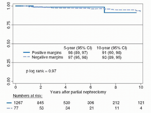
FIGURE 44C.12. Recurrence-free and metastasis-free survival based on positive or negative surgical margins. (Adapted from Yossepowitch O, Thompson HR, Leibovich BC, et al. Positive margins at partial nephrectomy: predictors and oncologic outcomes. J Urol 2008;179:2152-2157.)


For tumors close to the renal hilar vessels or projecting into the renal sinus as an endophytic extension from the renal cortex, a troubling preoperative image may look far worse than what is encountered in the operating room. Fox Chase surgeons reported a series of 36 patients with central renal tumors who underwent successful PN. In their series, six patients had benign tumors (17%), all tumors were pT1, and 34/36 (94%) were considered “low oncological risk” (80). The complete mobilization of the kidney upon the renal artery, renal vein and ureter, ligation and division of restraining polar vessels, and the use of the renal parenchymal elevating Gil Vernet type maneuver may allow ready access to the renal mass and its complete resection with minimal loss of healthy renal cortex. Also during PN, an intrarenal vein tumor thrombus in a branched renal (T3b) may be encountered. If the thrombus does not extend to the main renal vein or inferior vena cava, complete resection can be achieved by adhering to the principles of vascular surgery and obtaining proximal and distal control with care to be certain that thrombus extraction in complete. The ultimate prognosis for these more complicated central tumors following complete resection depends upon tumor histology, size, and grade. Optical loupes and liberal use of intraoperative ultrasound can aid the surgeon in these challenging cases (81).

Bilateral renal tumors are reported in between 1% and 5% of patients (82). In a study of 1,082 nonmetastatic renal tumors managed from 1989 to 2001, MSKCC investigators identified 46 patients with bilateral tumors (4.25%) of which 33 (71.7%) were synchronous and 13 (28.3%) were asynchronous. Median tumor size for the synchronous group was 3.9 cm (range 1.0-12.5 cm). The first tumor in the asynchronous group had a median tumor size of 4.75 cm (range 2.5-12.5 cm) and the second asynchronous tumor had a median tumor size of 2.25 cm (range 1-4.0 cm) occurring at a median time of 84.5 months (range 28-240 months) from the contralateral tumor. A total of 92 tumors were identified with a histological concordance rate of 76% between kidneys. Surgical management in this series was 42% RN and 58% PN with seven patients undergoing bilateral PN. The most common histological subtypes were conventional clear cell in 66% and papillary in 14%. With a median follow-up of 74 months, 72% of patients were disease free and seven patients had recurrence (two local, five metastatic). When this series of bilateral tumors, either synchronous or asynchronous, were compared to patients with unilateral disease, there was no difference in disease-specific survival (83). For patients with bilateral synchronous tumors, controversy exists among urological oncologists regarding what operation to do and in what sequence. In a follow-up study from MSKCC that focused only upon bilateral synchronous tumors, MSKCC investigators identified 73 (3%) out of 2,777 patients with bilateral synchronous tumors from 1989 to 2008. Three patients underwent bilateral RN (all before 2003), 28 patients (38%) underwent a RN followed by a PN, and 32 (44%) underwent bilateral PN. As this team became increasingly committed to maximally preserving renal function, the use of bilateral PN increased (34% from 1995-2004 to 92% from 2004-2008). Forty-five patients (62%) had the larger tumor removed first with a histological concordance rate of 70% between the kidneys (84). In this study too, there was no difference in overall survival between patients with unilateral and bilateral synchronous tumors. In general, the larger tumor is pursued first since it is associated with greater metastatic potential.

Although multifocal and bilateral tumors are part of hereditary and familial tumor syndromes such as von Hippel-Lindau disease, hereditary papillary renal cancer, and Birt-Hogg-Dube syndrome, and may account for 3% to 5% of all renal cancers (85), multifocal RCTs can also occur in sporadic renal tumor patients. MSKCC investigators evaluated 1,071 RN specimens from 1989 to 2002 and found 57 (5.3%) with pathological evidence of tumor multifocality including 6 (11%) that occurred in the bilateral synchronous setting. Preoperative imaging detected multifocality in 19 patients (33%) and therefore occult multifocality was detected 38/1071 RN (3.5%). Primary tumors in the multifocal group were conventional clear cell (51%) followed by papillary (37%) and 74% had the same tumor histology in all lesions. Multivariate analysis demonstrated that bilaterality, papillary histology, advanced tumor stage, and lymph node metastases were associated with multifocal tumors. After a median follow-up of 40.5 months, disease-free survival was not significantly different between multifocal and unifocal renal tumors (86). When faced with an index renal tumor and surrounding minute satellite tumors detected visually or with the aid of intraoperative ultrasound, the smaller tumors should be resected first without the use of cold ischemia by using a No. 15 blade on a long knife handle followed by argon beam coagulation of the renal cortical resection bases. When faced with larger tumors (>2 cm), formal PN with necessary collecting system or vascular repairs with or without cold ischemia, depending the tumor location, is performed. Multifocal tumors that can be resected completely, even those requiring numerous excisions in the same kidney, should not trigger an automatic RN.

Only gold members can continue reading. Log In or Register to continue

Stay updated, free articles. Join our Telegram channel

Jul 15, 2016 | Posted by in ONCOLOGY | Comments Off on Management of Localized Renal Cell Carcinoma

Full access? Get Clinical Tree

Get Clinical Tree app for offline access