The Myelodysplastic Syndromes


The Myelodysplastic Syndromes


Guillermo Garcia-Manero


INTRODUCTION

The myelodysplastic syndromes (MDS) are a very heterogeneous group of myeloid malignancies that result in bone marrow failure and peripheral blood cytopenias.1 MDS is diagnosed based on the presence of dysplastic features in the bone marrow. Diagnosis is often supported by the presence of cytogenetic alterations and, more recently, genetic mutations. The natural history of patients with MDS is also very heterogeneous, with a small subgroup of patients surviving for long periods of time with minimal intervention and other patients with very poor prognosis that succumb early to the disease either from complications of infections or bleeding or from transformation to acute myelogenous leukemia (AML).2,3 Over the last 15 years, the field of MDS has transformed from a rarely studied condition, often considered to be a preleukemia, to the focus of the work of multiple investigators around the world. These efforts have resulted in significant improvements in our understanding of MDS as well as in our ability to treat patients with this group of disorders. Here I will summarize this progress and provide concepts for future work in this area.


Clinical Presentation and Complications of Myelodysplastic Syndrome

The clinical presentation of most patients with MDS is not specific. At the present time, most patients with MDS are diagnosed after a routine examination of peripheral blood. MDS is suspected based on the presence of one or more peripheral blood cytopenias. Although there is no formal data to prove this point, it is my experience that is uncommon to diagnose patients with MDS because of critically symptomatic anemia, bleeding, or neutropenic fever.4 That said, symptoms of anemia, thrombocytopenia, fever, other constitutional symptoms, or unexplained infectious processes can lead to a diagnosis of MDS. The presence of more than one cytopenia should prompt a diagnostic bone marrow evaluation, as will be discussed below. Because the initial clinical differential diagnosis of MDS is broad and includes potentially clinically significant conditions, it is important to perform a thorough evaluation to rule out other conditions that may exclude a diagnosis of MDS or may contribute to the severity of the disease. Common examples include iron deficiency anemia from gastrointestinal sources, hemolysis, and immunemediated cytopenias. Inflammatory clinical syndromes, that can be seen, for instance, in the context of connective tissue disorders,5 are also important to be investigated in patients with suggestive clinical manifestations. Drug-induced cytopenias are not uncommon and need to be excluded.6 It is also important to realize that all these conditions can overlap. Patients with MDS can have gastrointestinal blood losses that may present with normal to mildly increased red cell volumes. A fraction of patients with MDS may also have evidence of a concomitant hemolytic process. Finally, the diagnosis of MDS should be confirmed after performing a diagnostic test that always includes evaluation of the morphology of a bone marrow specimen.

The natural history of patients with MDS has not been studied in detail. In the earlier phases of the disease it is likely that most of the complications are derived from persistent anemia, thrombocytopenia, and neutropenia. Also, a fraction of patients will progress to either higher-risk MDS or AML. The natural history of patients with higher-risk MDS is probably not different from that of patients with AML. We performed an analysis of the cause of death of patients with lower-risk MDS.3 In this study, we retrospectively analyzed the cause of death in a cohort of 273 deceased patients with lower-risk MDS that presented to MDACC from 1980 to 2004. Patients had received supportive care only. The cause of death was MDS-related in 230 of 273 patients (84%) with the most common events being infection (38%), transformation to AML (15%), and hemorrhage (13%)3 (Fig. 79.1).


Diagnostic Evaluation

A diagnosis of MDS is based on evidence of the presence of dysplastic features on examination of a bone marrow aspirate. At the present time, most investigators use criteria proposed by the World Health Organization (WHO).7 This classification has by and large replaced the prior French-American-British system (FAB) that was commonly used before.8 By FAB criteria, patients with up to 30% blasts were considered to have MDS. This percentage was decreased to 20% by the WHO classification. It should be noted that WHO should not strictly be used to make treatment decisions, as several of the drugs used in MDS, including decitabine9 and azacitidine,10 were approved based on FAB criteria. WHO classification includes the percentage of blasts in both the bone marrow and peripheral blood, the number of dysplastic lineages, and the presence of ring sideroblasts. It is recommended that 500 nucleated bone marrow cells and 200 from peripheral blood be evaluated. Dysplasia should be present in more than 10% of cells evaluated for each specific lineage. Erythroid dysplasia can affect both the nucleus and the cytoplasm, including the presence of sideroblasts, cellular vacuolization, and PAS positivity. Dysgranulopoiesis can be represented by alterations in size, nuclear hypolobation, or irregular hypersegmentation. Dysmegakaryopoiesis is often represented by the presence of micromegakaryocytes, hypolobation, or multinucleation. Examination of a bone marrow biopsy specimen, in conjunction with bone marrow aspirate, helps in evaluating bone marrow cellularity and the presence of significant fibrosis, and in making a diagnosis of hypoplastic MDS versus aplastic anemia.11

Based on morphologic criteria, MDS is subclassified into 10 categories (Table 79.1). Bone marrow and peripheral blood findings are also summarized in Table 79.1. A photomicrograph of a representative case of MDS is shown in Figure 79.2. Although morphology is fundamental for diagnosis, and morphologic subsets have different natural histories, morphology by itself is insufficient to make prognostic predictions or to select therapy. Because the diagnosis of MDS can be subjective, several investigators have noted a significant degree of discrepancy in the diagnosis of
patients with MDS.12 In a series of 915 patients with MDS referred to a tertiary care center, discordance in diagnosis using very strict criteria was documented in 12% of patients. A majority of patients were reclassified as having higher-risk disease by the International Prognostic Scoring System (IPSS).2 This has obvious implications, as most patients with higher-risk disease will be candidates for some form of therapy. Therefore, it is fundamental for the clinician to have proper documentation of the final morphologic diagnosis both for proper prediction of survival and for therapy selection.


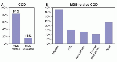
FIGURE 79.1. Cause of death in patients with myelodysplastic syndromes (MDS). A retrospective analysis was performed to determine the cause of death in patients with MDS. As shown in panel A, most patients succumb to causes intrinsic to the disease. B, The most common causes of death are infectious complications, transformation to AML, and bleeding. Adapted from Dayyani F, Conley AP, Strom SS, et al. Cause of death in patients with lowerrisk myelodysplastic syndrome. Cancer 2010;116:2174-2179.


TABLE 79.1 WORLD HEALTH ORGANIZATION (WHO) CLASSIFICATION OF MYELODYSPLASTIC SYNDROME (MDS)


Disease


Blood Findings


Bone Marrow Findings


Refractory cytopenias with unilineage dysplasia (RCUD)


Unicytopenia or bicytopenia


Unilineage dysplasia: ≥10% of the cells in one myeloid lineage


Refractory anemia (RA); Refractory neutropenia (RN)


No or rate blasts (<1%)


<5% blasts


Refractory thrombocytopenia (RT)


<15% of erythroid precursors are ring sideroblasts


Refractory anemia with ring sideroblasts (RARS)


Anemia


No blasts


≥15% of erythroid precursors are ring sideroblasts


Erythroid dysplasia only


<5% blasts


Refractory cytopenia with multilineage dysplasia (RCMD)


Cytopenia(s)


Dysplasia in ≥ 10% of the cells in ≥ two myeloid lineages (neutrophil and/or erythroid precursors and/or megakaryocytes)


No or rare blasts (<1%)


No Auer rods


<1 × 10⁹/L monocytes


<5% blasts in marrow


No Auer rods


±15% ring sideroblasts


Refractory anemia with excess blasts-1 (RAEB-1)


Cytopenia(s)


<5% blasts


No Auer rods


<1 × 10⁹/L monocytes


Unilineage or multilineage dysplasia


5%-9% blasts


No Auer rods


Refractory anemia with excess blasts-2 (RAEB-2)


Cytopenia(s)


5%-19% blasts


Auer rods ±3


<1 × 10⁹/L monocytes


Unilineage or multilineage dysplasia


10%-19% blasts


Auer rods ±3


Myelodysplastic syndrome—unclassified (MDS-U)


Cytopenias


Unequivocal dysplasia in less than 10% of cells in one or more myeloid cell lines when accompanied by a cytogenetic abnormality considered as presumptive evidence for a diagnosis of MDS (see Table 5.04 )


≤1% blasts


<5% blasts


MDS associated with isolated del(5q)


Anemia


Usually normal or increased platelet count


Normal to increased megakaryocytes with hypolobated nuclei <5% blasts


Isolated del(5q) cytogenetic abnormality


No or rate blasts (<1%)


No Auer rods


A number of diagnostic tools can help the hematopathologist confirm the diagnosis of MDS and classify the disease. The most important assay, which is mandatory in the evaluation of a patient with MDS, is cytogenetic analysis. There is no specific cytogenetic pattern diagnostic of MDS. Typically, 20 metaphases are required for optimal analysis. That said, it is not uncommon to obtain fewer cells in patients with MDS. The presence of cytogenetic alterations can have both a diagnostic and a prognostic value. For instance, in patients with profound marrow hypocellularity, the presence of
a cytogenetic alteration will allow the differentiation of hypoplastic MDS from aplastic anemia.11 Finally, and more significantly, specific cytogenetic alterations have different prognostic values. A number of cytogenetic classifications have been proposed in patients with MDS. The most recent one is a 5-subgroup classification that forms the basis of the Revised International Prognostic Scoring System (IPSS-R).13,14 This cytogenetic scoring system is summarized in Figure 79.3. It is of interest as it further delineates rare cytogenetic alterations with favorable prognostic impact, such as alterations of chromosome 11, and also further defines the weight of alterations of chromosome 7 or complex karyotypes such as those with 3 or more abnormalities. Finally, the presence of specific cytogenetic alterations may help in the selection of specific forms of therapy. A classic example is the presence of deletions of chromosome 5 in patients with the so-called 5q-syndrome.15 Other alterations, such as chromosome 7 alterations or complex karyotypes may aid the clinician in selecting different forms of therapy with different intensities, such as hypomethylating agents.10 This is discussed later. A number of groups have proposed the use of fluorescence in situ hybridization (FISH) techniques to aid in the clinical work-up of patients with MDS.16 At the M.D. Anderson Cancer Center (MDACC), we do not routinely use FISH in MDS because it evaluates only a limited number of chromosomes, and the sensitivity and specificity of the different probes used is not fully understood or standardized. The frequency of cytogenetic alterations depends on risk. For instance, patients in the lower-risk categories by IPSS are diploid in over 50% of cases.17 This figure increases in patients with more advanced forms of the disease,18 and over 70% of patients with therapy-related MDS will have a cytogenetic alteration.19


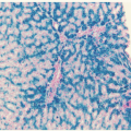
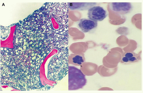
FIGURE 79.2. Representative example of morphologic alteration in MDS. Panel A shows a view of a bone marrow biopsy with almost complete replacement of marrow space with cellular element. Panel B shows a higher magnification of bone marrow aspirate, demonstrating nucleated red cell and micromegakaryocytes characteristic of this disease.


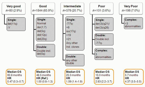
FIGURE 79.3. New 5-subgroup cytogenetic classification of myelodysplastic syndromes (MDS). This new cytogenetic classification divides patients into 5 categories based on their characteristics. Impact on survival is shown for each subset at the bottom of each subgroup. Adapted from Schanz J, et al.14 J Clin Oncol 2012;30:820-829.

Flow cytometry can help in the confirmation of a diagnosis of MDS, and specific phenotypes may have prognostic value.20 At the present time, there is no flow cytometry panel that is diagnostic of MDS, and flow cytometry cannot replace morphologic examination.21 It is not uncommon that clinicians may try to quantitate the percentage of blasts by annotating the number of CD34+ cells. Although the number of CD34+ cells has been proposed to have prognostic value,20,22 it is not an appropriate tool to estimate percentage of blasts and it should be considered only complementary.

Newer genomic technologies are currently being developed that allow the analysis of multiple genetic events in MDS and other
cancers. These include next-generation gene sequencing23 and analysis of single nucleotide polymorphisms.24 Although these assays are of great interest, they are not currently integrated into clinical practice.


Specific Diagnostic Subgroups of Myelodysplastic Syndrome-Related Syndromes: Chronic Myelomonocytic Leukemia and Overlap Myelodysplastic Myeloproliferative Syndromes

Chronic myelomonocytic leukemia (CMML) is considered a distinct clinical entity by the WHO classification and is grouped in the subset of patients with myelodysplastic myeloproliferative neoplasms. This group also includes BCR/ABL negative chronic myelogenous leukemia (CML), MDS/MPN unclassified, juvenile myelomonocytic leukemia, and potentially refractory anemia with ring sideroblasts and thrombocytosis (RARS-T). Traditionally, CMML has been considered 1 subtype of MDS. IPSS2 included patients with CMML if the white cell count was less than 12 × 103/L. From a practical and therapeutic perspective, most clinicians still consider CMML as a subtype of MDS. The natural history of patients with CMML is distinct from that of patients with classic MDS. Patients tend to have higher frequency of B symptoms and extramedullary manifestations of the disease. Tissue infiltration causing hepatic or renal dysfunction is not uncommon. Diagnosis is established by the presence of persistent monocytosis (>1 × 10⁹/L) in the peripheral blood without evidence of BCR/ABL fusion genes or PDGFR alterations. Blasts, that include promonocytes, should be less than 20% and dysplasia is routine, although often less pronounced that in other MDS categories. CMML is further divided into CMML-1 and CMML-2 based on the percentage of bone marrow and peripheral blood blasts (CMML-1 is less than 10% bone marrow blasts or less than 5% in peripheral blood; whereas CMML-2 will include cases with more blasts). Cells usually express markers of myelomonocytic differentiation that include CD33 and CD13. Cytogenetic alterations occur as in other cases of MDS, and the presence of RAS mutations can be observed in up to 40% of patients.25 Another group of disorders are the MDS/MPN unclassified (MDS/MPN-U) disorders. These are of particular interest at this time because of the advent of agents that inhibit JAK2,26 a common molecular alteration in MPN that may explain the proliferative feature of the disease. Although the natural history of patients with MDS/MPN-U is not fully understood, it appears that specific subsets of patients such as those with RARS-T may have a more benign prognosis.27


EPIDEMIOLOGY AND ETIOLOGY

Because of the heterogeneity of MDS and a prior concept that MDS was a preleukemic condition of unclear significance affecting older individuals, there are no robust mature registries of MDS, at least in the US. A number of studies have tried to delineate the incidence and prevalence of MDS. Data from the North American Association of Central Cancer Registries (NAACCR) and the Surveillance, Epidemiology and End Results (SEER) program indicate that the average annual age-adjusted incidence rate of MDS for 2001 through 2003 was 3.3 per 100,000. This translates to approximately 9,700 patients with MDS in the US per year. Of interest, incidence rates increased per year in that analysis. Of importance, only a minority of patients were reported to registries by physicians’ offices.28 After this initial study, several other groups have reported higher incidence rates in MDS. For instance, investigators using a claims-based algorithm have reported the incidence of MDS being close to 75 individuals per 100,000 in people over 60 years of age.29,30 More recently, Cogle et al. constructed 4 claims-based algorithms to assess MDS incidence and applied them to the 2000 to 2008 SEER-Medicare database.30 Using this approach, the annual incidence of MDS in the US was projected to be 75 per 100,000 persons 65 years or older, much higher than previously estimated. MDS is a disease of aging. The incidence of the disease sharply increases in patients older than 60 years of age, and the median age is 70 years.28 As the age of the population increases, it is expected that MDS may become a major medical problem, at least in developed countries.

The cause of MDS is not known, but there is strong data that suggest that MDS can also be the result of toxic exposure of bone marrow stem cells. A prototypical example is cases secondary to exposure to prior chemotherapy or radiation therapy, i.e., therapy-related (t)-MDS.19,31 Therapy-related MDS often is characterized by complex karyotypes and can occur in younger patients. Depending on the type of chemotherapy exposure, different patterns of disease evolution and cytogenetic abnormalities can be documented.31 The prognosis of patients with t-MDS is very poor, but it is unclear if this is the result of characteristics intrinsic to the disease or because a large majority of these patients have very complex karyotypes that are associated with poor prognosis. For instance, in therapy-related AML (t-AML), it has been shown that common functional p53-pathway variants such as the MDM2 SNP309 and the TP53 codon 72 polymorphism may be associated with an increased risk of developing t-AML.32 This data is of significance as it suggests that predisposing molecular features are involved in the development of therapy-related myeloid malignancies.33 At MDACC, we evaluated the characteristics of patients with t-MDS. We studied 281 patients with MDS that had received prior chemotherapy and/or radiotherapy for prior malignancy. Multivariate Cox regression analysis identified 7 factors that independently predicted short survival in t-MDS: age ≥ 65 years (HR = 1.63), ECOG performance status 2 to 4 (HR = 1.86), poor cytogenetics (-7 and/or complex; HR = 2.47), WHO MDS subtype (RARS or RAEB-1/2; HR = 1.92), hemoglobin (<11 g/dl; HR = 2.24), platelets (<50 × 109/dl; HR = 2.01), and transfusion dependency (HR = 1.59). These risk factors were used to create a prognostic model that segregated patients into three groups with distinct median overall survival: good (0 to 2 risk factors; 34 months), intermediate (3 to 4 risk factors; 12 months), and poor (5 to 7 risk factors; 5 months) (p < 0.001); and 1-year leukemia-free survival (96%, 84%, and 72%, respectively, p = 0.003). This model also identified distinct survival groups according to t-MDS therapy.

A number of epidemiologic studies have suggested that environmental factors play a role in the development of MDS. Recently, a pooled analysis studied the effects of benzene exposure in oil workers. Exposure to benzene was associated with MDS. High benzene exposure (>3 ppm) was associated with a risk of MDS (OR = 6.32, 95% CI = 1.32 to 30.2). Of interest, no association was observed with AML.34 In a hospital-based case-control study of 354 adult de novo MDS cases and 452 controls, a family history of hematopoietic cancer (odds ratio [OR] = 1.92), smoking (OR = 1.65), and exposure to agricultural chemicals (OR = 4.55) or solvents (OR = 2.05) were associated with MDS risk. For patients with lower-risk disease (RA/RARS) only smoking (OR = 2.23) and agricultural chemical exposure (OR = 5.68) were identified. For patients with higher-risk disease (RAEB/RAEBT), a family history of hematopoietic cancer (OR = 2.10), smoking (OR = 1.52), and exposure to agricultural chemicals (OR = 3.79) or solvents (OR = 2.71) were independent risk factors. Drinking wine reduced risk for all FAB types by almost 50% (OR = 0.54). A joint effect between smoking and chemical exposure was observed, with the highest risk among smokers exposed to solvents/agricultural chemicals (OR = 3.22).35

Finally, a number of genetic syndromes associated with bone marrow failure were recently associated with the development
of MDS.36 Of importance, several of these disorders are ribosomopathies characterized by altered ribosome biogenesis and function.37 Syndromes in this category include Diamond-Blackfan anemia, Schwachman-Diamond syndrome, dyskeratosis congenita, cartilage hair hypoplasia, and Treacher Collins syndrome.37 Haploinsufficiency in ribosomal genes, such as RPS14, are also implicated in the pathogenesis of the 5q-syndrome, thus providing further linkage between these conditions.38 Patients with Fanconi anemia are also at increased risk of developing MDS.39 Mutations in Runx1 have been described in MDS of patients with Fanconi anemia.40

A number of rare familial syndromes have been reported. For instance, germline mutations in Runx1 have been shown to occur in families characterized by thrombocytopenia and increased risk of developing MDS and AML.41 Mutations are more common in the DNA binding domain or N-terminus of the gene. The median incidence of MDS/AML among carriers of RUNX1 mutation was 35%.42 It should be noted that not all family members with the mutation had low platelet counts.42 It should also be noted that allogeneic stem cell transplantation (SCT) was associated with a high rate of complications.42 Therefore SCT cannot be recommended in all patients at risk. Because mutational analysis of Runx1 is not commonly performed in clinical practice, it is possible that there are more individuals and families affected by this type of familial syndrome. Younger patients with thrombocytopenia or MDS should be screened for Runx1 mutations. Germline mutations in GATA-2 have also been involved in a familial syndrome of MDS/AML, MonoMAC, and lymphedema.43 MonoMAC is an autosomal dominant syndrome associated with monocytopenia; B and NK cell lymphopenia; and mycobacterial, fungal, and viral infections. This syndrome is also associated with pulmonary alveolar proteinosis.44


Molecular Pathogenesis

The cause of MDS is not known but remains strongly linked to senescence. Over the last 5 years we have gained significant knowledge in both genetic and epigenetic alterations that characterize MDS. This information is of great significance and is going to aid not only in understanding the molecular bases of MDS but also in developing molecularly based classifications of MDS, as well as in developing new targeted interventions for patients with MDS.

The molecular analysis of MDS has been revolutionized by the advent of powerful new sequencing techniques. Using these technologies, several groups have reported a large number of genetic mutations in patients with MDS.23,45 A list is shown in Table 79.2. The most frequent events are genes involved in control of gene splicing46, 47 and 48 and epigenetic regulators49 such as TET245,50 or ASXL145,51 or EZH2.45,52 At the present time, it is not known why splicing mutations are so prevalent in MDS and what the downstream effects of these mutations are. The mutations on epigenetic regulators are of special interest. TET2 mutations were first identified in myeloid leukemia but their functional relevance or clinical impact was unknown.50,53 The presence of mutations in the TET family was rapidly confirmed by several groups.53 TET2 is located on chromosome 4q24 and has been shown to have a role in the control of DNA hydroxymethylation.54,55 Therefore it is likely that patients with mutations in TET2 will have abnormal DNA methylation patterns that could broadly impact gene expression patterns in MDS. It has been shown that TET2 has a role in the homeostasis of hematopoietic stem cells.56,57 Although the prognostic impact of TET2 is not clear at this time, data from several groups has suggested that the presence of TET2 mutations may be associated with response to azacitidine.58 EZH2 is located on chromosome 7 and is a member of the Polycomb group family. It is a histone 3 k27 methylase and therefore is also involved in the control of epigenetic gene repression. In contrast with EZH2 mutations described in lymphoma59 that are activating, EZH2 mutations in MDS inactivate the gene. Mutations in EZH2 are associated with a poor prognosis, particularly in patients with lower-risk MDS.60 Although the analysis of current genetic data in MDS is in flux, it is becoming apparent that specific molecular pathways may separate different subsets of patients. For instance, in the analysis of Bejar et al.,45 patients could be separated into 2 major subgroups: those with p53 mutations and complex cytogenetics, and those without p53 mutations45 (Fig. 79.4). These results should be considered as preliminary, as it is likely that ongoing studies using whole genome sequencing technologies will uncover additional mutations that will provide a deeper insight into the biology of MDS.


TABLE 79.2 REPORTED FREQUENCY OF GENETIC LESIONS IN MDS


Gene


%


Location


Function


SF3B1


28


2q33


Splicing factor


TET2


21


4q24


Control of cytosine hydroxymethylation


ASXL1


14


20q11


Epigenetic regulator


SRSF2


12


17q25


Splicing factor


RUNX1


9


21q22


Transcription factor


TP53


8


17p13


Transcription factor


U2AF1


7


21q22


Splicing factor


EZH2


6


7q36


Polycomb group protein


NRAS


4


1p13


Signal transduction


JAK2


3


9p24


Tyrosine kinase


ETV6


3


12p13


Transcription factor


CBL


2


11q23


Signal transduction


IDH2


2


15q26


Cell metabolism, epigenetic regulation


NPM1


2


5q35


Phosphoprotein


IDH1


1


2q33


As IDH1


KRAS


<1


12p12


Signal transduction


GNAS


<1


20q13


G protein


PTPN11


<1


12q24


Protein phosphatase


BRAF


<1


7q34


Raf kinase


PTEN


<1


10q23


Phosphatase


CDKN2A


<1


9q121


Cell cycle control


Together with genetic alterations, epigenetic lesions, in particular aberrant DNA methylation of promoter CpG islands, have been reported in MDS. Aberrant DNA methylation is common both in AML61 and MDS.62 This observation has promoted significant interest in the use of hypomethylating agents in MDS, which is discussed below. Although aberrant DNA methylation is common in MDS, whether specific methylation patterns are associated with response to these agents or with overall outcome is not fully understood. In a study by Shen et al.62 patients with higher methylation scores had worse survival. Further studies correlating methylation and hydroxymethylation patterns with genetic alterations are needed in MDS and other leukemias to clarify these important concepts.


Prognostic Classifications

A number of clinical and variable characteristics are associated with prognosis in MDS. These include percentage of marrow blasts captured by the FAB classification, cytogenetics (discussed above), age, molecular alterations, presence of bone
fibrosis, number of marrow CD34+ cells, LDH, ferritin, and beta 2 microglobulin, to name a few. Prognostic stratification has an important role in MDS. One can consider this as a static concept helping the clinician predict survival and risk of transformation at the time of initial presentation without including the impact of any therapy. Other systems may allow prognostic calculation in a dynamic fashion by permitting sequential application during the life of the patient. And finally prognostic models may incorporate calculations of the impact of responses and or survival and response durations for a specific form of therapy. A number of classifications exist that fulfill one or more of these criteria. Until 2012, the standard prognostic classification system for patients with MDS was the IPSS.2 This model was developed in 1997 by Greenberg et al. and included a cohort of 880 patients that had not received prior therapy. This model has been the basis of most clinical research performed in the field over the last 20 years and therefore is of significant importance. Because IPSS is based on FAB morphologic criteria, in particular the percentage of marrow blasts up to 30%, and most currently approved drugs use either IPSS or FAB for their approval, IPSS still is of significant practical importance. IPSS is summarized in Table 79.3. IPSS has several limitations, the most important being that it underestimates the importance of the severity of cytopenias and it places too much weight on the percentage of blasts at the expense of cytogenetic alterations. Because of these limitations, a number of newer classifications have been developed by several groups. Examples include the WHO-based prognostic scoring system (WPSS) system63 and the Global MD Anderson Cancer (MDACC) model.18 That said, neither of these latter two models have been formally accepted
by all groups. Because of this, a very large international effort was initiated approximately 4 years ago to develop a new international MDS scoring system. This system is known as IPSS-R,13 or revised IPSS, and was recently published.13 IPSS-R is summarized in Table 79.4. The major differences between IPSS and IPSS-R is that the latter includes the new 5-subgroup cytogenetic classification discussed above and different cut-offs of cytopenias and percentages of marrow blasts, paralleled by 5 prognostic categories. IPSS-R has not been formally evaluated in a prospective fashion and has not yet been tested in patients receiving active therapy. Also, IPSS-R does not include molecular data and therefore is likely to be revised in the near future once large-scale mutational analyses are incorporated into routine clinical practice.

Only gold members can continue reading. Log In or Register to continue

Stay updated, free articles. Join our Telegram channel

Oct 21, 2016 | Posted by in HEMATOLOGY | Comments Off on The Myelodysplastic Syndromes

Full access? Get Clinical Tree

Get Clinical Tree app for offline access