The Epidemiology and Control of Malaria


The Epidemiology and Control of Malaria


William J. Moss

Richard H. Morrow


BACKGROUND, HISTORY, AND PUBLIC HEALTH IMPORTANCE


History of Malaria Control

Vertebrates, mosquitoes, and plasmodia have been interacting and evolving together for tens of thousands of years, and humans have been afflicted with malaria as long as there have been humans. Documentation of what is certainly malaria dates back to 2700 bce in China, and malaria is featured in the writings of Homer, Plato, Aristotle, Chaucer, Pepys, and Shakespeare. It has been more than 100 years since the discovery that malaria is caused by a protozoan parasite that infects red blood cells and is transmitted by mosquitoes from human to human. In 1902, Ronald Ross was awarded the Nobel Prize in medicine for his work on malaria and its transmission cycle. Since then, major scientific advances have continued to understand the parasite, its life cycle within anopheline mosquitoes, and its pathogenesis in humans.1

The Greeks had known of the relationship between fever and swamps and low-lying water since the sixth century bce. Control of mosquito breeding through drainage and environmental control was a key aspect in the development of the Panama Canal and continued to be the major approach to malaria control until after World War II. During World War II, two major biochemical and pharmaceutical advances in unrelated fields revolutionized malaria control and its treatment: dichlorodiphenyltrichloroethane (DDT) as an insecticide was found to be highly effective against anopheline vectors, and chloroquine replaced quinine as the principal antimalarial drug.

With these new tools, plans for the reduction and control of malaria were envisioned, but beyond that the prospect of total eradication of this horrendous disease was proposed. Fundamentally, achieving this goal required the elimination of every parasite of all human malaria species, principally by stopping the transmission by the vector from one human to the next. Residual spraying of DDT on the walls of households was the major weapon employed and, in nearly all early trials, proved remarkably effective in killing mosquitoes that had just taken a blood meal from a sleeping household resident. The availability of chloroquine—a rapidly and uniformly curative drug with a wide margin of safety and very low cost—for treatment provided an additional tool.

Following World War II, the newly formed World Health Organization (WHO) formulated a malaria eradication plan at the 8th World Health Assembly in 1955. It was estimated that eradication using DDT residual spraying could be accomplished at a cost of less than 25 cents per person per year; the total cost for the first 5 years would be $500 million.2

Plans were put into action in many areas of the world, and truly dramatic success was achieved in some regions. By the late 1950s, the most inspirational, ambitious, complex, and costly health campaign ever undertaken was well under way.3 Early campaign efforts in many countries in Europe, Asia, and Latin America were enormously successful. Indeed, in Malta the anopheline vector was completely eliminated. In contrast, little effect was seen in many continental tropical countries of Asia and South America. In Africa, where malaria was by far of greatest importance, virtually nothing was even attempted. Unfortunately, the emphasis on logistics and organization was accompanied by a comparable de-emphasis on scientific research.3 Over a period extending for 20 years, virtually no innovative research on malaria was undertaken—the general opinion, frequently and loudly expressed, was that
“We know what has to be done; let us get on with it!” As a consequence, an entire generation of malaria researchers was lost.

Even by the mid-1960s, it was clear that the eradication campaign would fail. The complex logistical and operational needs were too much for the weak infrastructures in most tropical countries; moreover, basic biological developments emerged, including anopheline resistance to insecticides and parasite resistance to antimalarials.4 Over time, the malaria eradication campaign came to be viewed as a major failure. In the wisdom of hindsight, the failure was a result of scientific arrogance and lack of foresight. Nevertheless, it should not be forgotten that large numbers of lives were saved in many countries, and major economic activities were spurred.

After the failed malaria eradication efforts, most countries greatly reduced their expenditures on malaria programs; in many areas, the result was an epidemic resurgence of malaria. However, with a shift in focus to reducing mortality and serious morbidity, rather than seeking eradication, came the gradual emergence of researchers seeking a fuller understanding of malaria parasites, the pathogenesis of malaria disease, and new approaches to control. By the end of the 1990s, major advances had been made in understanding the molecular biology of the malaria parasite, parasite and vector genomics and proteomics, vector control methods, the immunological responses to malaria infection, the rational development of novel antimalarial agents and vaccines, and a variety of innovative strategies for malaria control.

WHO, UNICEF, the United Nations Development Programme, and the World Bank came together to provide a coordinated international approach to fighting malaria when they launched the Roll Back Malaria (RBM) Partnership in 1998. The RBM Partnership is a global initiative of more than 90 partners whose activities include coordinating the many stakeholders; working toward an in-depth understanding of the ecology, biology, and epidemiology of malaria, particularly in Africa; developing comprehensive and cohesive planning; and raising funds and political support. Other important efforts to reduce malaria morbidity and mortality include the Multilateral Initiative on Malaria (MIM), the Malaria Vaccine Initiative (MVI), the President’s Malaria Initiative (PMI), and the Global Fund to Fight AIDS, Tuberculosis, and Malaria. The Bill and Melinda Gates Foundation has challenged the public health community by calling for renewed efforts to achieve malaria eradication and committed enormous resources to this goal.

The terms “control,” “elimination,” and “eradication” for malaria are now defined5 as follows:


  • Malaria control: reducing the disease burden to a level at which it is no longer a public health problem.


  • Malaria elimination: interrupting local mosquito-borne malaria transmission in a defined geographical area—that is, zero incidence of locally contracted cases, although imported cases will continue to occur. Continued intervention measures are required.


  • Malaria eradication: permanent reduction to zero of the worldwide incidence of malaria infection.

Although the goal of global eradication per se remains contentious, prospects have been brightened such that organizations can now realistically work toward malaria elimination in many parts of the globe. It is vital that such malaria elimination and eradication efforts be based on detailed understanding of local malaria epidemiology and transmission dynamics, effective use of current tools and strategies, and, likely, new tools, particularly with the threat of the spread of drug and insecticide resistance.6


Public Health Importance

The impact of malaria in human populations varies greatly in different parts of the world. Wherever there is Plasmodium falciparum, there will be dire consequences, but the public health consequences will vary according to the intensity of transmission. Traditionally, geographical areas are classified into four levels of endemicity (intensity of transmission) as set forth in Table 27-1. 
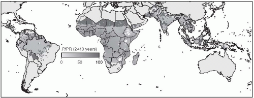
Figure 27-1 shows a map of the distribution of Plasmodium falciparum malaria created by the Malaria Atlas Project.7

Although P. vivax malaria is a major cause of morbidity in parts of China, Southeast and Central Asia, Latin America, and the Caribbean, the overwhelming burden of disease occurs in those countries subjected to P. falciparum malaria, especially in sub-Saharan Africa. Most of the discussion of this type of malaria in this chapter will focus on issues related to tropical Africa, where malaria is of greatest importance and where approaches to control have had, until recently, the least success.

The public health significance of a disease depends on its incidence and resulting disability and mortality. In addition, this impact depends on epidemiological characteristics such as those related to person (age, sex, and other demographic variables such as occupation, education, and socioeconomic group), place (urban and rural, or particular
ecological zones), and time (including seasonal or other cyclical variation or secular trends). In infectious diseases, the incidence of a disease is commonly expressed in terms of episodes per time period or the number of persons affected per time period. In many places where malaria is hypoendemic or mesoendemic, the incidence of malaria has meaning and can be expressed as the number of episodes per thousand persons per year or as the number of persons having episodes per thousand persons per year. In holoendemic areas, however, with an entomological inoculation rate ranging from dozens to hundreds of infectious bites per person per year, everyone is infected all the time and is reinfected every few days. The very concept of incidence, or indeed prevalence, has little meaning in this context. Instead, the health status of an individual in these situations results from a balance between the parasite and host immunity. Clearance of parasites from the host occurs with reasonable certainty only when an individual is given an effective antimalarial drug. Reinfection and, therefore, “a new incident” case occurs as soon as the level
of the antimalarial drug drops below the effective therapeutic level and recently injected sporozoites develop into blood-stage parasites. Further discussion of different measures of malaria transmission is found in the “Malaria Metrics” section later in this chapter.


Table 27-1 Malaria Endemicity


Transmission


Children 2-9 Yrs with
Enlarged Spleens &
Parasites


Parasitemia


Entomologic Infectious
Rate (EIR)
Bites/Person/Year


Holo-endemic


Intense, everyone has malaria
parasites all the time.*


>75%


>75%


>20**
SouthAmerica/Asia = 20-50,
very rare >50
Africa >50 -100s


Hyper-endemic


Regular, often seasonal
transmission.


50-75%


50-75%


5-20


Meso-endemic


Regular, but much lower than
hyper. Danger of occasional
epidemics with fairly high
morbidity and mortality.


10-50%


10-50%


>10


Hypo-endemic


Population will have little or
no immunity. Danger of severe
epidemics involving all age
groups.


<10%


<10%


Not detected except
when epidemic


* detection of parasites may be very difficult because of high levels of immunity, but sufficient search will reveal presence of parasites


Figure 27-1 The Spatial Distribution of P. falciparum Malaria Endemicity. Reproduced from Hay et al. A World Malaria Map: Plasmodium falciparum Endemicity in 2007. PLOS Medicine: San Francisco, CA.

Since rates were first reported by WHO in the 1950s, childhood malaria mortality has been estimated at 1-2 million children per year. In the past decade, however, two important trends have taken place: the quality of malaria mortality data has improved and, even more notably, increased effectiveness of antimalarial efforts has become evident. In a 2004 WHO report, global deaths from malaria were estimated to have dropped to 1,272,000 (with 1,136,000 in Africa).8 A continuing decline in mortality is supported by the estimates for 2009 of 781,000 deaths from malaria worldwide (including 709,000 in Africa).9

An indirect indicator of the importance of malaria mortality is the relative frequency of sicklecell trait (AS) in tropical Africa. This deleterious trait is maintained in the population because of the protection it provides against malaria. The high proportion of adults with the AS genotype in West Africa is a reflection of the selective mortality, 15-20%, among children with the AA genotype (see the section on “Human Genetic Factors”).10


THE BIOLOGY OF MALARIA PARASITES AND ANOPHELINE VECTORS


Malaria Parasites and Their Life Cycle

Five species of protozoan parasites of the genus Plasmodium infect humans: P. falciparum, P. vivax, P. ovale, P. malariae, and P. knowlesi. P. knowlesi, whose natural vertebrate host is the monkey Macaca fascicularis, was recently recognized as a zoonotic infection of humans in Malaysia, Indonesia, and other parts of Southeast Asia.11, 12 Although P. vivax is the most widespread form of malaria infection in the world, P. falciparum causes the most severe disease and is responsible, by far, for most deaths and serious morbidity due to malaria (Table 27-2).

The complex life cycle of the malaria parasite is shown in Figure 27-2. The parasite undergoes multiple transformations within the mosquito and human host; at least a dozen separate steps have been defined. The parasite is transmitted to humans as the sporozoite form in the saliva of an infected female anopheline mosquito taking a blood meal (1). Sporozoites enter the venous blood system from the subcutaneous tissues through the capillary bed, and within minutes those that avoid the defending reticuloendothelial system invade liver cells (2). Over the next 5 to 15 days, each sporozoite nucleus replicates thousands of times to develop into a hepatic schizont within the liver cells (3). When released from the swollen liver cells, each schizont splits into tens of thousands of daughter parasites called merozoites (4). Merozoites attach to specific erythrocyte surface receptors (Duffy blood group antigen for P. vivax, glycophorins for P. falciparum)13 and penetrate the erythrocyte (5). Each intraerythrocytic merozoite differentiates into a trophozoite that ingests human hemoglobin, enlarges, and divides, into 6 to 24 intraerythrocytic merozoites, forming a schizont. The red cell swells and bursts, releasing the next batch of approximately 20 merozoites (6), which then attach and penetrate new erythrocytes to begin this cycle again.

Along with the liberation of merozoites, the resultant hemolysis and release of pyrogens from infected red cells and the host’s response to these toxins correspond with clinical paroxysms of fever and chills; when synchronous, the simultaneous release from many red cells accounts for the periodicity of these symptoms in some patients. This second stage of asexual division takes approximately 48 hours for P. falciparum, P. vivax, and P. ovale, and 72 hours for P. malariae. A single P. falciparum merozoite potentially can lead to 10 billion new parasites through these recurrent cycles.

After a number of cycles within red cells, some merozoites differentiate into sexual forms called gametocytes (7)—macrogametocytes (female) and microgametocytes (male)—that are then available
to be ingested by an anopheline mosquito during its blood meal (8). Factors related to gametogenesis include the species of parasite, length of infection or number of intraerythrocytic cycles, density of parasitemia, drug treatment, and age or immune status of the infected individuals.14


Table 27-2 Malaria Parasites of Humans


Species


Intra-RBC Schizont Period


Type of RBC


Relapse (hypnozyte)


Global Distribution


P. vivax


48 hours


Reticulocytes


Yes


Everywhere except Africa


P. ovale


48 hours


Reticulocytes


Yes


Africa


P. malariae


72 hours


Older RBCs


No


Everywhere


P. falciparum


48 hours (±)


All


No


Tropical regions


Figure 27-2 Life cycle of the malaria parasite. Reproduced from the Centers for Disease Control and Prevention. Laboratory Identification of Parasites of Public Health Concern. Life cycle of malaria at: http://www.dpd.cdc.gov/dpdx/images/ParasiteImages/M-R/Malaria/Malaria_LifeCycle.gif. Last modifided July 20, 2009. Accessed January 24, 2012.


Sporogonic Development

Once in the mosquito, the red cells are digested, freeing the gametocytes, which then begin sexual reproduction leading to sporogonic development.15 The male and female gametes fuse, providing for genetic recombination, to form a zygote (9). Over the next 12 to 14 hours, the zygote elongates and forms an ookinete (10), which in turn penetrates the wall of the mosquito’s stomach and becomes an oocyst (11). During the next several days, the oocyst enlarges, forming more than 10,000 sporozoites. After the oocyst ruptures into the coelomic cavity of the mosquito (12), the sporozoites migrate to the salivary glands (1), ready to be injected back into the human host to complete their life cycle. Once infected with malaria, a female anopheline remains infected for life and can transmit sporozoites with each blood meal.

The phases of parasite development—from ingestion of gametocytes to when sporozoites in the salivary glands are poised for reinoculation into the human host—constitute the extrinsic cycle or sporogonic phase, which generally takes 7-12 days. The time required depends on the species of parasite and the ambient temperature. For example, under optimal conditions with the temperature at 30°C, P. falciparum requires 9 days to complete the sporogonic phase; at 20°C, however, it takes 23 days—a
difference of 14 days for a temperature differential of 10°C. With an average life span for most anophelines of less than 3 weeks, the ambient temperature is critical to transmission of the malarial infection.

During sporogonic development, each female- male pair of gametocytes potentially can produce more than 10,000 sporozoites for inoculation. Because dozens to thousands of gametocytes can be ingested with one blood meal, the potential exists for millions of sporozoites to be injected with one bite. Perhaps related to damage to the mosquito from such heavy loads or insufficient nutrients and metabolites available to support these levels of parasitemia, such high inocula counts are not observed. Limited studies of naturally infected mosquitoes have found sporozoite loads (the number of sporozoites in the salivary glands of an anopheline) to range from 10 to more than 100,000.16

In vitro studies using experimentally infected mosquitoes have shown that most infected mosquitoes transmit fewer than 25 sporozoites per bite, but approximately 5% of these mosquitoes can transmit hundreds.17 Epidemiological studies comparing sporozoite rates (the proportion of anopheline females with sporozoites in their salivary glands) with infant infection have demonstrated that less than 20% of sporozoite inoculations result in infection. However, great variation in the ratio of the infant conversion rate (ICR—the rate at which infants acquire malaria) to the entomological inoculation rate (EIR—roughly the number of infectious bites per person) has been found between places, between seasons, and for evaluation of vector control.18, 19 In general, far lower numbers of sporozoites are delivered than are found in the salivary glands. Nevertheless, some bites do transmit high numbers of infective sporozoites, and although speculative, high inocula may lead to more severe disease.


Biological Differences Among Malaria Species

Important species-specific differences are found in the generic life cycle described in the preceding discussion. With P. vivax and P. ovale, some sporozoites entering hepatic cells do not immediately proceed to tissue schizogony, but rather become hypnozoites and lie dormant for months to years.20 Later, these hypnozoites can differentiate into hepatic schizonts, leading to the cycle of erythrocytic schizogony and consequent relapse of symptoms. This biologic capability accounts for the relapses characteristic of P. vivax and P. ovale and the need for specific drug treatment targeted to the hypnozoite stage (primaquine). Different strains of P. vivax from diverse areas of the world are known to have characteristic relapse patterns. In general, following acute infection with either P. vivax or P. ovale, patients are at risk of having a relapse for as long as 3 to 4 years. No diagnostic test is available to determine whether individuals have hepatic hypnozoites.

P. falciparum, P. malariae, and P. knowlesi (despite the phylogenetic relatedness of the latter to P. vivax) do not produce hypnozoites and do not lead to relapse, but untreated or inadequately treated infections may cause persistent low-grade parasitemia leading to recrudescent clinical disease. The term “relapse” refers to renewed infection from survival of the parasite in hepatic cells as hypnozoites, whereas “recrudescence” refers to renewed infection from surviving erythrocytic forms.

The different species of human malaria parasites have affinities for particular types of erythrocytes. P. vivax and P. ovale parasites invade young reticulocytes; thus the density of peripheral parasitemia in these infections rarely exceeds 3%. In contrast, P. malariae is limited to older red cells. However, P. falciparum infects erythrocytes of all ages, and for this reason it is able to produce high-density parasitemias with serious morbidity and high mortality. P. knowlesi also infects erythrocytes of all ages, completes its intraerythrocytic life cycle in 24 hours (faster than other human malaria parasites), and can reach high levels of parasitemia, resulting in severe disease similar to P. falciparum.21

Important differences in gametocyte production are also noted among species.22 After infection with P. vivax, infective gametocytes appear in the peripheral blood almost as soon as the asexual blood-stage schizonts form. Gametocytes are usually present when P. vivax malaria is first diagnosed and before antimalarial treatment has been started. P. vivax can be transmitted prior to symptomatic disease, and its gametocytes will not have been exposed to drug pressure that would select for drug-resistant mutants; as a consequence, drug-sensitive parasites are not at a disadvantage in competition with the drug-resistant strains. In contrast, following infection with P. falciparum, gametocytes appear only after several intraerythrocytic cycles, first appearing at least 10 days after the onset of clinical symptoms. Early treatment of P. falciparum with an effective drug will kill bloodstage schizonts, preventing gametocytes from developing and blocking transmission. However, those gametocytes that do develop will be derived from malaria parasites that survived drug treatment and may carry drug resistance genes. This pattern strongly facilitates the selection and spread of drug-resistant parasites. The difference in timing of gametocyte emergence may be a factor accounting for the much higher rate of drug resistance in P. falciparum as compared to P. vivax.


Aspects of the Molecular Biology of the Malaria Parasite

Understanding of the molecular biology of malaria parasites was advanced by the publication of the genome sequence of P. falciparum in 200223 and the P. vivax genome in 2008.24 Functional and comparative genomics and proteomics of the malaria parasite during different stages of its life cycle will lead to improved understanding of Plasmodium biology and pathogenesis, and the identification of potential new drug and vaccine targets.25

The genome of P. falciparum consists of 14 chromosomes containing more than 5000 predicted genes. Several aspects of the molecular biology of P. falciparum are relevant to malaria epidemiology and control. A large proportion of identified genes are involved in immune evasion and host-parasite interactions. P. falciparum contains three families of highly variable genes, the most important of which is the var gene family, comprising approximately 60 genes encoding for the P. falciparum erythrocyte membrane protein 1 (PfEMP1). Transcriptional switching between different var genes allows the parasite to evade immune responses directed against PfEMP1. These genes may also be modified by the exchange of material between chromosome ends, where these genes are located. PfEMP1 is located on the surface of infected red blood cells and mediates adherence to endothelial cells. One var gene encodes a protein that mediates adherence to chondroitin sulfate A in the placenta and is partly responsible for the severe disease observed in pregnant women.26 Specifically, a conserved parasite var gene (var2csa) is associated with placental malaria and mediates adherence of parasitized erythrocytes to the placenta.27 Thus this gene family encodes for an important virulence factor responsible for the sequestration of infected red blood cells in various organs of the infected human.28

Genome sequencing of P. falciparum has led to the identification of plasmodia-specific metabolic pathways. These pathways are ideal targets for novel antimalarial drugs. For example, enzymes involved in fatty acid synthesis or protein degradation in food vacuoles may be ideal drug targets. Genomic research may also identify antigens for vaccine development, particularly conserved sequences expressed on cell surfaces during different stages of parasite development.29 However, P. falciparum geneticists caution, “genome sequences alone provide little relief to those suffering from malaria,” and work must be done to convert this knowledge into effective interventions.23

The malaria genome project focused on a single clone of P. falciparum, and further work is needed to define the genetic diversity found among parasites from different geographical regions. Improved understanding of strain genetic differences will provide insights into transmission, pathogenesis, and drug resistance.30


Anopheline Mosquitoes and Their Life Cycle

Malaria is transmitted only by the genus Anopheles mosquitoes. More than 70 species of Anopheles are known to be capable of transmitting malaria to humans, with approximately 40 considered important vectors. The female anopheline requires protein derived from host blood for egg production; therefore, only the female feeds on blood, and she is the only vector for malaria.

There is great variation among different species in host feeding preference, biting, and resting behavior, and in selection of larval habitat for laying the eggs, and these differences determine the local epidemiology of malaria. Some anophelines are opportunistic feeders on a variety of vertebrates (zoophilic), whereas others are very particular and take blood meals only from humans (anthropophilic). Some feed only indoors (endophagic), whereas others may occasionally or exclusively feed outdoors (exophagic). Whether they rest indoors (endophilic) or outdoors (exophilic) after feeding is critical to understanding the potential impact of indoor insecticide spraying. Nearly all anophelines prefer clean water in which to breed, but some have very specific preferences for the aquatic environment in which they lay their eggs. For example, Anopheles stephensi breeds in tin cans and in confined water systems, whereas Anopheles gambiae— the most important malaria vector in Africa—prefers small, open, sunlit pools. Knowledge of this variation is critical for effective vector control.

The mosquito goes through four stages of growth during its life cycle, from egg to larva to pupa to adult. Shortly after emerging as an adult (eclosion) and before the first blood meal, adult anopheline females mate. They usually mate only once and store the sperm, laying a total of 200-1000 eggs in 3 to 12 batches over their lifetime. A fresh blood meal is required for development of each egg batch. After hatching, an anopheline larva feeds at the water’s surface and develops over 5-15 days before pupation. Within 2-3 days, an adult mosquito emerges from the pupal case. The entire cycle requires a total of 7-20 days, depending on the anopheline species and the environmental conditions. Under favorable conditions of high humidity and moderate temperatures, female anophelines can survive at least one month—time enough for the parasite to go through the sporogonic cycle of 7-12 days needed to develop
sporozoites in the salivary glands for injection with the next blood meal. Thereafter, the mosquito is capable of transmission with each subsequent human blood meal, often taken every 2-3 days for the remainder of its life. Therefore, the longevity of the anopheline mosquito is critically important in determining the efficiency of transmission.

Mosquitoes are able to seek out their host in response to a combination of chemical and physical stimuli, including carbon dioxide, body odors, warmth, and movement. The odorant receptors of A. gambiae have been characterized, and this knowledge could lead to strategies to block the ability of mosquitoes to track human hosts.31 Most anopheline mosquitoes feed at night, but some species may feed in late afternoon or early morning. During feeding, the mosquito injects salivary fluid containing enzymes into the subcutaneous tissue. These enzymes diffuse through the surrounding tissue and increase blood flow, facilitating both the blood meal and the transfer of sporozoites to the capillary bed. Anophelines generally feed on people sleeping indoors. Some species, however, bite outdoors, especially those that are forest dwellers, such as Anopheles dirus. After feeding, the engorged female seeks a resting place on a nearby wall or in a secluded spot outdoors. Some species alter their behavior in the presence of DDT or other insecticides, becoming irritated and flying outside to seek refuge. Engorged mosquitoes usually rest for 24-36 hours to digest the blood meal before they search for an oviposition site.

Vector identification involved in malaria transmission serves as the starting point for entomological investigation of local malaria epidemiology. Measurement of the entomological inoculation rate (EIR; described later in this chapter) for each of the potential vectors is important for determining the relative contribution of each species to transmission as well as for determining the intensity of transmission in the area.

Enormous spatial and temporal variation in vector species is evident. Ecological change induced by human activities such as urbanization, deforestation, and irrigation may modify mosquito habitats and vector distribution. A major obstacle to species identification is the existence of species complexes, in which genetically distinct sibling groups are morphologically indistinguishable. Sibling species may vary in their potential as vectors due to differences in susceptibility to parasitic infection, resistance to insecticides, breeding habitat, geographic distribution, host preference, or biting and resting habits. For example, the A. gambiae complex consists of seven species, including two of the most important vectors in sub-Saharan Africa: A. gambiae and A. arabiensis. In contrast to A. gambiae, A. arabiensis often feeds on cattle, rests outdoors, and is tolerant of low humidity. Another member of this species complex, A. quadriannulatus, feeds only on animals and is not a vector of human malaria. Even within A. gambiae, genetic heterogeneity occurs in conjunction with geographic and seasonal variations. Further advances in understanding local ecological, vector, host, and parasite factors should translate into improved approaches to malaria control.

Concurrent with the publication of the genome sequence of P. falciparum, a first draft of the genome sequence of A. gambiae was published in 2002.32 Identification of polymorphisms within the A. gambiae genome will aid in the detection of insecticide resistance (e.g., detoxifying enzymes), genetic factors responsible for transmission efficiency of malaria parasites, and gene flow within mosquito populations. Genetic studies should lead to a better understanding of the determinants of anopheline behaviors, including the mechanisms by which the mosquito identifies human hosts as well as metabolic targets for insecticide development. Components of the mosquito’s innate immune system have been identified that allow for a better understanding of the coevolution of parasite and vector, and potential reasons for different transmission efficiencies.33 Interestingly, bacteria within the mosquito midgut, including Enterbacter34 and Wolbachia,35 inhibit P. falciparum oocyst development.


MALARIA METRICS

Understanding the dynamics of malaria transmission is fundamental to understanding how best to reduce transmission through vector control and other antimalarial measures.36 Sir Ronald Ross (1857-1932) and George Macdonald (1903-67) pioneered the quantification of variables related to the transmission dynamics of malaria. Their work laid the foundation for the worldwide malaria eradication described in the “History of Malaria Control” section; these measures and their use in defining critical points of action for malaria control are now of renewed interest as countries are strengthening their control efforts and are reconsidering the prospects of malaria elimination and eradication.5, 36

The starting point for Ross and Macdonald in understanding the determinants of malaria transmission was to consider the basic reproduction rate (R, or Z0 in their nomenclature) as the number of new cases of malaria generated by a single case of malaria
in a given period of time. For malaria to be eliminated, R must be reduced to less than 1.

The critical variables that determine R are those detailed later in this section for the entomological inoculation rate (EIR) and vectorial capacity (VC). These basic indices of malaria transmission are closely related; both are applied to a defined ecological zone or geographical area, and both vary greatly in place and time.37 The EIR is the number of infected bites that each person receives per night (or time period) and is calculated by multiplying the human landing rate (HLR) by the sporozoite rate (SR) and usually expressed on an annual basis as the annual EIR. The HLR, previously termed the “human biting rate,” is obtained by capturing all mosquitoes that land on a person (the “bait”) during a night and is expressed as the number of mosquitoes landing (bites) per person per night. The SR is determined by microscopic examination of dissected salivary glands to detect sporozoites in these captured mosquitoes, expressed as the ratio of infected anophelines to the total anophelines collected. More recently, serologic and molecular techniques have been developed to measure the SR, including rapid dipstick methods that are easily used under field conditions to detect circumsporozoite proteins.38 Even with these advances, measuring the HLR and the SR is difficult, tedious, and costly. Only a fraction (perhaps 20%) of the sporozoites inoculated are infectious. The EIR provides a direct measure of malaria transmission and the risk of human exposure to the bites of infected mosquitoes.

In contrast, the VC measures the rate of potentially infective contact, meaning the potential for malaria transmission, and is based solely on key vector parameters in a particular area. In theory, it is independent of whether humans are actually present (to obtain variable a in the VC formula below, of course, requires that some humans have been bitten). The VC is the number of potentially infective contacts an individual human could acquire in an area, through the vector population, per unit of time.39 The formula for its calculation is as follows:


where m is the density of vectors in relation to humans (obtained by standardized sampling methods to give the number of female anophelines caught in a collection per person per night); a is the human biting habit (the proportion of blood meals taken from humans to the total number of blood meals taken from any animal), so that a person is bitten by ma vectors in 1 day; p is the daily survival probability of the vector; and n is the extrinsic incubation (or sporogonic) period of the vector measured in days, so that a fraction pn of the vectors survive the extrinsic cycle (incubation period). The vectors still have an expectation of life of 1/-log p (the expectation of life is assumed to be independent of age): each of the surviving vectors bites a persons per day. In principle, the VC can predict the extent to which anopheline populations must be reduced to reduce transmission.

It is important to understand the nonlinear relation among these variables. In Figure 27-3, the prevalence of parasitemia in the human population is charted against the average annual VC.40 Note that at low levels of VC, small increases result in a rapid rise in parasite prevalence; thereafter, a long plateau
is reached, where large changes in the VC do not change the parasite prevalence. This is the situation in much of tropical Africa. In these areas, reduction of several 100-fold or even 1000-fold in the VC will not change the prevalence of malaria (although it may change the frequency and nature of severe malaria).


Figure 27-3 Prevalence of parasitemia as related to vectorial capacity. Data from Garki Project Molineaux and Gramiccia, WHO, Geneva, 1980, p. 281.

Current antimalarial interventions reduce transmission (i.e., the reproductive rate) as follows: effective treatment of people with parasitemia reduces human infectivity (SR in the EIR, no effect on VC); indoor residual spraying reduces the daily survival rate of the mosquito (SR in the EIR, ma in the VC); and insecticide-treated mosquito nets reduce the human biting rate of the mosquito (HLR in the EIR, ma in the VC) and its daily survival rate (SR in the EIR, expectation of vector life in VC).


Geographical Areas According to Intensity of Transmission

As indicated in the section on the public health importance of malaria and outlined in Table 27-1, traditionally geographical patterns of transmission have been classified into four broad categories according to the intensity of transmission: holoendemic, hyperendemic, mesoendemic, and hypoendemic. Further classification schemes have been devised in efforts to simplify the complex epidemiological factors of malaria and to better target strategies for malaria control.36


PATHOGENESIS IN INDIVIDUAL HUMANS


Infection and Disease

The distinction between infection and disease is particularly important in malaria. Infection with the malaria parasite does not necessarily result in disease, especially in highly endemic areas. In these regions, children may have parasitemia prevalence rates of 50% or more, yet few will have symptoms.

Disease is the result of the combination of parasite multiplication and the host reaction to the parasite. The classic description of periodic shaking chills, severe fever, and drenching sweats every two or three days can be seen in non-immune adults infected with P. vivax (every other day) or P. malariae (every third day). The symptoms in these cases result from the host response to synchronous lysis and release of pyrogens from infected red cells. However, with falciparum malaria, clinical manifestations, particularly in children, range from asymptomatic parasitemia to severe overwhelming disease and rapid death.41 Children may present with drowsiness, coma, convulsions, or simply listlessness and fever with nonspecific symptoms. Abdominal cramping, cough, headaches, muscle pains, and varying levels of mental disorientation are common. Severe and complicated malaria due to falciparum malaria is a medical emergency.


Host Response

Malaria, on a population basis, is the most intense stimulator of the human immune system known. Many immunologic defense systems are activated in response to malaria infections, including the reticuloendothelial system with enhanced phagocytosis in the spleen, lymph nodes, and liver to remove infected RBCs; an intense production of antibodies (humans can develop several grams per liter of immunoglobulin directed against malaria); and a range of cellular immune responses and cytokine cascades. Some of these responses are protective, whereas others contribute to pathology; often they have both effects. For example, pro-inflammatory cytokines, severe metabolic acidosis, and the classical sequestration of infected RBCs that causes cerebral anoxia all may contribute to the pathogenesis of cerebral malaria; at the same time, however, this pro-inflammatory response and acidosis evidently account for why some patients survive without neurological complications.42

The host response to malaria can go wrong in several ways. For example, the pathogenesis of “big spleen disease” (tropical splenomegaly) is caused by an excessive and inappropriate host response to malaria. Tropical splenomegaly is fairly common among relatively non-immune populations who, because they move to a malarious area or experience a change in climate, are exposed to more intense transmission. The disease starts in childhood, progressing through adolescence to young adulthood with severe anemia, high levels of immunoglobulin M (IgM) and antimalarial antibodies, a decrease in platelets, and a huge spleen. Malaria parasites are rarely detected. If untreated, tropical splenomegaly is often fatal, usually from a secondary infection. Two lines of evidence indicate this outcome is directly the result of an abnormal reaction to malaria: (1) those persons with sickle trait (hemoglobin SA) do not develop big spleen disease,43 and (2) individuals with tropical splenomegaly who take long-term antimalarial prophylaxis have gradual reduction in spleen size and anemia, and return to normal health after many months.

Malaria parasites have evolved complex mechanisms to evade host immune responses and establish persistent or repeated infections. Understanding the basis of effective immune responses that either prevent infection or reduce disease severity is important for vaccine development. Protective immunity following
natural infection takes years to develop.44 However, no immunodominant response has been identified, and an effective immune response is likely the sum of cellular and humoral responses to multiple parasite antigens. Antibodies to the circumsporozoite protein can prevent the binding of sporozoites to liver cells. Cellular immune responses—specifically interferon-γ secreting T cells—are important in killing infected liver cells.45 Antibodies play a role in killing parasitized red blood cells and can protect the host from sporozoite challenge, although cell-mediated immunity contributes to blood-stage protection as well. The great antigenic variability of P. falciparum, as described previously, is in large part responsible for the ability of the parasite to evade effective host immune responses.


Human Genetic Factors

As humans, mosquitoes, and malaria parasites have evolved, many human genetic characteristics that provide partial protection against malaria have emerged. These genetic polymorphisms mostly involve the red blood cell, and include structural variants in the β globin chain of hemoglobin such as sickle-cell trait (hemoglobin S) and hemoglobins C (West Africa) and E (Southeast Asia); altered α and β globin chain production leading to the α and β thalassemias (Mediterranean anemia); erythrocyte enzyme deficiencies including glucose-6-phosphate dehydrogenase (G6PD); red cell cytoskeletal abnormalities such as ovalocytosis; and changes in the red cell membrane such as the absence of the Duffy blood group factor (West Africa).46 The mechanism that provides protection seems clear for sickle-cell trait—when red cells are invaded, they sickle and are preferentially removed by the reticuloendothelial system, thereby reducing parasite density levels47—but mechanisms have not been fully elucidated for many other genetic polymorphisms. In addition to genetic changes involving hemoglobin, red cell membrane proteins, or red cell metabolism, polymorphisms in human immune response genes may affect infection and disease, and polymorphisms in cytokine genes or their promoters have also been associated with disease susceptibility and severity.48

Many polymorphisms place a heavy burden on the homozygous individual, such as hemoglobin SS or sickle-cell disease. Accounting for the frequency of sickle-cell trait (AS) in tropical Africa (ranging from 15% to 30% AS hemoglobin in adults), the historical case-fatality rate attributable to malaria is approximately 10-20% of all children with AA hemoglobin genotype.10 For example, if 28% of the adult population has sickle trait, then the S gene allele frequency in the adult gene pool would be half of 28%. The Hardy-Weinberg law gives a selection coefficient for the AA genotype of 0.14/(1.00 — 0.14) = 0.1628. The ratio of AS-genotype individuals to AA-genotype individuals for survivorship to adulthood would be 1/(1.00 — 0.1628) = 1.194, which is equivalent to nearly a 20% excess death rate for those with the AA genotype before adulthood. Because the only advantage of AS over AA is protection from severe malaria, the case-fatality rate due to malaria in those persons with AA hemoglobin is 19.4%.

Another way to calculate the mortality rate attributable to malaria among AA children is to compare the prevalence of difference genotypes in adults and children. The sharp age-specific rise in prevalence of sickle-cell trait in West Africa from 20-24% in newborns to 26-29% in adults indicates that many infants born without the “protective” sickle-cell trait die before adulthood. This differential survival can be expressed as the ratio of the proportion of AS in adults to the proportion of AS in newborns, or as the proportion of AA in adults divided by the proportion of AA in newborns. In the Garki study area, which is characterized by very high malaria transmission and sickle-trait rates, adults were found to be 28.96% AS and 70.2% AA (0.84% were classified as “other,” including those with AC and SC hemoglobin), whereas newborns were 23.6% AS and 73.78% AA (2.62% were “other,” including 2.1% SS and 0.5% AC), giving a ratio of 3.86/2.99 = 1.29. This ratio indicates that there is a 29% case-fatality rate in persons with the AA genotype because of malaria.19


Undernutrition and Micronutrient Deficiencies

Malaria is prevalent in regions where childhood undernutrition is common, and nutritional deficiencies interact with malaria infection in complex ways. Early observational studies suggested that undernourished children suffered lower morbidity and mortality than adequately nourished children. These early reports often involved selected groups or were conducted during famine, and refeeding severely malnourished children may worsen apparent disease severity. However, more recent studies have not confirmed this association. Pooled analyses of two cohort studies that examined the relationship between underweight and the severity of malaria found that malnourished children were more likely to die from malaria than adequately nourished children.49 Malaria chemoprophylaxis should be provided to famine victims or severely malnourished child in malarious areas when treating severe protein-energy malnutrition.

Iron deficiency is the most common micronutrient deficiency and is associated with defects in
immune responses and a number of poor health outcomes. Early observations suggested that infants with iron deficiency had less severe malaria than children without iron deficiency, and that providing iron supplementation might increase disease severity.50 A more recent systematic review concluded that oral iron supplementation for preventing or treating anemia among children in malaria-endemic areas did not increase the risk of clinical malaria or death when adequate malaria surveillance and treatment were in place.51 However, the increase in hemoglobin was variable.

Vitamin A is essential for proper immune function in response to malaria. A trial of vitamin A supplementation in preschool children in Papua New Guinea found that vitamin A reduced clinical episodes of malaria, splenic enlargement, and parasite density, particularly in children 1 to 3 years of age.52 However, vitamin A may not affect severe malaria or mortality. Extrapolating from this single trial, the fraction of malaria morbidity attributable to vitamin A deficiency was estimated to be 20% worldwide.49 Complicating interpretation of such studies is the fact that low serum retinol levels are commonly found in children with malaria, but this condition may be due to preexisting vitamin A deficiency, a contribution of malaria to vitamin A deficiency, or merely an acute effect of malaria on retinol metabolism or binding.53

Zinc is another micronutrient essential for both cell-mediated and humoral immunity. In randomized trials in Papua New Guinea and the Gambia, the effects of zinc supplementation on malaria morbidity and mortality were investigated.54, 55 The fraction of malaria morbidity attributable to zinc deficiency was estimated to be 20% worldwide—similar to that for vitamin A.49 In Papua New Guinea, zinc supplementation reduced malaria-attributable clinic attendance by 38% and had its greatest effect on attacks having high-density parasitemia. In the Gambia, the effect of zinc supplementation was less pronounced, but the treatment group did have fewer clinic visits for malaria compared to the placebo group. In contrast, a recent review of five clinical trials concluded that zinc had no effect on malaria incidence or mortality.56


THE DIVERSITY OF FALCIPARUM MALARIA DISEASE

Advances have been made in differentiating distinctive forms of severe falciparum malaria and in understanding underlying pathogenic factors— for example, inoculum size, differing strains of falciparum, differing immunologic responses, and various host factors. These discoveries, in turn, have contributed to an appreciation of distinctive epidemiological features of different forms of clinical disease, based on an understanding of the relation of disease manifestations to intensity and constancy of transmission due to ecological factors, genetic characteristics of the parasite, and genetic and acquired immune mechanisms in humans.


Severe Malaria

In areas of Africa with very high transmission rates (EIR >50), severe disease in children does not progress from mild or moderate illness; instead, it strikes abruptly without warning. Mothers are frequently unable to get their infants and young children to a health center in time to provide treatment before the child dies, even when facilities with trained health workers are readily available. If they do manage to reach a hospital, many patients die within 24 hours of admission despite treatment efforts.

Rapid progression to severe disease is not characteristic in areas with less intense transmission. In southeast Asia, malaria typically slowly progresses in severity over several days in both children and adults, and does so in both major types of severe disease that occur there: cerebral malaria, which has a median time of 5 days from onset to cerebral symptoms, and multiple organ dysfunction syndrome (MODS), which is somewhat slower in development and has a median time of progress of 8 days.59 Early effective treatment before the onset of the severe phase is the key to reducing mortality in these circumstances. Although these slowly progressive types are also seen in Africa, they are less common in high-transmission areas than the rapid severe forms in children.

Only gold members can continue reading. Log In or Register to continue

Stay updated, free articles. Join our Telegram channel

Jul 8, 2016 | Posted by in INFECTIOUS DISEASE | Comments Off on The Epidemiology and Control of Malaria

Full access? Get Clinical Tree

Get Clinical Tree app for offline access