Surveillance of Patients following Primary Therapy


Surveillance of Patients following Primary Therapy


Robert W. Carlson


The success of therapy for breast cancer results in an increasing number of breast cancer survivors being monitored for the development of recurrent disease. It is estimated that there are approximately 3 million breast cancer survivors in the United States alone. The magnitude of the follow-up of this large population requires efficient, timely, and cost effective monitoring. The optimal monitoring for recurrence of disease requires knowledge of the risk for recurrence, common sites of recurrence, accuracy of methods of detection of recurrence, and potential benefits and risks of detection of early disease recurrence.


ASSESSMENT FOR RISK OF RECURRENCE OF DISEASE

Assessment for risk of recurrence of disease may be performed by the integration of the anticipated natural history of a breast cancer based upon anatomic and biologic prognostic factors and the anticancer treatment delivered. The hazard rates for recurrence of disease have been studied retrospectively among 3,585 patients enrolled in seven large clinical trials (1). The peak for annual hazard for recurrence occurred in year 1 to 2 and then decreased consistently to 5 years, and then declined slowly through year 12. The hazard for recurrence was especially high for those with 4 or more involved axillary lymph nodes during the first 5 to 6 years of follow-up, but thereafter was similar to those with fewer nodes involved. The hazard for recurrence was higher in those women with estrogen receptor negative versus receptor positive breast cancer during the first 3 years of follow-up, and then similar or lower thereafter.

Long-term follow-up studies have documented that the most common sites of recurrent disease are local soft tissue, bone, lung, liver, and brain. Multiple sites are often involved at the time of detection of first recurrence, and almost always during the course of the metastatic disease. Tumors that are estrogen receptor positive, progesterone receptor positive, low or intermediate grade, and with low mitotic rate are more likely to metastasize to bone than viscera when compared to tumors without those. In contrast, menopausal status, tumor size, and nodal status do not impact the frequency of bone versus visceral site of metastatic disease. Further, many factors associated with overall prognosis at diagnosis of early breast cancer retain prognostic significance for survival following first diagnosis of metastatic breast cancer. Long-term survival after recurrent breast cancer is relatively unusual, and apparent cures of disease are uncommon except for patients with ipsilateral in-breast tumor recurrences (2).

The goals of surveillance are to detect recurrence at a time that allows initiation of therapy to improve survival and to maintain a high quality of life. There is little high-level evidence that these goals are achieved by any surveillance program.


THE CONTRALATERAL BREAST


Frequency of Contralateral Disease

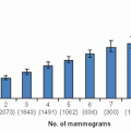
The occurrence of breast cancer in the contralateral breast of women with a known history of breast cancer may represent either a new primary tumor or a metastasis from the originally diagnosed breast cancer. While the determination of a new primary versus a metastasis may be difficult, a contralateral breast cancer represents a new primary if the cancer is of a different histology (e.g., ductal vs. lobular) or is associated with an in situ component. Metachronous second primary breast cancers are more likely to be in situ cancer, small size, and node negative (3). The risk of a metachronous, contralateral, second primary breast cancer is generally estimated at 0.5% to 1.0% per year (3, 4). Recent data suggests that the frequency of metachronous, contralateral, estrogen receptor positive second primary breast cancer is declining, probably because of the risk reduction provided by adjuvant endocrine therapies (5). Factors that increase the risk include a known BRCA1 or BRCA2 mutation, young age at first primary, family history of breast cancer, lobular histology for first primary breast cancer, and prior radiation exposure. Factors that decrease the risk include prior chemotherapy or endocrine therapy (6). While there are no randomized studies of screening mammography of the contralateral breast, contralateral second primary breast cancers in women undergoing routine screening mammography are smaller, and more likely to be in situ and node negative than those in women not undergoing routine mammography (7). The occurrence of metachronous contralateral breast cancer has modest impact on overall survival (7).


Screening for Contralateral Disease

Conceptually, the monitoring for a new primary breast cancer in the contralateral breast may be viewed as monitoring in a high risk for breast cancer population with an increased risk of competing mortality secondary to the initial primary breast cancer. In the general population of women age 40 and older, the use of screening mammography has been demonstrated to decrease breast cancer mortality. It is thus likely that routine screening mammography would decrease breast mortality from a second primary tumor, although the mortality rate for second primary breast cancers is low.

Breast magnetic resonance imaging (MRI) screening has high sensitivity, moderate specificity, and high cost as an adjunct to the performance of mammography. No randomized clinical trials of breast MRI as screening or surveillance are available in any clinical setting. Non-randomized trials in women without known breast cancer but at high risk of breast cancer document a higher detection rate of breast cancer with the use of breast MRI scanning (8). To date, no similar prospective trial has been reported in the follow-up of women following diagnosis of breast cancer. The American Cancer Society currently recommends screening MRI as an adjunct to screening mammography, based upon non-randomized screening trials and observational studies, in women with known BRCA mutation, who have a first degree relative with a known BRCA mutation, who have a lifetime risk of greater than 20% to 25% based upon family history, who had radiation exposure to the chest between age 10 and 30 years, who have known Li-Fraumeni syndrome themselves or in first degree relatives, or who have Cowden and Bannayan-Riley-Ruvalcaba syndromes themselves or in first-degree relatives (9). The American Cancer Society makes no recommendation for or against screening MRI in women with a personal history of breast cancer. A study of breast MRI screening in women at risk of breast cancer and with negative mammograms included 245 subjects with a personal history of breast cancer (10). Breast cancers were documented in 4% of the subjects with a prior history of breast cancer, and the positive predictive value of MRI recommended biopsy was 32%. A retrospective study of women screened by MRI who had a personal, but no family, history of breast cancer documented 17 cancers in 144 subjects (11). Ten of the 17 cancers were detected by MRI only. The positive predictive value of MRI was 39%. The non-randomized nature of all of these studies prohibits assessment of the impact of breast MRI screening on breast cancer mortality.

The value of clinical breast examination has not been adequately studied, although the performance of clinical breast examination is a generally accepted part of routine healthcare of the adult female. Breast self-examination was found to be of no advantage in early detection or mortality reduction in a large, randomized clinical trial in a population of factory workers in China (12).

Based on the increased risk of a second primary breast cancer in the contralateral breast, it appears prudent to perform regular clinical breast examinations and mammography as a routine part of surveillance programs. The role of breast MRI screening is yet to be defined, but would appear reasonable in those with very high contralateral risk. Such high risk patients are those with a known, or a high risk for, genetic mutation conferring risk for breast cancer or a prior history of thoracic radiation. For other populations, insufficient information exists to allow for specific recommendations regarding the use of screening MRI. The value of breast self-examination has not been demonstrated.


LOCOREGIONAL RECURRENCES

Most patients with a locoregional recurrence of breast cancer following either breast conserving therapy or mastectomy present with symptoms. Approximately 40% of isolated locoregional recurrences are detected during routine examinations in asymptomatic patients and a similar number are diagnosed outside of routine follow-up examinations in symptomatic or asymptomatic patients (13).


Ipsilateral Breast Tumor Recurrence

Ipsilateral breast tumor recurrence following breast conserving surgery is experienced by 5 years in approximately 7% of patients with whole breast irradiation and 26% of patients without whole breast irradiation (14). The addition of a radiation boost to the tumor bed decreases in-breast recurrence rates by approximately 41% compared with whole breast irradiation alone (15). Most recurrences occur in the prior tumor bed, and positive pathologic margins, younger age, higher grade tumor, larger tumor size, negative estrogen receptor status, and involvement of axillary lymph nodes have all been reported to increase the risk of ipsilateral breast tumor recurrence (14, 15 and 16). Approximately 70% of ipsilateral breast tumor recurrences occur within the first 5 years of primary diagnosis (14, 16). Breast recurrence during the first 5 years of follow-up is associated with a substantially worse overall prognosis than are in-breast recurrences that manifest later.

Detection of ipsilateral breast tumor recurrence is often difficult because of post-surgical, post-radiotherapy changes in the breast. The sensitivity of mammography for ipsilateral breast tumor recurrences is approximately 50% to 70% and ultrasonography 80% to 85%. Overall, approximately two thirds of local recurrences are detected by the patient or on clinical examination (17, 18).Detailed reviews of studies of screening mammography in women with a personal
history of breast cancer generally document that patients diagnosed with mammography detected only in-breast recurrences have superior survival to those with symptomatic recurrences (19, 20). These studies all suffer from being relatively small in sample size and are non-randomized. Thus, it is impossible to correct for confounding by lead-time and length-time biases.

Breast MRI scanning has high sensitivity for detecting inbreast recurrences, but is expensive and is associated with highly variable specificity.


Local-Regional Recurrence Postmastectomy

Local-regional recurrence following mastectomy is experienced by 5 years in approximately 6% of patients with postmastectomy regional irradiation and 23% of patients without postmastectomy irradiation (21). In the overview analysis, axillary lymph node status strongly predicted for absolute risk for local-regional recurrence (21). In women with axillary lymph node-negative disease, the 5-year local recurrence risk following surgery alone was 6%, and this was reduced to 2% with the use of local-regional irradiation. In women with axillary lymph node-positive disease, the 5-year local recurrence risk following surgery alone was 23% and this was reduced to 6% with the addition of local-regional irradiation. Increasing tumor grade, tumor size, and number of involved axillary lymph nodes increases the risk of local-regional recurrence.

Detection of local-regional recurrences following mastectomy with or without radiation is typically the result of either patient identification or of a routine clinical examination. Local-regional recurrences are rarely detected by radiographs or other screening studies.


Distant Recurrences

Well established prognostic factors allow the estimation of risk for development of systemic disease following treatment for stage 0, I, II, and III breast cancer. Known prognostic factors include histologic subtype of breast cancer, tumor grade, tumor size, involvement of skin or chest wall, extent of involvement of regional lymph nodes, hormone receptor status, HER2 level of expression or amplification, and multigene array expression profile.

Breast cancer metastases occur in a generally predictable pattern, with synchronous multiple sites of recurrence being common. Bone is the most common site of disseminated disease, and represents approximately 40% of first recurrences. The most commonly involved bones are the spine, ribs, pelvis, skull, femur, and humerus. Breast cancer metastasis to bone distal to the elbow or knee is rare. Other common sites for metastatic disease include lung, liver, lymph nodes, and soft tissue. The site of first metastasis from breast cancer is influenced by estrogen receptor status (Table 67-1). Estrogen receptor-positive breast cancer is more likely to spread to bone, while receptor-negative breast cancer is more likely to spread to viscera and soft tissues and is associated with a higher rate of early recurrence (Table 67-1) (22, 23). Even in those patients undergoing routine surveillance during follow-up, most recurrent disease is symptomatic at time of diagnosis (24, 25).

Infiltrating lobular breast cancer has a propensity for recurrences in intra-abdominal and retroperitoneal sites including stomach, intestine, peritoneum, and ureter (often bilateral) (26).

Currently available treatment of recurrent or metastatic breast cancer is rarely curative, even when the recurrence is limited (2). Further, the amount of tumor burden in asymptomatic or minimally symptomatic patients does not predict disease response to systemic treatment, ability to palliate symptoms, or overall survival. Thus, there is no advantage to diagnosing asymptomatic, early, subclinical disease.


Routine Blood Tests

The routine performance of blood tests for alkaline phosphatase, aspartate aminotransferase, γ-glutamyl transferase, bilirubin, calcium, and creatinine was studied by the International Breast Cancer Study Group in 4,105 women
with invasive breast cancer (27). At the time of analysis, 2,140 patients had experienced a relapse, 93 had a second non-breast primary tumor, and 111 had died without relapse during 10-years median follow-up. In this analysis, only alkaline phosphatase was abnormal in at least 20% of patients with recurrent disease, and was abnormal in 32% of patients with bone metastasis and 71% of patients with liver metastasis. Aspartate aminotransferase and γ-glutamyl transferase were elevated in 62% and 75% of patients with liver metastasis. Bilirubin, calcium, and creatinine were of no value in detecting recurrent disease. Thus, while alkaline phosphatase was the most reliable of the blood tests, it was of low sensitivity for bone or liver disease. In another study of 1,371 patients with node positive breast cancer, serial alkaline phosphatase determinations were found to have low sensitivity and specificity for bone recurrence (28). Thus, monitoring of routine blood tests as a part of breast cancer surveillance is not recommended.


TABLE 67-1 Location of First Recurrence in Randomized Trials of Intensive versus Routine Surveillance


GIVIO Trial (24)


National Research Council Project on Breast Cancer (46)


Type of Recurrence


Intensive Monitoring (n = 201 Recurrences or Deaths)


Control (n = 196 Recurrences or Deaths)


Intensive Monitoring (n = 219 Recurrences)


Control (n = 174 Recurrences)


Local regional recurrence alone


32 (15.9%


36 (18.4%)


55 (25.1%)


49 (28.2%)


Contralateral breast alone


12 (11.4%)


13 (6.6%)


Not stated


Not stated


Distant metastases


127 (63.1%)


127 (64.8)


164 (75.9%)


125 (71.8%)


Bone


52 (25.9%)


55 (28.1%)


84 (38.3%)


53 (30.5%)


Liver


13 (6.5%)


12 (6.1%)


Lung/pleura


24 (11.9%)


21 (10.7%)


28 (12.8%)


18 (10.3%)


Other sites


19 (9.4%)


27 (13.8%)


22 (10.0%


21 (12.1%)


Multiple sites


19 (9.4%)


12 (6.1%)


30 (13.7%)


33 (19.0%)


Second Primary (not breast)


8 (4.0%)


11 (5.6%)


Death without recurrence


11 (5.5%)


9 (4.6%)

Only gold members can continue reading. Log In or Register to continue

Stay updated, free articles. Join our Telegram channel

Jul 9, 2016 | Posted by in ONCOLOGY | Comments Off on Surveillance of Patients following Primary Therapy

Full access? Get Clinical Tree

Get Clinical Tree app for offline access