Breast Cancer Screening


Breast Cancer Screening


Christoph I. Lee

Joann G. Elmore


INTRODUCTION

Until breast cancer can be prevented, regular screening programs are widely recommended for asymptomatic women. The rationale behind such programs is that early diagnosis through screening can possibly reduce mortality and morbidity more effectively than no screening at all. Nevertheless, even though breast cancer screening has been more thoroughly studied than any other type of cancer screening, questions remain about its overall benefits and potential harms.

Evaluating any screening program is challenging, and breast cancer screening in particular has been a topic of many controversies over the years. In this chapter, we review the general principles of cancer screening that women, clinicians, and health system administrators need to consider before undertaking a breast cancer screening program. We then describe the many modalities that have been studied for possible inclusion in screening programs, including screening mammography, ultrasound, magnetic resonance imaging, breast examination by clinicians, and breast self-examination by individual women. We also review published data from randomized, controlled trials, population studies, and meta-analyses regarding the effects of screening programs on mortality and their potential harms, including false positive and false negative results, overdiagnosis, radiation exposure, anxiety, and economic cost. We then look at data on screening issues in special populations, such as elderly women and women with genetic mutations. We conclude with suggestions on informed decision-making as individual women are the ones who must decide whether to undergo medical screening of any kind.


PRINCIPLES OF CANCER SCREENING

The goal of breast cancer screening is early detection of disease, to be followed by appropriate treatment. Screening tests differ from diagnostic tests insofar as screening identifies subgroups of people who have a high probability of
asymptomatic disease, while diagnostic tests are obtained in symptomatic patients. A positive screening result in an individual rarely provides direct evidence of disease; screening tests must be followed by diagnostic tests to determine whether disease is truly present.

When an apparently healthy population undergoes regular screening, medical professionals have an obligation to show that the benefits of screening outweigh the costs. As we discuss in more detail later in this chapter, a positive screening test result and a diagnosis of breast cancer brings anxiety and treatments with associated morbidities and costs. Screening tests should therefore be safe, with minimal side effects. The minimum requirements for establishing a safe, ethical, and cost-effective screening program involve three areas: the disease targeted by the program, the screening tests needed to detect the disease, and the features of the health care system needed to support the program (1). All the requirements for each of these areas are reviewed in further detail in the following material. If these requirements are not at least partially met, population-wide screening may be ineffective.


Disease Requirements

First, the disease must be serious, with significant morbidity or mortality. Second, an effective therapy for the disease must be available if it is detected as screening would obviously have no value if subsequent treatment would not be beneficial. Third, the natural history of the disease must be understood clearly enough to identify a significant window of opportunity during which the disease is detectable and detection would probably lead to a cure, or at least an effective treatment with less morbidity than the disease itself. Finally, the disease must not be too rare; if it is rare, we can expect an excess of false positive test results, which increases the cost and effort necessary to detect true positives.


Screening Test Requirements and Characteristics

First, a screening test must be reasonably easy and inexpensive to perform. Otherwise, the costs of large-scale screening in terms of time, effort, and money will be prohibitive. This is an important point to remember when considering the relative utility of the many screening modalities now available, as we will discuss in the next section. Second, the screening test must be safe and acceptable both to the individuals undergoing the screening and to their physicians. Finally, the level of accuracy of the screening test must be known and acceptable to the health care system, the physician, and the patient. Its sensitivity, specificity, positive predictive value, and other operating characteristics require careful assessment.

It is critical to understand the characteristics of a given screening test, as well as the interplay of its characteristics with those of the population screened and the clinicians who perform and interpret the test. We present a standard 2 × 2 table (Table 10-1) comparing the results of screening tests with the disease status of the individuals screened, along with a series of formulas to measure the sensitivity, specificity, and other performance features of the test. The next three paragraphs explain Table 10-1 in more detail.

A positive test result for a person who does not have the disease assessed by a test is called a false positive result while a negative result for a person who actually has the disease is called a false negative result.

Sensitivity refers to the ability of a screening test to detect a disease when it is present and is calculated as a/(a + c).

If a test is not sensitive, it will fail to detect disease in some people who actually have the disease; they appear in cell c.

Specificity refers to the ability of a screening test to indicate the absence of disease when no disease is present and is calculated as d/(b + d). If a test is not specific, it will falsely indicate the presence of disease in some people who do not have the disease; they appear in cell b.

Another important parameter of a screening test is its predictive value, which may be either positive or negative. If a test result is positive, what is the probability that the person tested actually has the disease (i.e., true positive)? If the result is negative, what is the probability that the person does not have the disease (i.e., true negative)? The answers to these questions depend on the sensitivity and specificity of the screening test, as well as on the prevalence of the disease in the underlying population that undergoes screening. Positive predictive value (PPV) is calculated as a/(a + b). Negative predictive value (NPV) is calculated as d/(c + d), indicating the proportion of people with negative test results who are truly free of disease.


Health Care System Requirements

A screening program divides results into positives and negatives. Follow-up within a health care system must be available for everyone who has a positive result to confirm or rule out the presence of disease. Some follow-up testing can be expensive, time-consuming, and painful; it may even entail a degree of risk for the people who receive it. For example, estimates indicate that for every $100 U.S. dollars spent on breast cancer screening, an additional $33 are spent on subsequent diagnostic evaluations stemming from false positive results (2).

Before screening is undertaken, treatment should be available, accessible, and acceptable to people with disease. If a country’s resources are too limited to provide treatment in an equitable manner, or if no effective treatment for a given disease is available, it makes no sense, either ethically or in terms of cost-effectiveness, to encourage screening when people in whom disease is actually detected must go untreated.


BREAST CANCER SCREENING MODALITIES


Screen-Film Mammography

Screen-film mammography (SFM) has historically been the standard modality used for breast cancer screening, and the technology studied in all major randomized controlled trials reporting mortality benefit from screening. SFM serves as both image receptor and display medium, thus, requiring images to be processed much like film-based photography prior to digital photography. SFM images need to be developed and fixed chemically, with an image rejection rate due to processing errors exceeding 20% (3). Repeat imaging due to processing errors results in increased examination time, increased patient exposure to ionizing radiation, and increased costs. Thus, full-field digital mammography (FFDM) has quickly replaced SFM, as it does not require chemical image processing and, by enabling real-time contrast and brightness correction, reduces the rate of image rejection due to processing errors.


Digital Mammography

Because SFM serves as both image receptor and display medium, the film must be processed before review, resulting in delayed interpretations and requiring additional resources
for image archiving. Full-field digital mammography (FFDM), in comparison to SFM, has been shown to have lower noise, higher contrast, and improved dynamic range (4). In addition, FFDM allows immediate display of digital images on a monitor without film processing, enabling more rapid interpretation (5). Moreover, FFDM makes the use of computeraided detection (CAD) software, which recognize suspicious image patterns, a possibility.


TABLE 10-1 Standard 2 × 2 Table Comparing Test Results and Disease Status of Subjects Tested, along with Formulas to Measure Test Characteristics


image


Cells:


a = subjects with true positive test results


b = subjects with false positive test results


c = subjects with false negative test results


d = subjects with true negative test results


a + b = all subjects with positive test results


c + d = all subjects with negative test results


a + c = all subjects with disease


b + d = all subjects without disease


a + b + c + d = all study subjects


Associated formulas:


a/(a + c) = sensitivity


d/(b + d) = specificity


b/(b + d) = false positive error rate (alpha error rate, type I rate)


c/(a + c) = false negative error rate (beta error rate, type II rate)


a/(a + b) = positive predictive value


d/(c + d) = negative predictive value


[a/(a + c)]/[b/(b + d)] = (a/b)/[(a + c)/(b + d)] = likelihood ratio positive (LR+)


[c/(a + c)]/[d/(b + d)] = (c/d)/[(a + c)/(b + d)] = likelihood ratio negative (LR-)


(a + c)/(a + b + c + d) = prevalence


Adapted from Jekel JF, Katz DL, Elmore JG. Epidemiology, biostatistics, and preventive medicine. 2nd ed. Philadelphia: W.B. Saunders, 2001.


Large clinical trials comparing FFDM to SFM have generally demonstrated similar accuracy overall, with slight improvements when FFDM is used in certain subpopulations(6, 7 and 8). For example, the Digital Mammography Imaging Screening Trial (DMIST) compared the performance of FFDM to SFM in asymptomatic U.S. women, finding similar accuracy for breast cancer detection(9). However, FFDM was more accurate than SFM in three subpopulations: preor peri-menopausal women, women younger than age 50, and women with mammographically dense breast tissue. Given the growing evidence that FFDM is at least as useful as SFM for screening purposes, FFDM has progressively been adopted over SFM, especially in light of the potential efficiencies inherent in digital image transfer, interpretation, and archiving=(10).


Computer-Aided Detection

Computer-aided detection (CAD) has been found effective for improving the detection of malignancy when it is used in conjunction with both SFM and FFDM (11, 12 and 13). CAD was initially developed to help radiologists identify small tumors that might otherwise be overlooked, and has been shown to detect 84% to 94% of small malignancies in SFM and FFDM, regardless of whether the malignancies present as masses or as calcifications (11, 14, 15). Moreover, CAD has been shown to have equal sensitivity for detecting malignancies in women regardless of breast density and histopathologic results (16). However, CAD is also associated with a high false positive rate of between 2 and 5 marks per screening case when the exams include four standard views (by either SFM or FFDM), which may potentially confound radiologic interpretation and lead to unnecessary diagnostic work-up (11, 14, 15, 17). It is currently uncertain what effect the addition of CAD has had on patient outcomes such as mortality. Despite our lack of knowledge, CAD capabilities have become a standard feature of the latest generation of digital mammography workstations.


Ultrasound

Ultrasound serves as a critical adjunct diagnostic modality after abnormalities are noted during mammographic screening, and is increasingly used as a primary diagnostic modality for evaluating focal breast symptoms in women younger than age 40 (18, 19). The use of ultrasound to screen asymptomatic women is also rapidly increasing, especially for women found to have extremely or heterogeneously dense breasts on mammography. Dense breast tissue reduces the sensitivity of screening mammography to detect malignancy, and is associated with an increased risk of breast cancer even after adjusting for associated risks such as age and body mass index (20, 21, 22, 23 and 24). As of early 2013, legislation has been passed in the states of Connecticut, Texas, Virginia, New York, and California to mandate that women with mammographically dense breasts be informed that they may be at higher than average risk for developing cancer, and that they may benefit from supplemental screening tests such as whole-breast ultrasound (25).

Given the wide availability and relatively low cost of ultrasound, it will likely become the most common adjunct screening modality for asymptomatic women with dense breasts. However, current evidence for the effectiveness of ultrasound in breast cancer screening is scarce. To date, the largest trial comparing the addition of screening ultrasound to mammography in women with dense breasts and at least one other risk factor demonstrated a detection rate of 4.3 additional cancers per 1,000 women screened (26). Known as ACRIN 6666, this trial also found that the increased yield of detections came with an unfortunate increase in biopsy rates, from 2% of women screened with mammography alone to 5% of women screened with both mammography and ultrasound. Of the additional biopsies, only 7.4% were positive for cancer, suggesting a very high false positive rate. Because the screening exams studied in ACRIN 6666 were performed by subspecialty radiologists, it is uncertain what the rates of false positives and true positives would be for screening performed by community radiologists under real-world conditions (25). Moreover, no studies have demonstrated the clinical effectiveness of ultrasound screening in asymptomatic women with dense breasts who lack other risk factors.

Recently, automated whole-breast ultrasound (AWBU) was approved for medical use by the U.S. Food and Drug Administration (FDA). It enables the acquisition of ultrasound images of the breast without the need for a hand-held ultrasound exam (27). Early pilot data suggests that AWBU may provide high diagnostic accuracy and operator independence in whole-breast evaluation (28). However, no data from any large trials are currently available to indicate the rates of cancer detection, false positive results, unnecessary diagnostic follow-ups, or image-guided biopsies associated with AWBU.


Magnetic Resonance Imaging

Magnetic resonance imaging (MRI) of the breast is more sensitive than mammography for identifying malignancies in women with a higher than average risk of breast cancer. Studies have found that the sensitivity of MRI for detecting cancer in this population ranges from 71% to 100%, in comparison to 16% to 40% for mammography (29, 30 and 31). Currently, annual MRI screening is recommended for women with the gene mutations known as BRCA1 and BRCA2, as well as women whose lifetime risk of breast cancer is higher than 25% (29). The latter group includes women with a strong family history of breast or ovarian cancer and women treated with radiation for Hodgkin disease. Using MRI to perform annual screening of women aged 35 to 54 years who carry the BRCA1 mutation has been shown to be cost-effective, requiring $55,420 per year of life gained (after adjustment for quality of life) (32).

Insufficient data are available to assess the efficacy of annual MRI screening for women who have either personal histories of breast cancer, previous diagnoses of high-risk breast lesions (e.g., atypical ductal hyperplasia or lobular carcinoma in situ), or extremely dense breasts (29). Because the specificity of screening MRI is lower than that of mammography, its use may result in an increase in false positives and associated unnecessary diagnostic workups and image-guided biopsies. Studies suggest that 8% to 15% of all women who receive screening MRI are recalled for additional imaging evaluation, while 3% to 15% of all recipients of screening MRI will ultimately undergo breast biopsies (33 to 35). MRI remains an expensive technology, so its use for screening in the general population is unlikely to be covered by health insurance and will therefore require substantial out-of-pocket expenses for individual patients.


Digital Breast Tomosynthesis

While the sensitivity of FFDM is comparable to that of SFM, FFDM suffers from a masking effect caused by fibroglandular tissue lying directly above and below tumors in twodimensional images. The masking problem can be partially overcome by digital breast tomosynthesis (DBT). This new technology, which has received approval by the FDA, uses a rotating camera that images the breast from various angles to create a three-dimensional view (36). DBT offers significant advantages over ultrasound and MRI in terms of cost, operation, and ease of use, as it has become an integrated component of the latest generation of digital mammography units (37).

Initial studies comparing DBT to FFDM demonstrated comparable sensitivity and specificity for detecting cancer (38, 39, 40 and 41). By eliminating the masking problem from screening mammography, adjunct DBT has been reported to produce a 30% to 40% reduction in call-back rates compared to FFDM alone (40, 41 and 42). Because about 10% of U.S. women are recalled for additional views after screening mammography, DBT may offer substantial cost savings by reducing unnecessary diagnostic workups. As DBT is associated with an additional radiation dose equal to that of routine mammography, the use of adjunct DBT effectively doubles the radiation dose received by patients (43). The clinical and cost-effectiveness of DBT is currently under study in large clinical trials, but no results are available to date.


Molecular Breast Imaging

Molecular breast imaging includes several modalities that use nuclear medicine techniques in combination with radiopharmaceutical agents. Among them are breast-specific gamma imaging (breast scintigraphy) and positron emission mammography, two new technologies that have been approved by the FDA. Some physicians have begun to use molecular breast imaging either as an adjunct diagnostic modality for evaluating abnormalities found on screening mammography or in place of diagnostic MRI for patients with metallic implants or other contraindications for MRI (44). Anecdotally, some facilities also offer molecular breast imaging for screening high-risk patients and women with dense breasts. However, data on the clinical effectiveness of these techniques remain sparse. Nevertheless, as nuclear medicine technologies evolve, these devices may play an increasing role in breast cancer screening and diagnosis.


Thermography

Breast thermography, or digital infrared imaging, is based on the belief that the tissue surrounding a developing breast cancer has higher metabolic activity and vascular circulation compared to normal breast tissue. Supporters of this technology claim that increased regional surface temperature can be imaged and used as a means for identifying breast cancer (55). However, there is no substantial scientific evidence to give credence to this theory and this unproven technology is not endorsed by leading medical societies for breast cancer screening.


BENEFITS OF SCREENING—EFFECT ON BREAST CANCER MORTALITY AND CHALLENGES OF STUDYING SCREENING PROGRAMS

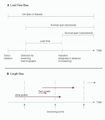
It is difficult to establish the value of a community-based screening program unless an RCT is conducted, as RCTs are less prone to bias than other types of study designs. An improvement in survival among women who have undergone breast cancer screening is often taken to imply that the test saves lives. However, association between receipt of screening and longer survival does not necessarily prove a cause-and-effect relationship, because a study might be limited by various forms of bias (Fig. 10-1) (1). Selection bias occurs when most participants in a screening program are healthier than average, so they will likely have a better overall rate of mortality. Lead-time bias occurs when screening detects a disease earlier in its natural history than would otherwise have happened, thereby lengthening the time between diagnosis and death. Nevertheless, having additional time during which the diagnosis is known seems unlikely to alter the natural history of the disease, so that no overall reduction in mortality will result. Length bias occurs when the full spectrum of a disease includes both indolent and aggressive cases, such that screening participants with less aggressive illness are likely to survive longer after diagnosis, regardless of the treatment they receive.


RANDOMIZED CLINICAL TRIALS OF SCREENING MAMMOGRAPHY


Summary of RCTs

Population-based randomized, controlled trials (RCTs) involving screening mammography have been conducted in North America and Europe with participation by nearly half a million women (46, 56, 57, 58, 59, 60, 61, 62, 63, 64, 65 and 66). These trials differ with regards to design, recruitment, participant characteristics, imaging protocols, management of control groups, compliance with assignment to screening and control group and analysis of outcomes (67). Almost all reported a mortality reduction for women screened by mammography.

Most randomized trials were not set up to specifically evaluate screening mammography for women less than 50 years of age, and the use of age 50 has been considered somewhat arbitrary. The recent AGE trial focused specifically on screening of women 40 to 49 years of age and found
a small reduction in breast cancer mortality from screening (65). However, the reduction did not reach statistical significance. Thus, the effectiveness of routinely screening women 40 to 49 years of age remains controversial, with concern regarding whether or not the magnitude of benefit from routine screening sufficiently outweighs the harms of false positives and overdiagnosis.


FIGURE 10-1 Lead-time (A) and length (B) bias. In part B, the length of the arrows represents the time required for the tumor to reach a palpable size. (From Institute of Medicine (IOM). Mammography and beyond: developing technologies for the early detection of breast cancer: a non-technical summary. The National Academies Press. http://www. nap.edu/openbook.php?record_id=10107&page=7)


Overview of Individual RCTs

The first RCT, the Health Insurance Plan of Greater New York (HIP), was met with great enthusiasm (45, 66). In this trial conducted from 1963 to 1966, women aged 40 to 64 years at entry were randomized to screening versus no screening. While there were slight imbalances in the distribution of women between assigned arms with regards to both menopausal status and education, these did not favor the screening nor the control group. The sample size was 30,239 women in the study group and 30,256 women in the control group with the intervention being two-view mammography annually and clinical breast examination (CBE) every 3 years. As in many of the other subsequent RCTs, noncompliance was an issue, with approximately 35% of the invitationto-screening cohort not attending the first screening. These women who did not attend their initial screening were not re-invited. In this early trial, screening mammography was not readily available outside of the clinical trial, as was often the case leading to contamination of the control groups in other subsequent RCTs. However, it is unclear whether CBE was performed with the same frequency in the two study arms. The follow-up duration for this study was 18 years with a relative risk of breast cancer death of 0.71 at 10 years, and at 0.77 at 15 years. Of note, the mammograms were performed with older equipment and may be of lower quality than current technologies (68). HIP also had differential exclusion between the intervention and control groups of women with a prior history of breast cancer.

The Malmo, Sweden study, which began in 1976, invited women aged 45 to 69 years for mammography screening (69, 70). This trial had 21,088 women in the intervention and 21,195 women in the control group, with 74% of women invited to screen attending their first screen, and 70% attending rounds 2 to 5. The intervention was two-view mammography every 18 to 24 months for nine rounds. The control group received mammography at the end of the study, after year 14. It is thought that about 24% of all control women had at least one mammogram. This study had 12 years of follow-up with a subsequent relative risk of breast cancer death at 0.81 (0.62-1.07). This Malmo study, which is often referred to as MMST1 Mammography Screening Trial 1, is often combined with the MMST2 trial for many analyses.

The Swedish Two-County Trial (71, 72 and 73), which began in 1977, enrolled women 40 to 74 years of age. The randomization was done through geographic clustering with
geographic units designed to be heterogeneous with regards to urban versus rural, population size, and socioeconomic factors. Women with preexisting breast cancer were excluded from both groups. This trial enrolled approximately 80,000 women to screening and just over 39,000 women in the control group from Ostergotland, Sweden, and approximately 39,000 women to screening and 18,000 in the control group from Kopparberg, Sweden. The intervention included one-view mammography every 2 years for women younger than 50 years and every 33 months for women 50 years and older. Contamination was much lower in this study compared to other RCTs; approximately 13% had mammograms as part of routine care, mostly in the later years of the study. The relative risk of breast cancer death for the screened population in the study was reported as 0.82 (0.64-1.05) in Ostergotland and 0.68 (0.52-0.89) in Kopparberg. Concerns have been raised about the randomization methods used as well as the analysis, which required correction for late performance of the control group mammography. However, the group from Sweden has performed subsequent meta-analysis that addressed many of these questions (70, 71, 74, 75 and 76).

Only gold members can continue reading. Log In or Register to continue

Stay updated, free articles. Join our Telegram channel

Jul 9, 2016 | Posted by in ONCOLOGY | Comments Off on Breast Cancer Screening

Full access? Get Clinical Tree

Get Clinical Tree app for offline access