Pathology of Benign Breast Disorders


Pathology of Benign Breast Disorders


Laura C. Collins

Stuart J. Schnitt


The term benign breast disorders encompasses a heteroge-neous group of lesions that may present as a palpable mass, a nonpalpable abnormality detected on breast imaging studies, or an incidental microscopic finding. Some are discrete lesions, such as fibroadenoma and intraductal papilloma, but a large number of benign breast biopsies exhibit a mixture of microscopic changes affecting the terminal duct lobular units. The two major goals in the pathologic evaluation of a benign breast biopsy are (a) to distinguish benign lesions from in situ and invasive breast cancer, and (b) to assess the risk of subsequent breast cancer associated with the benign lesion(s) identified.


BENIGN BREAST DISEASE AND BREAST CANCER RISK: NONPROLIFERATIVE LESIONS, PROLIFERATIVE LESIONS WITHOUT ATYPIA, AND ATYPICAL HYPERPLASIAS

It has been known for many years that some benign breast lesions are more highly associated with breast cancer than others. Two types of studies have evaluated this relationship. In the first type, the prevalence of benign alterations
in breasts with cancer was compared with their prevalence in breasts without cancer (1). While these studies demonstrated that some benign lesions are more common in cancer-containing breasts, the histologic coexistence of certain benign breast lesions with breast cancer is not sufficient to establish that those benign lesions impart an increased cancer risk.


TABLE 9-1 Categorization of Benign Breast Lesions According to the Criteria of Dupont, Page, and Rogers (3)


Nonproliferative


Cysts


Papillary apocrine change


Epithelial-related calcifications


Mild hyperplasia of the usual type


Proliferative lesions without atypia


Moderate or florid ductal hyperplasia of the usual type


Intraductal papilloma


Sclerosing adenosis


Fibroadenoma


Radial scar


Atypical hyperplasia


Atypical ductal hyperplasia


Atypical lobular hyperplasia


More recent studies have evaluated the subsequent risk of developing breast cancer in patients who have had a benign breast biopsy and for whom long-term follow-up is available (2, 3, 4, 5 and 6). In these studies, histologic sections of the benign biopsies were reviewed, and the type of benign lesions present were recorded and related to the risk of breast cancer. In some of these studies, it was also possible to study the interaction of the histologic findings with other factors, such as family history of breast cancer, time since biopsy, menopausal status, and other factors in determining cancer risk. The results of these studies have provided important information regarding the risk of breast cancer associated with benign breast lesions and this information is useful in patient treatment, counseling, and follow-up. These studies have further indicated that terms such as fibrocystic disease, chronic cystic mastitis, and mammary dysplasia are not clinically meaningful because they encompass a hetero-geneous group of processes, some physiologic and some pathologic, with widely varying cancer risks.


TABLE 9-2 Relative Risk of Breast Cancer According to Histologic Criteria of Benign Breast Disease in Four Studies Using the Criteria of Dupont, Page, and Rogers (3)


Histologic Category


Study


Study Design


Nonproliferativea


Proliferative without Atypia”


Atypical Hyperplasiaa


Nashville (3)


Retrospective cohort


1


1.9 (1.9-2.3)


5.3 (3.1-8.8)


Nurses’ Health Study (2)


Case-control


1


1.5 (1.2-2.0)


4.1 (2.9-5.8)


Breast Cancer Detection Demonstration Project (4)


Case-control


1


1.3 (0.8-2.2)


4.3 (1.7-11.0)


Mayo Clinic (5)


Retrospective cohort


1.3 (1.15-1.41)


1.9 (1.7-2.1)


4.2 (3.3-5.4)


a Numbers in parentheses represent 95% confidence intervals.


The seminal study evaluating benign breast disease and cancer risk is the retrospective cohort study of Dupont et al. (3, 7). In their study, the slides of benign breast biopsies from more than 3,000 women in Nashville were reviewed, and the histologic lesions present were categorized using strictly defined criteria (3, 7) into one of three categories: nonproliferative lesions, proliferative lesions without atypia, and atypical hyperplasias (Table 9-1). The risk of developing breast cancer was then determined for each of these groups. This system provides a pragmatic, clinically relevant approach to benign breast lesions and has been supported by a consensus conference of the College of American Pathologists (8). Studies from other groups have largely confirmed the initial observations of the Nashville group and have extended these findings by providing important new information regarding benign breast disease and breast cancer risk (2, 3, 4, 5 and 6) (Table 9-2).


Nonproliferative Lesions

Nonproliferative lesions, as defined by Dupont and Page (3), include cysts, papillary apocrine change, epithelial-related calcifications, and mild hyperplasia of the usual type.

Cysts are fluid-filled, round-to-ovoid structures that vary in size from microscopic to grossly evident (Fig. 9-1). Gross cysts, as defined by Haagensen (9), are those which are sufficiently large to produce palpable masses. Cysts are derived from the terminal duct lobular unit. The epithelium usually consists of two layers: an inner (luminal) epithelial layer and an outer myoepithelial layer. In some cysts, the epithelium is markedly attenuated or absent; in others, the lining epithelium shows apocrine metaplasia, characterized by granular eosinophilic cytoplasm and apical cytoplasmic protrusions (“snouts”).

Papillary apocrine change is characterized by a proliferation of ductal epithelial cells in which all of the cells show apocrine features as described above. Epithelial-related calcifications are frequently observed in breast tissue and may be seen in normal ducts and lobules or in virtually any pathologic condition in the breast. It should be noted that calcifications may also be seen in the breast stroma as well as in blood vessel walls. Mild hyperplasia of the usual type is defined as an increase in the number of epithelial cells within a duct that is less than four epithelial cells in depth. In this type of hyperplasia, the epithelial cells do not cross the lumen of the involved space.


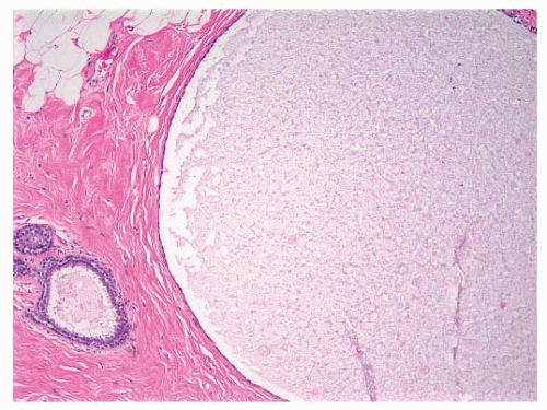
FIGURE 9-1 Cyst characterized by a large, dilated space filled with secretory material and lined by a flattened epithelial cell layer.

In the original study of Dupont and Page (3), 70% of the biopsies showed nonproliferative lesions. The risk of subsequent breast cancer among these patients was not increased, compared with that of women who have had no breast biopsy (relative risk [RR] 0.89), even in patients with a family history of breast cancer (in a mother, sister, or daughter). The only group of patients in the nonproliferative category with an increased risk of developing breast cancer was that with gross cysts plus a family history of breast cancer. The relative risk with gross cysts alone was 1.5, but was 3.0 in patients with gross cysts and a family history. It should be noted that, although Dupont and Page initially included fibroadenomas among the nonproliferative lesions, the results of a subsequent study by these investigators indicated a higher relative risk for breast cancer among patients with fibroadenoma than for patients with nonpro-liferative lesions (10). As a result, fibroadenomas are now included among the proliferative lesions without atypia (see the section on fibroadenomas).


Proliferative Lesions without Atypia

Included within the group of proliferative lesions without atypia are usual ductal hyperplasia (11) (also known as moderate or florid hyperplasias of the usual type), intraductal papillomas, sclerosing adenosis, and radial scars (3). As noted above, fibroadenomas are now included in this category as well. Women who have had a benign breast biopsy showing proliferative lesions without atypia, as defined previously, have a mildly elevated breast cancer risk, approximately 1.5 to 2.0 times that of the reference population (intraductal papillomas, radial scars, and fibroadenomas are discussed elsewhere in this chapter).

Usual ductal hyperplasias are intraductal epithelial proliferations more than four epithelial cells in depth. They are characterized by a tendency to bridge and often distend the involved space. The proliferation may have a solid, fenestrated or papillary architecture. If spaces remain within the duct lumen, they are irregular and variable in shape. These spaces are often slit-like and arranged around the periphery of the proliferation, with their long axes parallel to the basement membrane. The cells comprising this type of proliferation are cytologically benign and variable in size, shape, and orientation, and they often are arranged in a “swirling” pattern (Fig. 9-2). It is sometimes possible to discern multiple distinct cell populations, including epithelial cells, metaplastic apocrine cells, and myoepithelial cells (11).


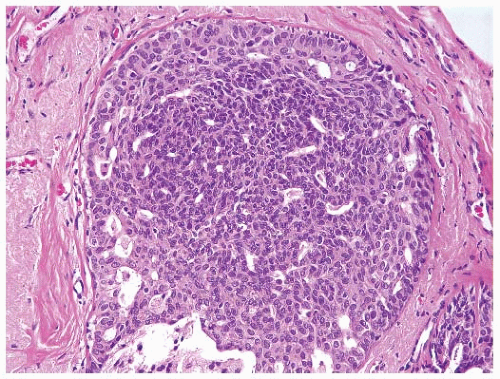
FIGURE 9-2 Usual ductal hyperplasia. A proliferation of cytologically benign epithelial cells fills and distends the duct. The nuclei vary in size, shape, and placement. The spaces within the duct are also variable in size and contour.

Sclerosing adenosis is usually an incidental finding, but may present as a mammographic abnormality (microcal-cifications, distorted architecture) or a mass lesion (also known as nodular adenosis or adenosis tumor). This lesion is composed of distorted epithelial, myoepithelial, and sclerotic stromal elements arising in association with the terminal duct lobular unit. This lobulocentric pattern is key to the correct diagnosis of sclerosing adenosis and its variants, and is best appreciated at low power microscopic examination (Fig. 9-3). The epithelium in sclerosing adenosis may undergo apocrine metaplasia, and is then referred to as apocrine adenosis. The apocrine metaplastic cells may show cytologic atypia, raising the differential diagnosis of invasive carcinoma if the lesion is examined at high microscopic power without accounting for the lobulocentric architecture appreciated at low power (12). Sclerosing adenosis may also be involved by atypical lobular hyperplasia, lobular carcinoma in situ, atypical ductal hyperplasia, or ductal carcinoma in situ (DCIS). Perineural “pseudoinvasion” may be present in approximately 2% of sclerosing adenosis cases and should not be confused with invasive carcinoma. Because of the distorted glandular pattern of sclerosing adenosis, this lesion may be confused with a lowgrade invasive carcinoma, particularly tubular carcinoma. In contrast to the lobulocentric pattern of sclerosing adenosis, tubular carcinoma is infiltrative in nature, however, and does not conform to the normal breast ductal and lobular microanatomy. Although sclerosing adenosis is composed of distorted, elongated, or obliterated glands and tubules, tubular carcinoma is composed of angulated tubules with open lumens. The stroma of sclerosing adenosis is fibrotic or sclerotic compared with the desmoplastic stroma of invasive carcinoma. Importantly, as opposed to tubular carcinoma, sclerosing adenosis contains myoepithelial cells, which may be highlighted by immunohistochemistry.


Atypical Hyperplasias

Atypical hyperplasias have been defined as proliferative lesions of the breast that possess some, but not all, of the features of carcinoma in situ and are classified as either ductal
or lobular type (3, 7). Atypical ductal hyperplasias (ADH) are lesions that have some of the architectural and cytologic features of low-grade DCIS, such as nuclear monomorphism, regular cell placement, and round regular spaces, in at least part of the involved space. The cells may form tufts, micropapillations, arcades, bridges, solid, and cribriform patterns (11). A second cell population with features similar to those seen in usual ductal hyperplasia is also typically present (Fig. 9-4).


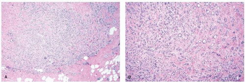
FIGURE 9-3 Sclerosing adenosis. (A) Low-power view demonstrates a lobulocentric proliferation of epithelial and stromal elements with scattered calcifications. (B) Higher-power view reveals glands and cords of epithelial cells entrapped in fibrotic stroma. The cells are cytologically benign, but the pattern simulates that of an invasive carcinoma.

Atypical lobular hyperplasia (ALH) is composed of cells identical to those found in lobular carcinoma in situ (LCIS). These cells are monomorphic, evenly spaced, and dyshesive, with round or oval, usually eccentric nuclei and pale cytoplasm often with intracytoplasmic vacuoles (Fig. 9-5). Although criteria for the distinction between ALH and LCIS differ among experts, we utilize the criteria proposed by Page and Anderson (13) and diagnose ALH when the characteristic cells are present but less than one-half of the acini of a lobular unit are filled, distorted, or distended. In addition to involving lobular units, the cells of atypical lobular hyperplasia may also involve ducts (14).


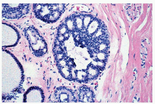
FIGURE 9-4 Atypical ductal hyperplasia. Near the center of this space is a proliferation of relatively uniform epithelial cells with monomorphic, round nuclei similar to those seen in low-grade ductal carcinoma in situ. However, these cells comprise only a portion of the proliferation within the space.

It is important to note that with the increasing use of mammographic screening, atypical hyperplasias are being diagnosed more frequently than in the past. For example, when a biopsy is performed because of a palpable mass, atypical hyperplasia is seen in only about 2% to 4% of cases (3). In contrast, atypical hyperplasia was identified in 12% to 17% of biopsies performed because of the presence of mam-mographic microcalcifications (15).

Women who have had a benign breast biopsy that demonstrates atypical hyperplasia are at a substantially increased risk for developing breast cancer, approximately 3.5 to 5.0 times that of the reference population. Some studies have suggested that the risk associated with ALH is greater than that associated with ADH (2, 7), but others have
not (16) (Table 9-3); at the present time this issue remains unresolved. Patients whose biopsies showed ALH involving both lobules and ducts had a higher relative risk of developing cancer (RR 6.8) than those with either ALH alone (RR 4.3) or those with only ductal involvement by atypical lobular hyperplasia (RR 2.1) (14).


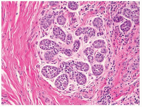
FIGURE 9-5 Atypical lobular hyperplasia. The acini of this lobule contain a proliferation of small uniform cells, which are dyshesive, and are identical to the cells that comprise lobular carcinoma in situ. However, the acini are not distended by this cellular proliferation.


TABLE 9-3 Relative Risk of Breast Cancer According to Type of Atypical Hyperplasia


Study/Reference


All Atypical Hyperplasia


Atypical Ductal Hyperplasiaa


Atypical Lobular Hyperplasiaa


Nashville (7)


5.3 (3.1-8.8)


4.7 (2.5-8.9)


5.8 (3.0-11.0)


Nashville (88)


3.1 (2.3-4.3)


Nurses’ Health Study (2)


4.1 (2.9-5.8)


3.1 (2.0-4.8)


5.5 (3.3-9.2)


Mayo Clinic (16)


3.9 (3.0-4.9)


3.8 (2.5-5.6)


3.7 (2.5-5.3)


a Numbers in parentheses represent 95% confidence intervals.


Columnar Cell Lesions and Flat Epithelial Atypia

Lesions of the breast characterized by enlarged terminal duct lobular units lined by columnar epithelial cells are being encountered increasingly in breast biopsies performed because of mammographic microcalcifications. Some of these lesions feature banal columnar cells in either a single layer (columnar cell change) or showing stratification and tufting, but without complex architectural patterns (columnar cell hyperplasia). In other columnar cell lesions, the lining cells exhibit cytologic atypia, most commonly of the low-grade, monomorphic type. Such lesions were included among lesions originally categorized by Azzopardi as “clinging carcinoma” (monomorphic type) (17), and were more recently included among lesions designated flat epithelial atypia (FEA) (11) (Fig. 9-6). The role of columnar cell lesions and, in particular, FEA in breast tumor progression is still emerging. FEA commonly coexists with well-developed examples of ADH, low-grade DCIS, and tubular carcinoma (11). These findings, in conjunction with the results of recent genetic studies (11), suggest that FEA is a neoplastic lesion that may represent either a precursor to, or the earliest morphologic manifestation of, DCIS. The few available clinical outcome studies suggest, however, that the risk of progression of FEA to invasive cancer is extremely low, lower even than that associated with ADH or ALH, supporting the notion that categorizing such lesions as clinging carcinoma and managing them as if they were fully developed DCIS will result in overtreatment of many patients (11). Additional studies are needed to better understand the biological nature and the level of subsequent breast cancer risk associated with these lesions.


Factors Modifying Breast Cancer Risk in Women with Biopsy-Proven Benign Breast Disease

A number of factors appear to modify the breast cancer risk associated with biopsy-proven benign breast disease, including a family history of breast cancer, time since biopsy, menopausal status, and the appearance of the background breast tissue.


Family History

There is general agreement that the presence of a family history of breast cancer in a first-degree relative (mother, sister, or daughter) is associated with a slight increase in the breast cancer risk in women with proliferative lesions without atypia (3, 4, 5, 6 and 7). The influence of family history on breast cancer risk in women with atypical hyperplasia is less clear, however. Dupont et al. (3, 7) reported that the risk among patients with both atypical hyperplasia and a family history of breast cancer was twice that of women with atypical hyperplasia without a family history. Similarly, in a study conducted by the Breast Cancer Detection Demonstration Project (BCDDP), the presence of a positive family history substantially increased the breast cancer risk among women with atypical hyperplasia (4). In a recent update of the Nurses’ Health Study (6) and in a recent study from the Mayo Clinic (5), the presence of a positive family history was not, however, associated with a further increase in breast cancer risk among women with atypical hyperplasia (Table 9-4). Additional studies are needed to clarify this important issue.


Time since Biopsy

Information regarding the relationship between time since benign breast biopsy and breast cancer risk is available from several studies. In the Nashville study, women with proliferative
lesions without atypia who remained free of breast cancer for 10 years after their benign breast biopsy were at no greater breast cancer risk than women of similar age without such a history. In addition, the breast cancer risk among women with atypical hyperplasia was greatest in the first 10 years after the benign breast biopsy (RR 9.8) and fell to a relative risk of 3.6 after 10 years (18). In contrast, in an analysis of data from the Nurses’ Health Study, the breast cancer risk among women with proliferative lesions without atypia was similarly elevated before and after 10 years following the benign breast biopsy (RR 1.4 and 1.6, respectively). In addition, the risk associated with atypical hyperplasia was higher after 10 years (RR 5.2) than in the first 10 years after the benign breast biopsy (RR 3.3) (2). Similarly, in the Mayo Clinic study, an excess breast cancer risk was seen among women with biopsy-proven benign breast disease for at least 25 years after the benign breast biopsy (5). Among patients with atypical hyperplasia, the relative risk was persistently elevated beyond 15 years (16). More data are needed to clarify further the relationship between time since biopsy and breast cancer risk for women with benign breast disease, particularly for women with atypical hyperplasia.


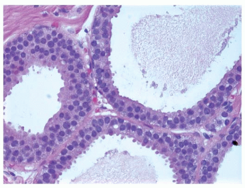
FIGURE 9-6 Flat epithelia atypia. The normal epithelial cells in the acini of this terminal duct are replaced by a population of columnar epithelial cells with round, monomorphic nuclei.


TABLE 9-4 Effect of Family History of Breast Cancer on Relative Risk of Breast Cancer


Proliferative without Atypia


Atypical Hyperplasia


Study


No Family Historya


Family Historya


No Family Historya


Family Historya


Nashville (3)


1.5 (1.2-1.9)


2.1 (1.2-3.7)


3.5 (2.3-5.5)


8.9(4.8-17.0)


Nurses’ Health Study (6)


1.5 (1.1-2.1)


2.5 (1.6-3.7)


4.3 (2.9-6.0)


5.4 (3.0-9.6)


Breast Cancer Detection Demonstration Project (4)


1.7 (0.9-3.2)


2.6 (1.0-6.4)


4.2 (1.4-12.0)


22.0 (2.3-203)


Mayo Clinic (5)


1.6 (˜1.4-2.0)b


2.2 (˜1.5-3.0)b


3.0 (˜1.7-4.9)b


4.0 (˜2.0-7.0)b


a Numbers in parentheses represent 95% confidence intervals.
b 95% confidence intervals estimated from Figure 2 in Ref. (5).


Menopausal Status

The risk of breast cancer among women with atypical hyperplasia appears to be influenced by the patient’s menopausal status. In the BCDDP study, premenopausal women with a biopsy showing atypical hyperplasia were at a substantially higher breast cancer risk (RR 12, 95% CI 1.0-68) than postmenopausal women with that diagnosis (RR 3.3, 95% CI 1.1-10) (4). In the Nurses’ Health Study, the breast cancer risk associated with atypical hyperplasia as a group was similar in premenopausal and postmenopausal women (RR 3.9 and 3.8, respectively). Among premenopausal women, however, the risk associated with ALH was greater than the risk associated with ADH (RR 7.3 and 2.7, respectively). In contrast, the risk associated with ALH and ADH were similar in postmenopausal women (RR 3.4 and 4.0, respectively) (2). Of note, in both the BCDDP study and the Nurses’ Health Study, the breast cancer risk among women with proliferative lesions without atypia did not vary according to menopausal status.

Another issue of clinical importance is the influence of postmenopausal hormone replacement therapy on the risk of breast cancer in women with biopsy-proven benign breast disease. Clinical follow-up studies have shown that women who take hormone replacement therapy are at increased risk for developing breast cancer (19). The use of hormone replacement does not, however, appear to further increase the risk in women with proliferative breast disease without atypia or in those with atypical hyperplasia. In an analysis from the Nurses’ Health Study among women with proliferative lesions without atypia, the relative risks of breast cancer were similar for those women who never used postmenopausal hormones, who were past users, and who were current users (RR 1.6, 2.1, and 1.9, respectively). Similarly, among women with atypical hyperplasia, the relative breast cancer risks were not significantly different for those who had not used hormone replacement, for past users, and for current users (RR 3.4, 3.0, and 2.5, respectively) (20). Thus, the available data suggest that the use of hormone replacement therapy does not further increase the breast cancer risk among women with a history of biopsy-proven benign breast disease, even among those with atypical hyperplasia.


Background Breast Tissue

A study from the Mayo Clinic has suggested that the presence of lobular involution in the background breast tissue of a benign breast biopsy is associated with a significant decrease in the risk of breast cancer. Furthermore, in that study the presence of lobular involution modified the risk in women with proliferative lesions without atypia and in those with atypical hyperplasia. For example, the relative risk for the development of breast cancer was 7.8 (95% CI 3.6-14.8) for women with atypical hyperplasia without involution in the background breast tissue and 1.5 (95% CI 0.4-3.8) for those with both atypical hyperplasia and complete involution of the background breast tissue (21). Similar results have recently been reported for women enrolled in the Nurses’ Health Study (22).

Only gold members can continue reading. Log In or Register to continue

Stay updated, free articles. Join our Telegram channel

Jul 9, 2016 | Posted by in ONCOLOGY | Comments Off on Pathology of Benign Breast Disorders

Full access? Get Clinical Tree

Get Clinical Tree app for offline access