Supportive Care in Hematologic Malignancies


Supportive Care in Hematologic Malignancies


Andrew J. Moore

Mary A. Vu

Stephen A. Strickland


Patients with hematologic neoplasms are at significant risk for complications both from the malignancy as well as the required therapy. The morbidity and mortality associated with the expected cytopenias, the therapeutic modalities, as well as the potential organ impairment present significant challenges to patients and health care providers alike. The frequency and severity of these complications vary among patients and according to the underlying disease process. Continued investigation and advances in antibiotics, antiemetics, and palliative therapies are allowing us to better meet the ever changing needs of our patients and ensure the provision of quality care. This chapter provides an overview of current recommendations and recent advances in the management of challenges encountered in the care of patients with hematopoietic malignancies.


INFECTIOUS DISEASE ISSUES RELATED TO HEMATOLOGIC MALIGNANCY


Deficits in Host Defense

Patients with hematologic malignancies are at increased risk for infection due to impaired host defense from the underlying malignancy along with concomitant illnesses or extrinsic factors including marrow-suppressive therapy. Hematologic malignancies are often associated with specific or global immune abnormalities that result in increased frequency of infections or opportunistic infections even in the absence of treatment. Understanding the different risk factors in the individual patient allows appropriate preventive strategies and treatments to be implemented more effectively. Defects in host defense mechanisms associated with specific malignancy are outlined in Table 69.1, whereas Table 69.2 identifies opportunistic infections by the host defects, such as those caused by impairment of phagocytosis (mainly neutropenia), defective production of circulating antibody (humoral immunity), and impaired cellular immunity. Concomitant illnesses, such as diabetes, nephrotic syndrome, cardiac disease, and liver disease contribute to infection risk and influence management decisions.1


Neutrophil Defects

Neutrophils play a critical role in the innate immune response, mediating both antimicrobial and inflammatory responses. They are produced in the bone marrow under the influence of an array of cytokines. Mature neutrophils circulate in the peripheral blood for only 3 to 6 hours, placing a demand on the marrow for an impressive capacity for constitutive neutrophil production that can be rapidly upregulated in response to acute bacterial, fungal, or inflammatory stresses.2 Deficits in neutrophil number and functional defects of mature neutrophils predispose to life-threatening infections, while disruption of the maturation sequence underlies the pathophysiology of myelodysplasia and leukemia.

Neutropenia, defined as a decrease of the peripheral blood neutrophil count below 0.1 × 10⁹/L, predisposes to bacterial and fungal infections. The severity (0.1 × 10⁹/L) and length of neutropenia (>2 weeks) contribute to the risk of serious infections.3 Neutropenia is usually caused by decreased production. Localizing signs and symptoms are often absent in the setting of severe neutropenia because of a lack of inflammatory response from absent granulocytes. Fever remains the most common sign of infection associated with neutropenia.

Neutropenia is a common complication of acute leukemia (AL) and is often prolonged during induction therapy.4 In chronic myeloid leukemia (CML), neutropenia typically occurs with the development of blast crisis, with the evolution of myelofibrosis (MF), or with therapy. Mild neutropenia is observed in patients with MF and multiple myeloma (MM), but it is uncommon during untreated phases. Neutropenia occurring in patients with Hodgkin lymphoma (HL) or non-Hodgkin lymphoma (NHL) is typically a result of marrow invasion with tumor or marrow fibrosis and occurs in conjunction with other cytopenias. Hairy cell leukemia patients may become neutropenic secondary to tumor cell invasion, splenomegaly, or both, but may also result from defects in cell-mediated immunity, monocytopenia, and decreased T cells after nucleoside analog therapy.5 In patients with T cell large granular lymphocytic leukemia, neutropenia may be the primary problem.

Functional defects in morphologically normal neutrophils have been described in hematologic malignancies, particularly myeloproliferative neoplasms (MPNs) and myelodysplastic syndrome (MDS).6 Such defects increase the susceptibility to infection.7 Neutrophils from untreated patients with CML may be mildly defective with respect to phagocytosis, oxygen consumption, and bactericidal capacity, and tend to have decreased concentrations of lactoferrin, elastase, collagenase, and peroxidase.8 Myeloblasts and lymphoblasts found in AL patients are of no benefit to the host against infection.


Deficient Immunoglobulin Production

The humoral immune response is one of the two main arms of the immune system. In this response, the immune system triggers specific B cells to proliferate and secrete their specific antibodies. Impaired humoral immunity is a major cause of frequent and severe infection in patients with hematologic malignancies. A decrease in Th2 CD4 T-lymphocyte-B-lymphocyte interaction results in decreased antibody production, complement-mediated damage, and phagocytosis. Diminished immunoglobulin synthesis is a major contributor to infection in patients with CLL, MM, and some B cell types of NHL.9,10,11 Myeloma and other plasma cell dyscrasias are often functionally hypogammaglobulinemic despite elevated total immunoglobulin. Splenectomized patients have impaired antibody response, reduced levels of tuftsin (natural activator of phagocyte cells), and are at increased risk for infections similar to those of patients with hypogammaglobulinemia, particularly encapsulated organisms (Streptococcus pneumoniae, Haemophilus influenzae, and Neisseria meningitidis).12


Defects in Cellular Immunity

Cellular immunity comprised of T lymphocytes, macrophages, and natural killer cells recognizes and combats pathogens that proliferate intracellularly. Cellular immune mechanisms are important in immunity to all classes of infectious agents, including most viruses and many bacteria (e.g., Mycoplasma, Chlamydophila, Listeria, Salmonella, and Mycobacterium), parasites (e.g., Trypanosoma, Toxoplasma, and Leishmania), and fungi (e.g., Histoplasma, Cryptococcus, and Coccidioides).13 T lymphocytes are activated by dendritic cells, macrophages, and B lymphocytes, which present foreign antigens in the context of the host’s own major histocompatibility complex antigen to the T cell receptor. Activated T cells then act in several ways to fight infection. Cytotoxic CD8+ T cells directly attack and lyse host cells that express foreign antigens. Helper CD4+ T cells stimulate the proliferation of B cells and the
production of immunoglobulins. Defects in cell-mediated immunity characterized by impaired Th1 CD4+ T lymphocytes and/or macrophage function results in increased risk of infections with intracellular bacteria, fungi, parasites, and viruses (Table 69.2).


TABLE 69.1 HOST DEFECTS PREDISPOSING TO COMPLICATIONS IN HEMATOLOGIC MALIGNANCIES


Disease


Host Defect


Complications


Acute myeloid leukemia


Neutropenia


Bacterial infections, including perirectal abscess, typhlitis, sinusitis; superinfections when hospitalized; increasing problem of methicillin-resistant Staphylococcus aureus and coagulase-negative staphylococci; Clostridium difficile colitis; aspergillosis with prolonged neutropenia; viral infections (herpes simplex)


Thrombocytopenia, disseminated intravascular coagulation


Hemorrhage


Hyperleukocytosis


Leukostasis, tumor lysis


Acute lymphoblastic leukemia


Neutropenia


Bacterial infections (see Acute myeloid leukemia)


Cellular immunity while on maintenance therapy


Pneumocystis jirovecii, disseminated varicella


Thrombocytopenia


Hemorrhage


Hyperleukocytosis


Tumor lysis, leukostasis


Chronic myeloid leukemia


Mild defects in neutrophil function


No increased risk of infections except in blast crisis


Thrombocytosis, platelet dysfunction


Increased risk of thrombosis and hemorrhage, similar to other myeloproliferative disorders


Chronic lymphocytic leukemia


Decreased immunoglobulins


Infections with encapsulated organisms (pneumococcus, Haemophilus influenzae, meningococcus)


Cellular immunity


Mycobacteria, fungal, viral (herpetic), Salmonella


Immune dysfunction


AIHA, ITP, red cell aplasia


Hodgkin lymphoma


Cellular immunity


Viral (herpes zoster, other), P. jiroveci, fungal, mycobacteria, listeriosis, Salmonella


Cytokine production


B symptoms, pruritus, eosinophilia


Splenectomized


Encapsulated organisms (above), increased risk of leukemia


Immune dysfunction


ITP, AIHA


Mediastinal disease


SVC syndrome


Non-Hodgkin lymphoma


Small B cell lymphoma


Decreased Igs


Similar infections to CLL


PTCL, particularly angioimmunoblastic and subcutaneous panniculitis like PTCL


Immune dysfunction


Cytokine production


AIHA


B symptoms, hemophagocytic syndrome, eosinophilia


Large B cell lymphoma


Mediastinal disease


SVC syndrome, pericardial disease


T-lymphoblastic lymphoma


Mediastinal disease


SVC syndrome, tumor lysis, CNS disease


Mantle cell lymphoma


Colonic polyposis


Gastrointestinal bleed


Burkitt lymphoma


Gastrointestinal primary


Obstruction, perforation, tumor lysis, CNS disease with advanced-stage disease


Multiple myeloma


Paraprotein


Hyperviscosity, hemorrhage


Decreased Igs


Similar infections to CLL


Osteoclast overactivity


Hypercalcemia


Waldenström macroglobulinemia


IgM paraprotein


Hyperviscosity, hemorrhage


Hairy cell leukemia


Neutropenia


Bacterial and fungal infections


Cellular immunity


P. jirovecii, atypical mycobacteria


Immune dysfunction


Periarteritis nodosa, lymphocytic vasculitis


Monocytopenia


T cell large granular lymphocyte leukemia


Neutropenia


Bacterial infections


Immune dysregulation, positive rheumatoid factor, antinuclear antibody


ITP, AIHA, red cell aplasia


Adult T cell leukemia/lymphoma


Cellular immunity


Opportunistic infections (Strongyloides stercoralis, P. jirovecii)


Parathyroid hormone-related protein


Hypercalcemia


AIHA, autoimmune hemolytic anemia; CLL, chronic lymphocytic leukemia; CNS, central nervous system; Ig, immunoglobulin; ITP, immune thrombocytopenic purpura; PTCL, peripheral T cell lymphoma; SVC, superior vena cava.


Multiple factors determine the severity and frequency of impaired cellular immunity. There may be differences in the stages of disease studied, the therapy used, and the sensitivity of the tests used to measure cellular immunity. Patients with HL often do not respond to new antigens and lose prior sensitivity as well.13,14 Patients with CLL usually show reduced or absent lymphocyte transformation with phytohemagglutinin but do not lose skin hypersensitivity to antigens such as old tuberculin.15 Patients with HL do not mount
either a primary or a secondary immune response, whereas those with CLL maintain secondary responses but cannot mount a primary response. Depressed cellular immunity is uncommon in AL16 except during maintenance therapy in ALL.17


TABLE 69.2 OPPORTUNISTIC INFECTIONS ASSOCIATED WITH DEFECTS IN IMMUNITY IN HEMATOLOGIC NEOPLASIA


Defect


Infections


Neutropenia


Gram-negative bacteremia (Escherichia coli, Pseudomonas, Klebsiella, Proteus)


Gram-positive bacteremia (methicillin-resistant Staphylococcus aureus, coagulase-negative staphylococci, Streptococcus viridans)


Fungemia (Candida species, aspergillosis)


Humoral immunity


Encapsulated organisms


Streptococcus pneumoniae


Haemophilus influenzae


Neisseria meningitidis


Cellular immunity


Bacteria


Listeria monocytogenes


Mycobacteria


Legionella species


Nocardia species


Salmonella species


Viruses


Herpes simplex


Varicella zoster


Parainfluenza, respiratory syncytial virus, cytomegalovirus


Fungi


Cryptococcus neoformans


Coccidioides immitis


Histoplasma capsulatum


Pneumocystis jirovecii


Parasites


Toxoplasma gondii


Strongyloides stercoralis


Approach to Infection in the Immunocompromised Host

The initial assessment of a febrile immunocompromised host is dependent on the underlying hematologic condition and other associated risk factors. It should focus on determining the potential sites and causative organisms and assessing the patient’s severity of illness. Although fever remains the most important clue to an infectious process, the characteristic signs and symptoms of infection may be absent in more than one-half of infected neutropenic patients, and routine cultures are often negative.3 It is estimated that 60% or more of neutropenic patients who become febrile have an established or occult infection.18 No known factors accurately predict which patients with fever and neutropenia are most likely to have bacteremia. As a result, a careful history and screening physical examination must be performed with special attention to the most common sites of infection: skin, oropharynx, nares, sinuses, lungs, GI tract (including perianal area), soft tissues, and indwelling catheter devices.

Risk assessment should be performed as part of the initial evaluation as it helps stratify the severity and facilitate goal-directed therapies. Assessing risk may determine the type of empiric antibiotic therapy (oral vs. intravenous), venue of treatment (inpatient vs. outpatient) and duration of antibiotic therapy. The most commonly used index for the stratification of risk for complications in febrile neutropenic patients is the Multinational Association for Supportive Care in Cancer (MASCC) index (Table 69.3).19 The following independent factors were found to be predictive of lower risk for complications: (1) burden of illness characterized by low or moderate symptoms, (2) absence of hypotension, (3) absence of chronic obstructive pulmonary disease, (4) presence of solid tumor or absence of previous fungal infection in patients with hematologic malignancies, (5) outpatient status, (6) absence of dehydration, and (7) an age less than 60 years. These variables predicting low risk were assigned an integer weight, and a risk index score consisting of the sum of these integers was derived. A score of 21 or greater identified low-risk patients with a positive predictive value of 91%, specificity of 68%, and sensitivity of 71%; whereas those with scores less than 21 are at higher risk for complications.19

In general, most experts consider high-risk patients to be those with anticipated prolonged (>7 days duration) and profound neutropenia (ANC < 0.1 × 10⁹/L) or significant medical comorbid conditions, including hypotension, pneumonia, new abdominal pain, or neurologic changes. High-risk patients warrant inpatient therapy with intravenous antibiotics. Lower-risk patients, including those with anticipated brief (<7 days duration), neutropenic periods, and few comorbidities, are candidates for empiric oral therapy.

Initial evaluation of neutropenic fever (Table 69.4) should include blood cultures, a urinalysis and urinary Gram stain, complete blood count with a differential, and blood chemistry tests to assess liver and renal function. Chest radiographs should be obtained for all patients with respiratory signs or symptoms, as well as viral swabs, and cultures of aspirated or biopsy material from accessible body sites that appear infected. At least two sets of blood cultures are recommended, with a set collected simultaneously from each lumen of an existing central venous catheter (CVC), if present, and from a peripheral vein site according to the updated guidelines from the Infectious Diseases Society of America (IDSA).3 An aggressive diagnostic workup is warranted if any localizing signs or symptoms are elicited. Patients who are unable to undergo invasive diagnostic procedures to determine an infectious etiology should be treated with empiric antibiotics until the appropriate diagnostic workup can be safely performed, or until the neutropenia resolves. Additional radiographic tests, such as computed tomography (CT) scans of the sinuses, chest, and abdomen, as well
as examination of cerebrospinal fluid, may be necessary in selected patients. Noninfectious causes such as drug reactions, mucositis, graft-versus-host disease (GVHD) should be considered in patients with persistent fever. Superinfection with fungi or Clostridium difficile needs to be considered as well. Frequent reassessment of the patient’s clinical status will help determine whether there is a need for additional coverage or changes to the ongoing antimicrobial regimen.


TABLE 69.3 THE MASCC RISK INDEX SCORE: DETERMINING THE RISK OF SERIOUS COMPLICATIONS IN FEBRILE NEUTROPENIA


Category


Weight


Burden of illness: no or mild symptoms


5


No hypotension


5


No chronic obstructive pulmonary disease


4


Solid tumor or no previous invasive fungal infection


4


Outpatient status


3


Burden of disease: moderate symptoms


3


No dehydration


3


Age < 60 y


2


MASCC, Multinational Association of Supportive Care in Cancer. The maximum theoretical score is 26 because the maximum favorable weight for burden of disease is 5.


Adapted from Klastersky J, Paesmans M, Rubenstein EB, et al. The Multinational Association for Supportive Care in Cancer risk index: A multinational scoring system for identifying low-risk febrile neutropenic cancer patients. J Clin Oncol 2000;18:3038-3051.


TABLE 69.4 EVALUATION OF FEVER IN IMMUNOCOMPROMISED PATIENTS


Cancer


Transplantation


Acquired Immunodeficiency Syndrome


Low Risk


High Risk


Bone Marrow


Solid Organ


Splenectomy


Children


Adults


History and physical examination


+


+


+


+


+


+


+


Hematologic


Complete blood cell and differential counts


+


+


+


+


±


±


±


Platelets


+


+


±


±


±


±


±


Coagulation studies


±


+


±


±


Microbiologic


Nose and throat


Sx


Sx


Sx


Sx


Urine


+


+


+


±


Sx


Sx


Stool


Sx


Sx


Blood


+


+


+


+


+


+


+


Cytomegalovirus PCR


+


+


Sx


Sx


Epstein-Barr virus PCR


Sx


Sx


Sx


Sx


Cerebrospinal fluid


±


±


Sxa


Radiologic


Chest


Sx


+


+


+


+


+


+


Sinus


±


±


±


Sx


±


Special studiesb


Sx


Sxc


Sxd


Sx


Sx


Sx


Sx


+, indicated; -, not necessary; ±, may be necessary; Sx, when symptoms are present. PCR, polymerase chain reaction.


a An evaluation of cerebrospinal fluid is especially important in patients with persistent fever.

b Special studies include computed tomography and magnetic resonance imaging.

c Abdominal computed tomography or magnetic resonance imaging to detect hepatosplenic candidiasis should be performed in patients recovering from neutropenia who have new or persistent fever.

d Lung computed tomography to detect pulmonary aspergillosis should be performed in patients with persistent fever and neutropenia who have had more than 1 wk of empirical therapy with antibiotics. Adapted from Pizzo PA. Fever in immunocompromised patients. N Engl J Med 1999;341:893-900.


Empiric Antibiotic Therapy: Current Guidelines and Regimens

The goal of initial empiric antibiotic therapy is to prevent serious morbidity and mortality due to bacterial pathogens until further culture results are available. In the early 1970s combination regimens incorporating two or three additive or synergistic drugs were used, such as antipseudomonal penicillin and an aminoglycoside. Subsequent regimens included beta-lactam and beta-lactamase inhibitor such as ticarcillin-clavulanic acid and piperacillin-tazobactam. Decades of well-studied clinical trials have not yet identified a superior empiric therapeutic regimen for the initial treatment of febrile neutropenic patients.20 Effective regimens (combination or monotherapy) share certain essential features, including bactericidal activity, antipseudomonal activity, and minimal toxicity. Initial choice of antimicrobials should be based on the risk status of the patient (low vs. high), on localizing signs or symptoms of infection, and on the epidemiology of pathogens and antibiotic susceptibility patterns in individual centers. Evidence-based guidelines are available for additional guidance on antibiotic therapy for neutropenic patients with fever.3,21,22 An algorithm for managing patients with febrile neutropenia is provided in Figure 69.1.


Initial Antibiotics for High-risk Patients

Monotherapy with selected broad-spectrum beta-lactams with activity against Pseudomonas species is as effective as combination antibiotic regimens (beta-lactam plus aminoglycoside) for empiric therapy of uncomplicated fever and neutropenia, and has fewer toxicities.23,24 Antibiotics recommended by the IDSA as appropriate monotherapy include the following: cefepime, imipenem-cilastatin, meropenem, and piperacillin-tazobactam.3 Many centers have found that ceftazidime is no longer reliable as empiric monotherapy for neutropenic fever because of decreasing potency against gram-negative organisms and poor activity against many gram-positive pathogens, such as streptococci.25, 26 and 27 Other antimicrobials (aminoglycosides, fluoroquinolones, and/or vancomycin) may be added to the initial regimen for management of complications such as hypotension and pneumonia, or there are specific clinical indications including suspected catheter-related infections, skin or soft-tissue infections, or if antimicrobial resistance is suspected or proven.

A meta-analysis of randomized trials involving cefepime reported that cefepime was associated with increased 30-day mortality when used for empiric therapy for neutropenic fever; however, no increase in infection-related mortality was noted.28 Subsequent analysis by the FDA failed to demonstrate a statistically significant increase in 30-day mortality for cefepime-treated
patients compared with controls, and thus concluded that cefepime remains a reasonable option for treating febrile neutropenia.28,29


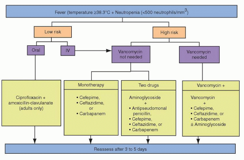
FIGURE 69.1. Empiric antibiotic regimens in febrile neutropenia.

Empiric combination therapy should be used in cases of severe sepsis or septic shock, or high prevalence of multidrug-resistant gram-negative bacilli. Effective antibiotic combinations include one of the aforementioned beta-lactams plus an aminoglycoside (choice based on local resistance, typically amikacin). Fluoroquinolones may be an acceptable alternative to aminoglycosides at institutions where prevalence of quinolone-resistant bacteria is low, or in patients with renal insufficiency. Febrile neutropenic patients with candiduria or oral thrush should also be covered by empiric antifungal therapy.


Role of Vancomycin and Other Agents in Gram-positive Coverage

Empiric vancomycin use in febrile neutropenia has led to considerable debate and concern, as the uncontrolled use of vancomycin facilitates vancomycin-resistant organisms. Randomized trial data provided by the European Organization for Research and Treatment of Cancer (EORTC) failed to demonstrate a true clinical advantage for empiric vancomycin in adults.30 It decreased the number of days the patient had fever, but did not improve survival and was associated with increased incidence of nephrotoxicity and hepatotoxicity. Empiric vancomycin should therefore be reserved for specific settings during neutropenia, including (1) hypotension or septic shock without an identified pathogen, (2) clinically apparent catheter-related infection, (3) positive blood cultures with a gram-positive organism prior to identification and susceptibility testing (linezolid or daptomycin are reasonable alternatives in environments with high prevalence of vancomycin-resistant enterococcus [VRE]), or (4) known colonization with methicillin-resistant Staphylococcus aureus (MRSA) or penicillin-resistant S. pneumoniae. Consideration should also be given to discontinuation of the empiric vancomycin after 2 to 3 days if the initial cultures are negative. Vancomycin plus aztreonam is an acceptable regimen in febrile neutropenic patients with allergies to beta-lactams.31


Lower-risk Patients with Fever and Neutropenia

Patients with fever and neutropenia can be stratified according to their risk of developing life-threatening infectious complications.21,22 Outpatient management with close monitoring and follow-up may be feasible for patients with MASCC scores of 21 or greater given the lower risk of complications. The IDSA, the American Society of Clinical Oncology (ASCO), and the National Comprehensive Cancer Network (NCCN) support the use of outpatient oral antibiotic therapy in carefully selected lower-risk patients with neutropenic fever. Ciprofloxacin plus amoxicillin/clavulanate is recommended for adult patients. Clindamycin can be substituted for amoxicillin/clavulanate in patients allergic to penicillins.3 Fluoroquinolones have an importance in the outpatient management of febrile neutropenic adults since they are the only class of oral antibiotics with activity against P. aeruginosa.


Evaluation of Response and Duration of Therapy

Modifications of the initial antimicrobial regimen are made based on new physical findings, microbiologic data, or persistent fever that indicates a resistant organism. It often takes 3 to 5 days to determine the efficacy of the initial antibiotic regimen (Fig. 69.2). Low-risk patients may become afebrile as early as 2 days, and high-risk patients may take as long as 7 days to have a response to the antibiotics. Antibiotics should be continued for a minimum of 7 days or until the documented infection has been eradicated. It is preferable to have neutrophil recovery (ANC > 0.5 × 10⁹/L) before discontinuing therapy. If the patient continues to be febrile 3 to 5 days after the initiation of empiric antibiotics, one of three choices can be made: continue the same antibiotics if the patient is stable and no source of infection has been found; change or add antibiotics if the patient develops a new complication, there are new findings on evaluation, or the patient appears to worsen clinically; or add an antifungal drug.3,32

Empiric amphotericin B has been replaced with the use of less toxic antifungals, such as caspofungin, micafungin, voriconazole, posaconazole, and the lipid formulations of amphotericin B. Risk of fungal infection (Aspergillus and Candida) rises precipitously in patients with profound neutropenia and persistent fevers after 7 to 10 days. Empiric antifungal therapy has been shown to reduce infectious mortality in patients with new or persistent fever occurring after 1 week of antibiotic therapy.33,34 Fluconazole is highly effective in the treatment of oropharyngeal and esophageal candidiasis and may be as effective as amphotericin B for systemic candidiasis in patients who are hemodynamically stable and have not been on antifungal prophylaxis. Broader spectrum antifungal agents are indicated in the setting of suspected Aspergillus,
atypical fungi, and several clinically relevant Candida species (C. krusei, C. tropicalis, C. lusitaniae, and Torulopsis glabrata) due to the absent or poor activity of fluconazole against these organisms.


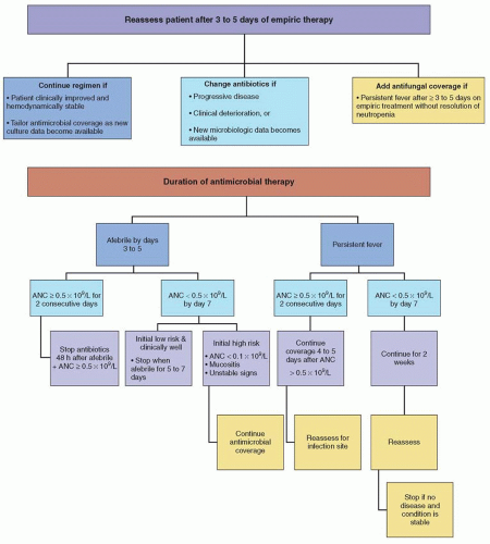
FIGURE 69.2. Common modifications of empiric antimicrobial therapy in the febrile neutropenic patient.

Echocardiography is recommended for S. aureus bloodstream infections to determine the presence or absence of endocarditis, and thus clarify the need for prolonged antibiotic therapy. Transesophageal echocardiography is more sensitive and preferred when compared with a transthoracic approach.35


Myeloid Colony-stimulating Factors

Prophylactic use of myeloid colony-stimulating factors (CSFs) is common in the setting of intensive chemotherapy regimens such as stem cell transplantation. Multiple randomized clinical trials of prophylactic recombinant granulocyte colony-stimulating factor (G-CSF) and granulocyte-macrophage colony-stimulating factor (GM-CSF) have shown benefits in reducing the time to neutrophil recovery and the duration of fever and hospitalization in patients with hematologic neoplasms.36,37 Prophylactic G-CSF and GM-CSF in autologous and allogeneic hematopoietic stem cell transplantation (HSCT) recipients have been associated with a small reduction in the risk of documented infections but do not appear to affect infection-related or treatment-related mortality.38 Empiric use of CSFs in the management of neutropenic fever is not standard practice, as no consistent benefit has been demonstrated in terms of morbidity or mortality among randomized controlled trials of their use in patients with febrile neutropenia.39,40 Neither ASCO nor EORTC recommend the routine use of growth
factors to treat episodes of febrile neutropenia unless patients are at high risk for infection-related complications, such as those older than 65 years, or those with prolonged (>10 days) and profound (<0.1 × 10⁹/L) neutropenia, active infection, hypotension, and/or multiorgan dysfunction39,40 (Fig. 69.3).


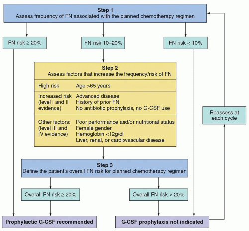
FIGURE 69.3. Algorithm to decide prophylactic G-CSF usage. FN, febrile neutropenia; G-CSF, granulocyte colony-stimulating factor. (With permission from Aapro MS, Bohlius J, Cameron DA, et al. 2010 update of EORTC guidelines for the use of granulocyte colony-stimulating factor to reduce the incidence of chemotherapy-induced febrile neutropenia in adult patients with lymphoproliferative disorders and solid tumours. Eur J Cancer 2011;47:8-32.)

G-CSF has a mild toxicity profile, with bone pain occurring 1 to 2 days prior to neutrophil recovery being the most common side effect. Recombinant yeast-derived GM-CSF reportedly has a better side effect profile than E. coli-derived GM-CSF (less fever, rash, and myalgias), although a randomized trial failed to reveal statistically significant differences.41 High-dose GM-CSF has been associated with hepatic transaminase elevation, serositis, fluid retention, venous thrombosis, and reactivation of autoimmune disease. A syndrome of hypoxemia, flushing, cardiovascular instability, musculoskeletal pain, nausea, and vomiting has also been described with intravenous GM-CSF.42

Pegfilgrastim offers once-per-cycle administration compared to the required daily doses of the nonpegylated CSFs, and a single dose of pegfilgrastim has been shown to exert a prolonged effect lasting for approximately 14 days or until neutrophil recovery.43 Two small phase II trials involving lymphoma patients demonstrated similar efficacy of pegfilgrastim versus filgrastim in regards to reduction of febrile neutropenia after chemotherapy administration.44,45


Granulocyte Transfusions

The role of granulocyte transfusions in the management of neutropenic fever is poorly defined. Although some small series and case report studies have attributed benefit to granulocyte transfusions in profoundly neutropenic patients with documented severe bacterial and fungal infections, others have failed to demonstrate significance.46,47 A Cochrane Review of 8 randomized controlled trials found the data inconclusive.46,48 The rationale for granulocyte transfusions is to provide support for the neutropenic patient with a life-threatening infection by augmenting the number of circulating neutrophils until autologous myeloid regeneration occurs. Rare adverse events associated with granulocyte transfusions include transmission of infections (commonly CMV), HLA alloimmunization, fever, potential transfusion associated graft-versus-host disease if granulocytes are not irradiated, and progressive platelet refractoriness. Appropriate patient selection is necessary prior to administration of granulocyte transfusions, given the risks and the marginal benefits reported.


CENTRAL NERVOUS SYSTEM INFECTIONS

Infection of the CNS can present with subtle nonspecific findings such as fever, headache, photophobia, changes in mental status, or more dramatic findings such as loss of consciousness and seizure. Prompt diagnosis and initiation of therapy are crucial to minimize negative clinical outcomes. CNS infections can be divided into surgical- and nonsurgical-related complications and infections. The IDSA has published guidelines on the management of CNS infections.95,96


Central Nervous System Infections Unrelated to Neurosurgery

Meningitis and encephalitis are part of a clinical spectrum of CNS disorders causing fever and meningismus. Encephalitis may manifest with signs and symptoms of meningeal inflammation, but is distinguished by the predominance of alterations of consciousness and neurologic deficits. Initial evaluation generally involves head CT to rule out intracranial bleeding in addition to brain MRI and lumbar puncture (assuming no contraindication). Cerebrospinal fluid (CSF) studies should be tailored to specific host factors, epidemiologic exposures, and clinical presentation, but should generally include a cell count with differential, glucose, protein, Gram stain, cryptococcal antigen, fungal culture, and bacterial culture. Noninfectious causes of meningitis include carcinomatous meningitis, nonsteroidal antiinflammatory medications, TMP/SMX, and serum sickness (e.g., associated with antilymphocyte gammaglobulin or intravenous immunoglobulin [IVIG]).

Empiric treatment for suspected bacterial meningitis should include antimicrobial agents that penetrate the blood-brain barrier and enter the CSF, such as ceftriaxone, ampicillin, and vancomycin.95 This regimen provides coverage against the most common causes of bacterial meningitis, including penicillin-resistant pneumococci and listeriosis. The combination of vancomycin and TMP/SMX may be used in patients allergic to penicillins.95 Cefepime or meropenem should be used instead of ceftriaxone in patients at risk for P. aeruginosa meningitis (e.g., neutropenia, neurosurgery within the past 2 months, allogeneic HSCT, prior history of P. aeruginosa infection). Meropenem should also provide appropriate coverage against Listeria. Conflicting results have been reported regarding the use of dexamethasone as an adjuvant therapy in the management of bacterial meningitis. Data
from a large meta-analysis found dexamethasone to not provide significant reductions in death or neurologic sequelae, although a statistically significant reduction in hearing loss was observed among surviving patients.97 ISDA guidelines for the management of bacterial meningitis support the incorporation of adjuvant dexamethasone in pediatric patients with H. influenzae type B meningitis and in adult patients with pneumococcal meningitis.95

Encephalitis in patients with cancer is most commonly caused by HSV. Intravenous acyclovir should be considered as empiric therapy for HSV in patients with suspected encephalitis (fever, mental status changes, CSF pleocytosis, and focal changes on EEG or MRI, especially in the temporal lobes). CSF studies should include PCR for HSV and CSF cytology. PCR for arboviruses should be considered in patients with exposure to endemic areas. The CSF should also be sent for nucleic acid amplification, adenosine deaminase level, and culture for tuberculosis in patients with known or suspected encephalitis. Patients with severe impairment of cellular immunity (e.g., allogeneic HSCT recipient, advanced AIDS) should be evaluated with PCR for CMV, VZV, HHV-6, and toxoplasmosis as well as culture for Nocardia. Most cases of encephalitis occur in HSCT patients and are due to reactivation of latent viral, bacterial, or parasitic infections: herpesviruses (HSV, VZV, CMV, EBV, human herpes virus [HHV]-6), adenovirus, mycobacteria, and Toxoplasma gondii.


Herpes viruses

HSV meningoencephalitis has been associated with older age, steroid therapy, and brain irradiation. The diagnosis is usually made by viral PCR from the CSF and enhancement of the temporal lobe on MRI. Intravenous acyclovir should be considered as empiric therapy for HSV.96 Other, less common herpesvirus infections can also be diagnosed by PCR of viral DNA in the CSF. Detection of high levels of EBV DNA should raise suspicion of EBV-related lymphoproliferative disorder, but primary EBV encephalitis and myelitis have been described.98,99 HHV-6 has been associated with a characteristic syndrome in HSCT patients consisting of confusion, lethargy, fever, rash, and hippocampal enhancement on T2-weighted MRI FLAIR images.100 The diagnosis is made by detection of HHV-6 DNA in the CSF by PCR. Ganciclovir or foscarnet may be used in treating HHV-6 infections.101 Adenoviral encephalitis in HSCT patients has been reported as part of disseminated adenoviral infection and treatment with cidofovir may be attempted.96

Other uncommon but important causes of meningoencephalitis are due to new primary infections rather than reactivation. Advanced age and cancer are risk factors for encephalitis from West Nile virus (WNV) transmitted by mosquitoes and for which there is no proven therapy.102


Brain Abscess

Brain abscesses that develop during neutropenia are typically caused by fungi (commonly Aspergillus and Candida).103 Bacterial abscesses may occur as a local extension of infection of the sinuses or dental caries and are often caused by a mixed aerobic and anaerobic flora (streptococci, Staphylococcus, Bacteroides). Other causes of CNS abscesses in patients with impaired cellular immunity include toxoplasmosis, nocardiosis, cryptococcosis, and mycobacterial infections.103

Noninfectious etiologies include CNS malignancies, including secondary lymphomas and EBV-associated posttransplant lymphoproliferative disease (PTLD) in patients with impaired cellular immunity. Given the broad differential diagnosis of new focal CNS lesions in the highly immunocompromised patient, a brain biopsy is recommended if feasible. Cultures and stains should include bacteria, fungi, mycobacteria, and Nocardia species. Serum galactomannan, CSF galactomannan, and beta-d-glucan testing are useful to facilitate a diagnosis of CNS aspergillosis.

Brain abscesses usually manifest with headache, focal neurologic findings, or seizures.104,105 MRI typically shows single or multiple lesions with edema and ring enhancement. Manifestations of CNS aspergillosis include focal seizures, hemiparesis, cranial nerve palsies, and hemorrhagic infarcts due to vascular invasion.106 Aspergillus brain abscesses are typically multiple, hypodense, and nonenhancing with little mass effect. CT scans with contrast enhancement may initially fail to reveal focal lesions, but subsequently evolve focal ring-enhancing or hemorrhagic lesions.107

Initial therapy with ceftriaxone plus metronidazole is advised in immunocompetent patients with a bacterial brain abscess.3,104,105,108 Patients with prolonged neutropenia should be treated with the combination of meropenem or cefepime plus metronidazole and voriconazole3,104 Adjustments may be needed for patients requiring certain antiseizure agents (e.g., phenytoin) due to the potential for significant drugdrug interactions with voriconazole (as well as itraconazole and posaconazole).109 Empiric high-dose TMP/SMX (trimethoprim component: 5 mg/kg every 8 hours) should be considered to cover toxoplasmosis and nocardiosis in allogeneic HSCT recipients and patients with severe T cell impairment. Retrospective analysis of CNS aspergillosis treated with voriconazole as either primary or salvage therapy indicated that 34% had a complete or partial response110 and compares favorably to previous reports in which frequency of successful responses to amphotericin B in CNS aspergillosis was close to zero.111


Cryptococcosis

Lymphoid malignancy and corticosteroid therapy are major risk factors for cryptococcal infection.112 Host defense against cryptococcal infection is dependent on T cell immunity. Isolated neutropenia is rarely associated with cryptococcal infection. The principal portal of entry of this organism is by inhalation. Spread to the blood and then to the central nervous system is a prerequisite for subsequent development of cryptococcal meningitis. Although meningitis is the most common presentation of cryptococcal infection, other manifestations include pneumonia, fungemia, cutaneous infections, and visceral dissemination. Visual loss may be a consequence of endophthalmitis (a space-occupying lesion in the visual pathway), direct invasion of the optic nerve, and elevated intracranial pressure.113

The IDSA recommends a regimen of amphotericin B (0.7 to 1 mg/kg daily) plus 5-fluorocytosine (100 mg/kg daily) for the first 2 weeks, followed by life-long maintenance fluconazole therapy in AIDS-associated cryptococcal meningitis.114 In the absence of modern randomized studies, the same induction regimen is recommended in non-AIDS-associated cryptococcal meningitis, followed by fluconazole for at least 10 weeks or until immunosuppressive agents have been discontinued.114 Reduction of the dosage of 5-fluorocytosine may be considered to minimize delay of myeloid recovery in neutropenic patients.


Toxoplasmosis

Reactivation of previously acquired infections is responsible for the majority of opportunistic CNS infections caused by T. gondii. The organism can be acquired by ingestion of undercooked meat or through contact with feline feces. CNS toxoplasmosis is typically associated with disseminated infection, and risk factors include therapy with corticosteroids, alemtuzumab, cytotoxic agents, and/or radiation therapy, and poorly controlled malignancy. Although toxoplasmosis is an uncommon complication of HSCT, nearly all occurrences are associated with seropositivity prior to transplantation.115 It tends to occur in the presence of moderate to severe GVHD with a median day of onset of disease at 64 days post-HSCT.116


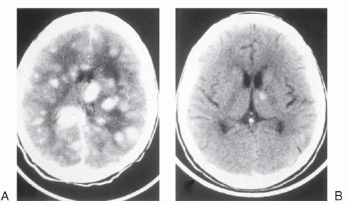
FIGURE 69.4. Central nervous system toxoplasmosis: magnetic resonance imaging study of the brain before therapy (A) and after therapy (B) with pyrimethamine and sulfadiazine.

Common clinical findings for CNS toxoplasmosis include altered mental status, coma, seizures, cranial nerve abnormalities, and motor weakness.117 MRI typically shows two or more lesions that may be ring-enhancing (Fig. 69.4). The differential diagnosis includes bacterial infection, invasive aspergillosis, nocardiosis, and malignancy. Differentiating toxoplasmosis from lymphoma is particularly difficult; [18F] fluorodeoxyglucose positron emission tomography typically shows increased metabolism in lymphoma.118 CSF in toxoplasmosis is usually normal but mononuclear pleocytosis and elevated protein levels may occur. In addition to CNS involvement, toxoplasma may also present with myocarditis, interstitial pneumonitis, culture-negative sepsis, and hemophagocytic syndrome. Definitive diagnosis of toxoplasmosis relies on demonstration of tachyzoites and cysts in histopathologic sections, but PCR testing of serum and CSF may facilitate earlier diagnosis.117,119 Oral sulfadiazine 1 to 1.5 g every 6 hours plus pyrimethamine (loading dose of 200 mg, followed by 75 mg daily) is the initial treatment of choice for toxoplasmosis. Folinic acid (10 to 20 mg daily) should be administered to reduce myeloid toxicity. At 4 to 6 weeks after resolution of symptoms and signs of infection and radiologic improvement, switching to a maintenance regimen (sulfadiazine 0.5 to 1 g four times daily plus pyrimethamine 50 mg/day) is reasonable. Maintenance therapy should be continued for the duration of immunosuppression and until radiologic resolution. Clindamycin and primaquine may be used instead, after verifying normal glucose-6-phosphate dehydrogenase activity in patients intolerant of sulfonamides. Atovaquone may also be used after exhausting other options.96


Dementias

Progressive multifocal leukoencephalopathy (PML) is a demyelinating disease associated with lytic infection of oligodendrocytes by the human polyomavirus JC virus.40 Patients may develop rapidly progressive dementia as well as focal motor neuropathy or cerebellar degeneration. This disease is most commonly seen in patients with advanced AIDS. However, PML is also seen in severely immunocompromised persons with hematologic malignancies and in HSCT recipients. Several reports have linked the use of rituximab with the development of PML and prompted an FDA advisory regarding a possible association.120 PML has also been reported with other immunosuppressants and biologics including belatacept,121 brentuximab,122 efalizumab,123 fludarabine,124 infliximab,125 and mycophenolate.126 Brain MRI typically reveals unilateral or bilateral white matter disease without mass effect or enhancement. Diagnosis is generally confirmed by detection of the JC virus in spinal fluid by PCR (90% sensitivity) or by brain biopsy. There is no established therapy for PML and the prognosis is poor.


Infections Related to Neurosurgical Procedures and/or Devices

Neurosurgical procedures such as resection of tumor, insertion of a shunt for hydrocephalus, or insertion of an Ommaya reservoir each carry a risk for infectious complications. Infection risk is further increased with use of steroids and brain irradiation. Early postoperative infections after placement of intraventricular devices are usually caused by skin flora: coagulase-negative staphylococci, S. aureus, streptococci, and Propionibacterium acnes. Enterobacteriaceae and P. aeruginosa account for approximately 10% of infections. Coagulase-negative staphylococci and P. acnes usually cause indolent late postoperative infections.

Infection of a shunt or an Ommaya reservoir may manifest with malfunction of the device, fever, or altered mental status. Overt signs of meningitis, such as meningismus and photophobia are often absent. CT imaging may suggest meningitis, ventriculitis, or a brain abscess if the device is infected at the proximal end. Evaluation of the CSF is required for diagnosis confirmation. Infections occurring in the distal region of the device may manifest as a soft tissue infection. In cases of ventriculoatrial shunts, distal infections may cause persistently positive blood cultures, thrombophlebitis, endocarditis, or septic pulmonary emboli. Distal ventriculoperitoneal shunt infections are associated with peritonitis and intraabdominal collections. Removal of the entire device plus systemic antibiotics is the most effective approach to eradicate infection. Use of parenteral and intraventricular instillation of antibiotics without hardware removal has demonstrated variable success, and recurrence of infection is common, particularly those caused by S. aureus.127 Antibiotic therapy should be tailored to the specific pathogen isolated. In acutely ill patients with meningitis suspected to be related to prior neurosurgery, empiric therapy with parenteral vancomycin should be administered to cover Staphylococcus, Streptococcus, and Propionibacterium species in combination with an agent with activity against Enterobacteriaceae and P. aeruginosa such as ceftazidime or meropenem.95


OROPHARYNGEAL AND SINUS INFECTIONS

The mucosal linings of the gastrointestinal and respiratory tracts constitute the first line of host defense against a variety of pathogens. During the course of treatment for hematologic malignancy these barriers often are compromised and expose patients to infections and invasions by local flora, resulting in bacteremia and candidemia. Chronic GVHD may further compromise mucosal immunity, including defective salivary immunoglobulin secretion.

Mucositis may extend into the esophagus, causing retrosternal chest discomfort. Mucosal lesions are usually caused by HSV and/or Candida species, but are occasionally caused by CMV, fungal (i.e., Histoplasma), or bacterial pathogens such as viridans group streptococcal infections and oral anaerobes (i.e., Pseudomonas species).

Oral mucosal candidiasis or thrush is common with T cell immunodeficiency. Cytotoxic chemotherapy, corticosteroids, and antibiotics predispose to oral candidiasis. The most common presentation is white adherent plaques on the palate, buccal mucosa, tongue, or gingiva. Pseudohyphae on a wet mount or Gram stain establish the diagnosis. Therapy includes local treatments such as clotrimazole troches or oral fluconazole.

Esophageal candidiasis may present as odynophagia and initial therapy with fluconazole is advised. Therapeutic options for fluconazole-resistant mucosal candidiasis include an echinocandin, voriconazole, posaconazole, or amphotericin B. Most fluconazole-resistant Candida isolates are susceptible to voriconazole and posaconazole, but cross-resistance may occur.


Congestion, sinus tenderness, and fever are common nonspecific signs of sinusitis. Respiratory bacterial pathogens, including S. pneumoniae, H. influenzae, and Moraxella catarrhalis predominate the etiology of sinusitis in immunocompetent patients. Common pathogens in those that are immunocompromised include P. aeruginosa, S. aureus, Enterobacteriaceae, and molds, and warrant investigation with sinus and facial imaging. Treatment of sinusitis in immunocompetent patients involves standard antibiotic regimens, such as amoxicillin-clavulanate, azithromycin, clarithromycin, or a cephalosporin with activity against respiratory pathogens. In addition to standard antibiotic therapy, neutropenic patients with symptoms or signs of sinusitis should have a CT scan of the sinuses and an otolaryngologist consultation and consideration of endoscopy to assess the possibility of invasive mold. Endoscopy with or without mucosal biopsy is often helpful in distinguishing these entities when there is no response to empiric antifungal or antiviral therapy or if clinical deterioration follows initial response to therapy. Systemic azole or amphotericin B therapy is preferred in patients with AIDS, neutropenic cancer patients, patients with recurrent perineal candidiasis, or those with refractory proven oropharyngeal candidiasis.87

The presence of a heterogeneous mass or bony erosion on CT is highly suggestive of invasive fungal sinusitis. Periorbital swelling and diplopia may be observed late in the course of infection. Aspergillus species are the most common isolate, but zygomycetes (e.g., Mucor, Rhizopus) as well as less common molds such as Fusarium are also being recognized. Aggressive surgical debridement in addition to antifungal therapy with two broad-spectrum antifungals is usually required to optimize the chances of recovery. Empiric therapy should include a lipid formulation of amphotericin B (5 mg/kg/d) to ensure treatment against Aspergillus species and zygomycetes.128 Antifungal therapy should be continued for weeks to months even if all of the visualized necrotic tissue is fully resected. Voriconazole may be substituted for many cases of fungal sinusitis, but should be avoided in zygomycetes (Mucor, Rhizopus) infections which are not susceptible to voriconazole. Posaconazole is an oral azole with activity against zygomycetes and may provide a useful alternative to an amphotericin B formulation for long-term therapy.129 Posaconazole therapy does require adequate food intake for optimal absorption to reach therapeutic levels.130


LUNG INFECTIONS

Pneumonia is the most common infectious cause of death in immunocompromised patients. Numerous noninfectious processes should also be considered in patients with pulmonary infiltrates: malignancy, its treatment (drug toxicity, radiation pneumonitis), congestive heart failure, pulmonary hemorrhage, pulmonary embolism with infarction, cryptogenic organizing pneumonia (also known as bronchiolitis obliterans organizing pneumonia), and acute respiratory distress syndrome. It is imperative that prompt empiric therapy should be initiated when an infectious cause is suspected. The differential diagnosis of pulmonary infiltrates in patients with hematologic malignancies is shown in Table 69.5. Noninfectious causes of interstitial pneumonitis (IP) in the febrile patient are diagnosed only after exclusion of infectious etiologies.


Pulmonary Infiltrates in Neutropenic Patients

Diagnosing the etiology of pulmonary infiltrates in neutropenic patients can be challenging for many clinicians. Physical findings of consolidation and sputum production may be absent as a result of the neutropenia. General work-up for suspected pneumonia should include routine blood cultures, a chest radiograph, and if possible, sputum culture and Gram stain. Respiratory viral pathogens should be considered during peak season and local pattern of infections. Rapid testing for influenza A and B may be performed using a throat or nasopharyngeal swab but viral culture is more definitive. Nasopharyngeal wash may also be used to diagnose respiratory viral infections. Legionellosis can be diagnosed based on urine antigen testing or sputum culture. While the urine antigen testing is more easily obtained, it only detects Legionella pneumophila type I.131 Broad-spectrum antibiotics should be initiated immediately and therapy tailored once culture data become available.


Initial Antimicrobial Therapy

In patients with less than 7 days of neutropenia, pulmonary infections are likely to be caused by Enterobacteriaceae, P. aeruginosa, and S. aureus. Community respiratory viruses should also be considered during winter months. If community-acquired pneumonia is suspected, a macrolide or fluoroquinolone should be given to cover for atypical pneumonia organisms. The addition of TMP/SMX should be considered if PJP is suspected. Vancomycin or linezolid should be added for pneumonia in patients colonized with MRSA and for nosocomial pneumonia. If clinical improvement occurs within 48 to 72 hours of therapy, the antibiotic regimen should be tailored based on available culture data and continued for at least 10 to 21 days and until neutropenia resolves. In the absence of clinical improvement, resistant bacterial and nonbacterial pathogens including filamentous fungi should be considered.


Pulmonary Infiltrates in Patients with Defects in Cell-mediated Immunity

In addition to the common bacterial causes of pneumonia, patients with defects in cell-mediated immunity are at risk for infections with P. jirovecii, Nocardia species, and viruses, as well as Legionella, mycobacteria, mold, and fungi. Invasive procedures, including bronchoscopy for bronchoalveolar lavage (BAL) and/or biopsy, should be considered to aid in diagnosis.


Fungal Pneumonia

Patients with allogeneic HSCT, prolonged high-dose systemic corticosteroids, immunosuppression with T cell suppressants and neutropenia for more than 10 days are at increased risk of fungal pneumonia. Chest CT imaging is recommended in patients failing to respond to initial empiric treatment or those in whom there is a high suspicion for fungal or mold infection.132 CT may disclose lesions missed by the chest radiograph as well as findings characteristic of invasive fungal disease: well-circumscribed, dense infiltrates, the “halo sign,” and/or cavitation.133 Other angioinvasive infections including P. aeruginosa may cavitate. The galactomannan assay is specific for invasive aspergillosis,134 whereas the beta-d-glucan assay detects aspergillosis and other invasive fungal infections including invasive candidiasis, PJP, and fusariosis.135 Galactomannan assay testing is associated with a sensitivity of 70% and specificity of 89% for invasive aspergillosis.136 False-positive results may be more common in children and allogeneic HSCT recipients with concomitant use of piperacillin/tazobactam.137,138 Rising serum galactomannan levels correlate with failure of antifungal therapy, whereas decreasing levels are associated with positive outcomes in patients with invasive aspergillosis.139,140

Aspergillus species are by far the most common invasive mold infections (Fig. 69.5). Voriconazole is currently the treatment of choice for invasive aspergillosis.34,137 Amphotericin B is active against Aspergillus species (with the exception of Aspergillus terreus), but may be inferior to voriconazole in aspergillosis.137 Initial antifungal therapy should be either with voriconazole or a lipid formulation of amphotericin B in patients at high risk for invasive mold diseases or with suspected pneumonia of unknown etiology. Echinocandins have not been evaluated as initial monotherapy for invasive aspergillosis, but have favorable response
rates in patients refractory or intolerant to triazoles or amphotericin B.141 Posaconazole has been used successfully as salvage therapy for a variety of invasive fungal infections refractory to standard therapy.142 Posaconazole is currently approved by the FDA for prophylaxis of invasive Aspergillus and Candida infections, and in the European Union is indicated for treatment of invasive aspergillosis and other invasive fungal infections refractory to standard antifungal agents.143


TABLE 69.5 DIFFERENTIAL DIAGNOSIS OF PULMONARY INFILTRATE IN THE IMMUNOCOMPROMISED PATIENT


Cause


Diagnosis


Infectious Causes


Bacterial pneumonia


Lung infiltrate: positive culture from blood, BAL, or pleural fluid; resolution with antibiotics.


CMV pneumonia


Intranuclear or intracytoplasmic CMV inclusions in pulmonary epithelial cells in TBB specimen or BAL fluid, or positive BAL culture for CMV. Quantitative PCR can be monitored for response.


Other viral pneumonias


May be preceded by rhinorrhea, sore throat, and cough; diffuse interstitial pattern. Most common are RSV, influenza, parainfluenza, and adenovirus. Diagnosis can be made by detection of virus or viral antigen on throat swabs, nasal washes, or BAL fluid; immunologic and molecular techniques (PCR) are available.


Legionella pneumonia


Positive culture of Legionella species from sputum, BAL fluid, or pleural fluid, or positive test for Legionella antigen by DFA test in BAL fluid, or a positive urinary Legionella pneumophila antigen or Legionella titer ≥1:256 with compatible clinical features.


Invasive pulmonary aspergillosis


Halo or crescent sign on high-resolution chest CT; positive galactomannan assay; pathologic evidence of invasion of lung tissue by acutely branching septate hyphae consistent with Aspergillus species, or isolation of Aspergillus species from BAL fluid or bronchial washing with consistent clinical and radiologic features.


Pneumocystis jirovecii Pneumonia


Fever and dyspnea may precede interstitial pneumonia. Detection of cysts or trophic forms of the organism in cytocentrifuge preparation in BAL fluid.


Candida and other fungal pneumonia


Histologic evidence of tissue invasion by Candida or other fungal species.


Mycobacterium tuberculosis


Positive culture of M. tuberculosis from sputum, bronchial washing, BAL fluid, or lung biopsy showing caseating granuloma with acid-fast organisms.


Nontuberculous mycobacteria (NTM) infection


Consistent clinical and radiologic findings with either: (a) isolation of NTM from sputum, bronchial washing, or BAL fluid and lung biopsy showing mycobacterial histopathology features; or (b) isolation of NTM from lung tissue, blood, or bone marrow culture.


Nocardia pneumonia


Presumptive diagnosis can be made if acid-fast filamentous rods are visualized on TBB or BAL fluid; also, Nocardia species are gram-positive rods. Requires 5-21 d for culture. Disseminated disease occurs in one-third of patients, with brain abscess as the most common site.


Noninfectious Causes


Diffuse alveolar hemorrhage


≥20% hemosiderin-laden macrophages in BAL fluid or hemosiderin-laden macrophages in lung tissue.


Chemotherapy-induced pneumonitis


History of receiving chemotherapeutic agent, with BAL or lung biopsy showing atypical type II pneumocytes with consistent clinical features and no evidence of lung infection.


Radiation pneumonitis


History of receiving radiation, with BAL lymphocytosis with consistent clinical and radiologic features and no evidence of lung infection.


Congestive heart failure (CHF)


Clinical diagnosis of CHF (bibasilar rales, left ventricular third sound, and radiologic evidence of pulmonary edema with resolution of symptoms with diuretics), or pulmonary artery occlusion pressure >18 mm Hg. Older age and prior chemotherapy, particularly with anthracyclines, are contributing factors.


Pulmonary emboli


Diagnostic confirmation strategy varies among institutions; suspected with clinical features, elevated D-dimer, and V/Q scan and confirmed by spiral CT pulmonary angiography.


Leukoagglutinin reaction


Sudden onset of respiratory distress after transfusion. The incidence of transfusion-related lung injury (TRALI) is 0.04%-0.1%; mortality is estimated at 5%-8%.


ARDS/diffuse alveolar damage


Acute-onset, bilateral lung infiltrates on chest radiography, pulmonary artery wedge pressure ≤ 18 mm Hg, or the absence of clinical evidence of left atrial hypertension, and PaO2/FiO2 ≤ 300 (acute lung injury) or ≤ 200 (ARDS).


Nonspecific pneumonitis


Lung biopsy showing nonspecific inflammation and variable fibrosis with no evidence of lung infection, and without alternative clinical diagnosis.


Pulmonary infiltrates of unclear etiology


Data from imaging, cultures, serology, and FB inconclusive for a firm diagnosis.


ARDS, acute respiratory distress syndrome; BAL, bronchoalveolar lavage; CMV, cytomegalovirus; CT, computed tomography; DFA, direct immunofluorescent antibody; FB, fiberoptic bronchoscopy; PCR, polymerase chain reaction; RSV, respiratory syncytial virus; TBB, transbronchial biopsy.


Endemic Fungi

Commonly known endemic fungi include Histoplasma capsulatum, Coccidioides immitis, and Blastomyces dermatitidis. These dimorphic fungi exist in nature in the fruiting mycelial stage and then convert to the yeast stage at body temperature. Endemic mycoses in the central United States include histoplasmosis and blastomycosis. Immunocompetent hosts are typically asymptomatic following inhalation of Histoplasma microconidia but may manifest acute fever, pulmonary infiltrates, and hypoxia. Immunocompromised patients have a higher risk of disseminated histoplasmosis involving the liver, spleen, lymph nodes, bone marrow, adrenal glands, mucocutaneous tissues, gastrointestinal tract, and CNS. Chest radiographs may show a miliary reticulonodular appearance similar to that seen with tuberculosis. Blood cultures may be positive in disseminated histoplasmosis. Antigen detection in blood, urine, and BAL is both
sensitive and specific.144 Antibody detection may also be useful, but false-negative results may occur in immunocompromised patients.145 Biopsy specimens showing small intracellular or narrow budding yeast are suggestive of the diagnosis and should be confirmed by culture. IDSA guidelines recommend amphotericin B for severe pulmonary or disseminated histoplasmosis.145 Prolonged therapy with itraconazole may be initiated after stabilization of disease, and should be continued for the duration of immunosuppression.145


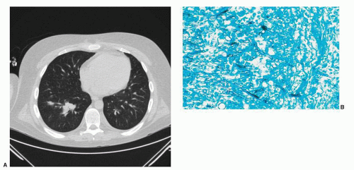
FIGURE 69.5. A: Chest CT of a hematopoietic cell transplant recipient who developed pulmonary Aspergillus infection after prolonged immunosuppression. B: Photomicrograph of the characteristic 45° angle branching of septate hyphal forms of Aspergillus. Gomori methenamine silver stain, × 400. (Courtesy of Margie Scott.)

Coccidioides immitis is endemic in the southwestern United States. C. immitis is more likely to be pathogenic in patients with compromised cell-mediated immunity. High rates of treatment failure and death have been reported in patients with hematologic malignancies.146 The diagnosis is most often established by finding the fungus in BAL, sputum, or biopsies. Serology is positive in only 55% of patients. Coccidioidomycosis can involve virtually any organ in disseminated disease but has trophism for bone and the CNS. Therapy for disseminated disease generally requires amphotericin B followed by maintenance fluconazole.147


Pneumocystis Jirovecii Pneumonia (PJP)

Pneumocystis jirovecii (formerly P. carinii) is classified as a fungus rather than a protozoan based on gene sequence data, although it lacks ergosterol, the main fungal cell-wall component. Defective T cell immunity and steroid use are risk factors for PJP. Pneumocystis jirovecii can have a fulminant course with rapid progression to respiratory failure in immunocompromised patients.148 Patients with pneumonia from P. jirovecii usually present with rapid onset of dyspnea, nonproductive cough, hypoxemia, and fever. Radiologic studies generally show diffuse bilateral interstitial infiltrates but can show focal infiltrates. Pleural effusion is uncommon. Diagnosis of PJP relies on visualization of the organism microscopically, as it does not grow in culture. BAL is the standard diagnostic modality for PJP, but induced sputum has acceptable yield in some institutions.149 Immunofluorescent staining with monoclonal antibodies is more sensitive than silver staining or Wright-Giemsa staining.150 PJP frequently results in positive serum beta-d-glucan testing.151 Treatment should be started based on clinical suspicion, and TMP-SMX (5 mg/kg IV every 8 hours) remains the treatment of choice.3 Prednisone should be added to the empiric treatment regimen if the pO2 is <70 mmHg.152 Alternatives for TMP/SMX-allergic/intolerant patients include dapsone, atovaquone, or clindamycin-primaquine for infections of moderate severity and IV pentamidine for infections of high severity.


Viral Pneumonia

Pneumonia due to respiratory viruses (respiratory syncytial virus [RSV], influenza, parainfluenza, adenovirus) is more common in patients with defects in cell-mediated immunity. Treatment with ribavirin (RSV, parainfluenza)153,154 with or without immunoglobulins or cidofovir (adenovirus)155 has not been shown to change outcome. Prompt treatment of RSV upper respiratory illnesses in HSCT recipients with a combination of aerosolized ribavirin and IVIG prevents progression to pneumonia in a pilot study.156 Oseltamivir and zanamivir are effective in HSCT and immunocompromised patients with influenza and are recommended in documented or suspected influenza infections.3,157

CMV pneumonia is a significant complication of allogeneic HSCT. It typically develops between 40 and 100 days posttransplant and presents with fever, dyspnea, hypoxemia, and diffuse interstitial infiltrates (Fig. 69.6). CMV pneumonia after day 100 is becoming more common and should be considered in patients with a history of previous CMV reactivation. In transplant patients the diagnosis is established by a compatible clinical syndrome with detection of CMV in BAL (by culture or cytopathology showing characteristic intracytoplasmic and intranuclear inclusions) or tissue (by culture or histologic diagnosis). CMV pneumonia is rare in nontransplant patients and the culture alone is not considered sufficient to make the diagnosis, as CMV can be shed from pulmonary secretions without causing invasive disease.158,159 Frontline treatment of CMV pneumonia typically consists of ganciclovir 5 mg/kg IV every 12 hours with IVIG 500 mg/kg every 48 hours for 3 weeks, although foscarnet (90 mg/kg every 12 hours) is an acceptable alternative to the ganciclovir.160


Hospital-Acquired Pneumonia

Hospital-acquired pneumonia (HAP) is considered “early” when it happens within 4 days of admission or “late” when it occurs 5 days or more after admission.70 Late HAP is more likely to be caused by multidrug-resistant pathogens. Risk factors for
multidrug-resistant pathogens include previous antimicrobials in the preceding 90 days, hospitalization for 2 days or more in the preceding 90 days, residing within a nursing home, chronic dialysis, home wound care, and exposure to others with multidrug-resistant pathogens.70 Initial therapy for HAP depends on severity of illness of the patient, previous antimicrobial exposures and hospitalizations, and institutional antibiogram data. Empiric therapy should include an antipseudomonal beta-lactam plus an antipseudomonal fluoroquinolone, or an aminoglycoside plus either linezolid or vancomycin to cover MRSA with subsequent tailoring of regimen once culture data becomes available.70


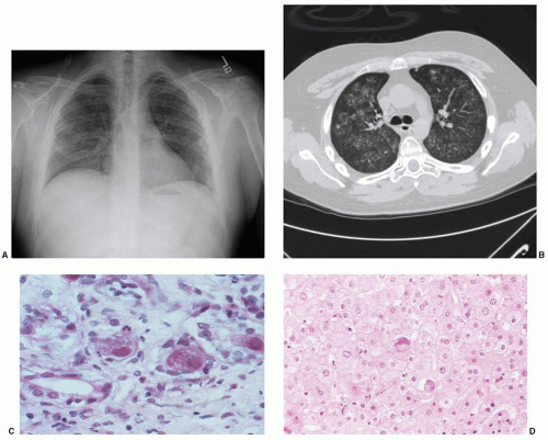
FIGURE 69.6. The spectrum of cytomegalovirus (CMV) disease in the abnormal human host. A: Chest radiograph demonstrating diffuse interstitial infiltrates in patient 60 days after cord blood transplant. B: Chest CT showing bilateral fine nodular infiltrates. C: CMV inclusion disease of the colon. Typical infected cells show cellular ballooning with dense primary nuclear inclusions surrounded by a thin, cleared rim; secondary inclusions appear as cytoplasmic granules after the nucleus has filled with virions. D: CMV hepatitis demonstrated on liver biopsy. Viral cytopathic effect may be difficult to establish, but rare viral inclusions with surrounding parenchymal changes are diagnostic of CMV. Hematoxylin and eosin stain ×400. (Photomicrographs courtesy of Margie Scott.)


GASTROINTESTINAL INFECTIONS


Neutropenic Enterocolitis (Typhlitis)

Typhlitis refers to inflammation of the cecum and results from a combination of neutropenia and defects in the bowel mucosa related to cytotoxic chemotherapy. When it extends beyond the cecum, the broader term neutropenic enterocolitis is used.110 Differential diagnosis for etiology of typhlitis includes C. difficile colitis, CMV enteritis, bowel ischemia, and GI tract GVHD.161 Typhlitis is pathologically characterized by ulceration and necrosis of the bowel wall, hemorrhage, and masses of organisms. Clinical signs include fever, abdominal pain and tenderness, and radiologic evidence of right colonic inflammation. Nausea, vomiting, and diarrhea (bloody or nonbloody) are the most common associated symptoms. Abdominal distention, tenderness, and a right lower quadrant fullness or mass reflect a thickened bowel. Bacteremia with bowel flora, P. aeruginosa, and polymicrobial sepsis may occur. Clostridium species are the most common anaerobic pathogens. Typhlitis should be suspected if right lower quadrant pain and bloody diarrhea are present. Surgical intervention may be required in the event of an intraabdominal catastrophe.

An abdominal and pelvic CT scan should be performed in patients with suspected typhlitis or undiagnosed abdominal pain in the setting of neutropenia. Positive CT scan findings are present in about 80% of cases162 and include a right lower quadrant inflammatory mass, pericecal fluid, soft tissue inflammatory changes, localized bowel wall thickening and mucosal edema,
and a paralytic ileus. All patients should be assessed for C. difficile infection. Treatment requires broad-spectrum antibiotics with activity against aerobic gram-negative bacilli and anaerobes (e.g., ceftazidime plus metronidazole, imipenem, meropenem, or piperacillin/tazobactam) and supportive care, including intravenous fluids or parental nutrition and bowel rest. The majority of patients will respond to antibiotic therapy and supportive care without the need for surgery. Indications for surgery often include (1) persistent gastrointestinal bleeding after resolution of neutropenia, thrombocytopenia, and clotting abnormalities; (2) intraperitoneal perforation; (3) uncontrolled sepsis despite fluid and vasopressor support; and (4) an intraabdominal process (such as appendicitis) that would require surgery in the absence of neutropenia.163


Clostridium Difficile Colitis

Pseudomembranous enterocolitis that is caused by Clostridium difficile may occur as a complication of antibiotic therapy, and stool should be assayed for the C. difficile toxin.164 The clinical presentation includes asymptomatic carriage, colitis without pseudomembrane formation, pseudomembranous colitis, and fulminant colitis with toxic megacolon. In severe C. difficile disease, paralytic ileus, toxic dilatation of the colon, and bowel perforation may occur. Abdominal radiographs may show nonspecific dilation of the colon with mucosal edema (“thumbprinting”). The mainstay of diagnosis is detection of C. difficile toxin A, toxin B, or both, in the stool with a cytotoxin test, enzyme immunoassay, or PCR for the toxin gene. Enzyme immunoassays have variable sensitivity, while DNA amplification testing for C. difficile has a reported sensitivity greater than 95%.165 Empiric therapy for C. difficile enterocolitis should be instituted despite a negative stool evaluation if suspicion remains high and other causes of diarrhea have been excluded. Traditional options for the treatment of C. difficile include oral or intravenous metronidazole and oral vancomycin.

Metronidazole is recommended as frontline therapy for uncomplicated cases, as its efficacy is similar to oral vancomycin, and the selection pressure on other flora is less dangerous.166 Oral vancomycin may be more efficacious in severe and refractory C. difficile colitis and should be considered for treatment in these cases.167,168 Fidaxomicin has also been found to be equivalent to oral vancomycin and is FDA approved for the treatment of C. difficile-associated diarrhea.169 Nitazoxanide and rifaximin are under investigation. Patients in whom oral agents cannot be administered should receive intravenous metronidazole. In cases involving toxic dilatation of the colon or perforation, subtotal colectomy, diverting ileostomy, or colostomy may be required.


GENITOURINARY INFECTIONS

Neutropenic patients with a urinary tract infection often do not show pyuria and are far more likely to become bacteremic compared with nonneutropenic patients. While treatment is typically reserved for symptomatic episodes in nonneutropenic patients, intervention should be considered in neutropenic patients with asymptomatic bacteruria. Candiduria may represent colonization in a patient with an indwelling urinary catheter, particularly in the setting of broad-spectrum antibiotics. Removal of the urinary catheter is frequently sufficient therapy. Patients with neutropenic fever and candiduria should receive systemic antifungal therapy over concerns of potential occult invasive candidiasis. Fluconazole 400 mg/day for 1 to 2 weeks is the treatment of choice. In the case of non-Albicans candiduria, another azole or amphotericin should be used. Echinocandins are minimally present in the urine, and they are not effective in the treatment of candiduria.170

Hemorrhagic cystitis is a common consequence of some cytotoxic regimens, particularly cyclophosphamide. A viral etiology should be considered in HSCT recipients and other immunocompromised patients with unexplained hematuria. Adenovirus, the polyomavirus BK, and CMV have been associated with hemorrhagic cystitis. Adenovirus hemorrhagic cystitis is usually self-limiting, but low-dose cidofovir (1 to 3 mg/kg/week, without probenecid) is occasionally used with the aim of preventing disseminated adenoviral disease.171 BK virus commonly reactivates after allogeneic HSCT, but only a minority of patients (typically those with high viral loads) develop hemorrhagic cystitis. Treatment consists of supportive care and reduction of immunosuppressants if possible.


SKIN AND SOFT TISSUE INFECTIONS

Skin eruption caused by infections must be distinguished from noninfectious ones such as drug reactions (including chemotherapy-induced hand-foot syndrome), Sweet syndrome, erythema multiforme, vasculitis, leukemia cutis, pyoderma gangrenosum, tumor, and GVHD. When evaluating the potential for a skin/soft tissue infection, careful examination of all line sites and perineal areas are essential. Early biopsy of skin lesions for histology and culture is recommended. Antimicrobial therapy should be tailored to the probable organisms: staphylococci and streptococci for catheter-associated processes and gram-negative and anaerobic organisms for perineal processes, respectively. Vancomycin may be considered for cellulitis, disseminated papules/lesions, and wound infections. Acyclovir, famciclovir, or valacyclovir should be considered for vesicular lesions after appropriate diagnosis is made for HSV or VZV.

VZV and herpes simplex virus (HSV) generally present as vesicular lesions and may be indistinguishable. Scrapings from the base of vesicles should be sent for direct fluorescent antibody (DFA) testing to diagnose VZV and for shell-vial culture to diagnose HSV. Intravenous acyclovir is the treatment of choice for VZV and HSV in the immunocompromised host.172 In immunocompetent patients, oral acyclovir, valacyclovir, and famciclovir have been used successfully.

Gram-positive bacteria that cause skin and soft tissue infections include Streptococcus (group A and B) and S. aureus. Gram-negative bacilli with propensity to cause dermatologic infections include P. aeruginosa, Stenotrophomonas maltophilia, Aeromonas hydrophila, and Vibrio vulnificus. The latter classically presents with septicemia and secondary cellulitis with hemorrhagic bullae following ingestion of contaminated seafood by patients with underlying liver disease. V. vulnificus may also present as a primary cellulitis with bacteremia when an open wound is exposed to seawater.

Ecthyma gangrenosum is typically a manifestation of P. aeruginosa or other gram-negative bacilli. It can be variable in its appearance but often presents as a dark, necrotic lesion in neutropenic patients. Antibiotic therapy with an antipseudomonal agent should be initiated and early surgical consultation for possible debridement is imperative.173

A rapidly progressive deep soft tissue infection with gas formation suggests clostridial myonecrosis (or polymicrobial necrotizing fasciitis) caused by Clostridium species. The lesions may become necrotic, bullous, and hemorrhagic. Systemic toxicity including fever, malaise, and mental status changes occur early. Extensive surgical debridement may be life-saving if initiated early, but the mortality rate remains high. Polymicrobial sepsis with enteric flora is commonly observed in association with clostridial bloodstream infection. In neutropenic patients, metronidazole plus an antipseudomonal cephalosporin or single-agent therapy with imipenem, meropenem, or piperacillin/tazobactam are recommended.174

Skin lesions of disseminated candidiasis are small, raised discrete erythematous papules.175 The lesions resemble those of heat rash early in their presentation. They are usually not tender.
Concurrent myalgias raise the possibility of Candida myositis. Biopsy and fungal staining of cutaneous lesions can provide an immediate clue to the diagnosis, prompting the early addition of antifungal therapy. Blood cultures are typically positive.


VIRAL INFECTIONS

Respiratory viruses include influenza, parainfluenza, respiratory syncytial virus, human metapneumovirus, adenoviruses, rhinoviruses, and coronaviruses. Most respiratory viruses typically cause self-limited infection in healthy persons, but cause significant morbidity and mortality in immunocompromised patients with hematologic malignancies and HSCT recipients.176 The role of empiric antiviral therapy in the treatment of neutropenic fevers is not well defined. Antiviral drugs are indicated in the management of neutropenic fevers only if there is clinical and laboratory evidence of active viral infection.3 Respiratory virus testing (including testing for influenza, parainfluenza, adenovirus, respiratory syncytial virus [RSV], and human metapneumovirus) and chest radiography are indicated for patients with upper respiratory symptoms such as cough and coryza. Although aerosolized and oral administration of ribavirin has been used, no antiviral agent has been proven to be effective against parainfluenza virus. Similarly, there is no clear indication for aerosolized or oral ribavirin or other antiviral against RSV pneumonia though modest effect has been observed in retrospective analysis.154


Influenza Virus

Influenza results in annual epidemics of respiratory viral illness during the winter. Unlike the mild and typically self-limited disease seen in immunocompetent patients, immunocompromised patients frequently have a more severe course. Several existing antiinfluenza agents are commercially available. Amantadine and rimantadine are active against influenza A, but not influenza B. Resistance to these agents may rapidly develop during therapy. The neuraminidase inhibitors, zanamivir and oseltamivir, are active against both influenza A and B. They are effective in reducing the duration of influenza illness if started early after onset of symptoms, and they have a prophylactic benefit during community outbreaks.177,178 Oseltamivir appears to be both safe and effective against influenza infections among HSCT recipients. If influenza is suspected, empiric therapy with an antiinfluenza agent (e.g., oseltamivir and zanamivir) should be initiated while awaiting test results3,178 Choice of drug should be based on the susceptibility patterns of the predominant influenza strain(s).


Respiratory Syncytial Virus

Respiratory syncytial virus (RSV) infection can cause high mortality in patients with acute leukemia and in HSCT recipients.179 Upper respiratory symptoms (sinusitis, coryza, rhinorrhea) usually precede lower respiratory tract involvement (dyspnea, wheezing) and pneumonia, but may be absent. Uncontrolled studies suggest that aerosolized ribavirin and IVIG containing high RSV neutralizing titers for at least 24 hours prior to respiratory failure prevent respiratory failure and result in much better outcomes.180 Inhaled ribavirin and immunoglobulin should be considered in patients with RSV infection at highest risk for severe complications, such as allogeneic HSCT recipients, patients with acute leukemia, and those with persistent neutropenia.3


Adenovirus

The spectrum of adenoviral infections in immunocompromised patients extends from asymptomatic shedding to fatal multisystem disease and includes upper respiratory tract infection, pneumonia, renal parenchymal disease, hemorrhagic cystitis, hepatitis, small and large bowel disease, and encephalitis. Viral shedding from throat secretions, urine, and stool is common, occurring in approximately 5% to 20% of HSCT recipients, and should not be equated with disease. Gastroenteritis and hemorrhagic cystitis are usually self-limited, whereas pneumonia and disseminated disease are associated with a high mortality rate. Adenoviral infection is more common in young transplant patients, those who receive T cell-depleted transplants, and those who receive stem cells from unrelated donors.155 Supportive care is important, though cidofovir may be beneficial.171


Parvovirus B19

Parvovirus B19 is a DNA virus that is transmitted via respiratory secretions and blood products. Infection in immunocompromised persons unable to mount a protective antibody response may cause prolonged fever, chronic pure red cell aplasia, thrombocytopenia, or pancytopenia. Predisposing conditions include acute and chronic leukemias, myelodysplastic syndrome, lymphoma, HSCT, potent antineoplastic chemotherapy, and systemic steroids. Diagnosis of parvovirus B19 infection in the immunocompromised patient relies on PCR detection of viral DNA from serum; these patients may be incapable of antibody responses. Treatment often consists of IVIG.181


INFECTIONS IN HEMATOPOIETIC STEM CELL TRANSPLANTATION PATIENTS

Autologous and allogeneic HSCT recipients have a unique set of infectious disease problems. Infection is reported as the primary cause of death in 8% of autologous HSCT patients and 17% to 20% of allogeneic HSCT recipients.182 High-dose chemotherapy or chemoradiotherapy preparative regimen causes short-term cytopenias and often mucositis, followed by profound immunosuppression with weeks to months of defective T cell-mediated immunity. All HSCT recipients experience profound immunosuppression at some point and the degree of immunosuppression experienced by individual patients varies greatly and is influenced by several factors. GVHD severity correlates with the degree of immunosuppression and infectious complications. This correlation is due to a variety of factors, including damage to lymphoid microenvironments, adverse effects of GVHD on homeostatic peripheral expansion, as well as the impact that chronic immunosuppression has on a reconstituting immune system. Recipient factors such as age, comorbidities, and infectious exposure prior to transplant contribute substantially to the risk for posttransplant infectious complications. Graft-associated factors also play an important role. Peripheral blood stem cell graft recipients show more rapid immune reconstitution,183 whereas umbilical cord blood transplantation in adults,184,185 and transplantation of profoundly T cell-depleted haploidentical grafts result in poor immune reconstitution and higher rates of infectious complications.

Most infections in autologous HSCT recipients occur during neutropenia or within the first few months after transplantation before reconstitution of cellular immunity. Recipients of CD34+ cell-enriched autografts appear to have a similar risk to allogeneic HSCT recipients for CMV and other opportunistic infections.186 Recipients of allogeneic peripheral blood stem cell grafts undergo more rapid immune reconstitution than those who receive marrow.187 Reconstitution of cellular and humoral immunity in allogeneic transplant patients occurs gradually over a period of 1 to 2 years. Duration of T cell immunity defects varies, depending on factors such as cancer type, manipulation of the stem cells preinfusion, and the age of the recipients.188 Infectious disease risk profiles vary depending on the type of transplant as well as the many variants associated with an HSCT (conditioning regimen,
degree of HLA matching, stem cell source, and GVHD prophylaxis). The spectrum of pathogens to which HSCT recipients are most susceptible follows a time line corresponding to the predominant immune defects as outlined in Figure 69.7.


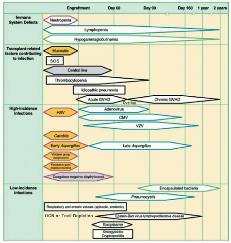
FIGURE 69.7. Phases of predictable opportunistic infections and complications among patients undergoing hematopoietic stem cell transplantation. CMV, cytomegalovirus; GVHD, graft-versus-host disease; HSV, herpes simplex virus; SOS, sinusoidal obstruction syndrome; UCB, umbilical cord blood; VZV, varicella zoster virus. Adapted from Van Burik J-AH, Freifeld AG. Infection in the severely immunocompromised host. In: Abeloff MD, Armitage JO, Niederhuber JE, et al., eds. Clinical oncology, 3rd ed. Philadelphia, PA: Churchill Livingstone; 2004:942.

Neutropenia is the principal host defect in the first few months after HSCT and predisposes patients to bacterial, fungal, and viral infections. The risk of bacterial infection, central venous catheter (CVC) infection, and reactivation of herpes simplex are highest in the preengraftment period. Prolonged neutropenia and antibiotic therapy leads to a steady rise in the risk of invasive fungal infection, which then decreases after engraftment. After myeloid engraftment, qualitative dysfunction of phagocytes persists because of immunosuppressive agents and corticosteroid. Cellular immune dysfunction peaks at approximately day 40 after engraftment and the typical onset of GVHD. The risk of opportunistic viruses and molds during this period is associated with the severity of GVHD and immunosuppressive regimens. Long-term venous access catheters continue to give rise to infection as long as the access is maintained. Continuation of prophylactic antimicrobials beyond 1 year depends on the individual patient’s infection history and the ability to wean immunosuppressive therapy.182

Preventing Early Infectious Disease (0 to 100 days after HSCT): Early after HSCT, neutropenia is the principal host defense defect, predisposing mainly to bacterial and fungal infections. Presence of a CVC remains as a risk factor for bacterial infection even after myeloid engraftment. Management of bacterial infections in the early neutropenic phase of the transplant is similar to the management of febrile neutropenia seen with any other myelosuppressive therapy. Bacterial infections account for over 25% of fevers in the preengraftment phase of allogeneic HSCT.189 The source of infection is usually the gut and oral flora due to mucositis, even though the increased use of CVCs has increased the risk of gram-positive bacteremias from skin flora. Streptococcal infections may be seen in patients with severe mucositis. The risk of late bacterial infections (those that occur after engraftment) depends on the immune status of the patient. Allogeneic HSCT recipients are at risk for bacterial infections with encapsulated organisms (Streptococcus pneumoniae, Haemophilus influenzae, and Neisseria meningitidis) if they develop chronic GVHD or profound hypogammaglobulinemia.190 Prophylactic fluoroquinolones are often maintained until marrow engraftment. This practice has been shown to reduce the incidence of infections, particularly
those due to gram-negative organisms, but has not been shown to definitively decrease infection-related mortality.81,176 Antibiotic prophylaxis has been found to significantly decrease the risk for all-cause mortality when compared with placebo or no treatment in neutropenic patients,81,191 IDSA and ASBMT guidelines do recommend prophylactic fluoroquinolone in high-risk patients with expected duration of neutropenia >7 days.182


Viral Infections


Cytomegalovirus

CMV infection has been a major source of morbidity and mortality in transplant recipients prior to the era of proper prophylaxis and monitoring for reactivation. Transplant recipients are at risk for reactivation (if CMV seropositive pre-HSCT) and also primary infection (from stem cells or blood products from CMV seropositive donors). Incidence of primary infection has decreased with the increasing use of leukofiltered blood products.

CMV infection is defined as the reactivation of the virus and the detection of the virus in the blood or other body fluids in the absence of organ-specific abnormalities (pneumonitis, hepatitis, colitis, and retinitis). CMV disease is defined as the isolation of the virus from body fluids or tissues in a symptomatic patient or the histopathologic evidence of CMV on tissue biopsy (see Fig. 69.6). Risk factors for the development of CMV disease include older recipient age, pretransplant seropositivity of the recipient or donor, or both, and severe acute GVHD.192,193 T cell depletion of the stem cell graft or treatment of the recipient with antithymocyte globulin for GVHD increases the likelihood of CMV reactivation.194

The incidence of CMV reactivation in allogeneic transplant recipients who are seropositive before transplant is 60% to 70%, as compared to a 10% to 30% incidence of primary infection in seronegative recipients.195, 196 and 197 Prior to the use of ganciclovir, CMV interstitial pneumonitis occurred in 15% to 30% of HSCT allograft recipients and the ensuing mortality was as high as 85%; however, mortality remained high at 30% to 50% even with the combined use of ganciclovir and CMV-specific immune globulin (Ig).194

Administration of CMV-safe or leukofiltered blood products is recommended to all seronegative autologous or allogeneic HSCT recipients to prevent primary CMV infection.195,198 CMV surveillance starting at the time of engraftment is recommended in instances of recipient and/or donor CMV seropositivity. Tests used for CMV surveillance include CMV pp65 antigenemia assay, DNA PCR detection methods, or CMV blood cultures.199 Detection of CMV in the blood is the strongest predictor of CMV disease, but 12% to 20% of patients with negative surveillance cultures still develop CMV disease. While high-dose intravenous acyclovir significantly reduces the incidence of all forms of CMV disease or delays the onset of CMV infection, it does not prevent CMV viremia.200,201

There are two recommended strategies for prevention of CMV disease. One strategy is prophylaxis with ganciclovir, valganciclovir, or valacyclovir, starting from engraftment until day 100, or longer if the patient remains at risk for CMV reactivation (active GVHD, high-dose steroids, low CD4 count).199,202 The other strategy is close surveillance and preemptive therapy with ganciclovir or foscarnet when CMV reactivation is detected. Both strategies reduce the incidence of CMV disease in the first 100 days; however, the median onset of CMV disease has shifted from 50-60 days to 160-176 days post-HSCT in the preemptive era.197

Preemptive therapy with ganciclovir for patients with positive CMV surveillance cultures (blood, urine, throat, or bronchoalveolar lavage fluid) has demonstrated improved survival at 100 and 180 days post-HSCT, but has failed to provide an overall survival advantage.203 Protracted ganciclovir prophylaxis can lead to emergence of resistant strains and the failure of natural immunity against CMV to develop, thus resulting in late recrudescence.196,204,205 Optimal preemptive therapy appears to be 1 to 2 weeks of twice-a-day induction followed by maintenance until PCR or antigen negativity.182

Ganciclovir is the drug of choice for CMV infection and disease, but its myelosuppressive effects may preclude its use in patients with significant cytopenias. Foscarnet is an equally effective alternative and can be used to treat ganciclovir-refractory CMV infections or in patients with significant cytopenias. Although it is not myelosuppressive, foscarnet is associated with renal toxicity and electrolyte imbalances.160 Cidofovir is another nephrotoxic antiviral with efficacy against CMV, but there are few data on its use in the stem cell transplant patient population.206 Maribavir is the latest antiviral in the armamentarium of drugs available for management of CMV and has been found to decrease rates of CMV infection when used as prophylaxis in the stem cell transplant setting,207 but has failed to prevent CMV disease in a randomized phase III study.208


Herpes Simplex Virus

HSV reactivation occurs in as many as 80% of seropositive allogeneic transplant recipients, causing mucocutaneous oral or genital lesions, esophagitis, and, occasionally, pneumonia or encephalitis. Testing all transplant recipients for herpes simplex virus exposure (HSV IgG) is recommended. Antiviral prophylaxis with acyclovir, valacyclovir, or famciclovir is recommended for all seropositive patients until the time of engraftment. Although its use is not recommended past 1 month after transplant, some patients with recurrent lesions might benefit from longer use of the prophylaxis.209,210


Varicella Zoster Virus

Impaired cellular immunity is the principal risk factor for VZV disease. Current recommendations are to test every transplant patient for varicella zoster virus serostatus (IgG). VZV reactivation may occur at any time after engraftment in autologous and allogeneic transplant recipients. Disseminated VZV is seen in as many as 30% of cases and is associated with a high mortality. Many centers administer oral acyclovir or valacyclovir for ˜12 months after transplant to VZV-seropositive patients.210 Seronegative patients should be given varicella zoster immune globulin within 96 hours of exposure to a VZV vaccine or upon contact with active infection.


Fungal Infections

Most fungal infections in the SCT population are due to Candida or Aspergillus. The etiology in the remaining <10% are uncommon fungi, such as Fusarium, Scedosporium, Blastomyces, and Histoplasma. Prevention of fungal infections in these patients is key. Fluconazole prophylaxis at a dose of 400 mg/day, beginning at the time of transplant and until the time of engraftment, provides adequate protection against invasive yeast infections.189,211,212 Failure of fluconazole prophylaxis against Candida is usually a result of the emergence of resistant yeast forms (C. krusei, C. glabrata). Empiric therapy for fungal infections in febrile neutropenic patients is considered the standard of care, and several studies are examining the preemptive approach.33,141 The toxicity of intravenous amphotericin B is substantial and it is no longer recommended for prophylaxis. Lipid formulations of amphotericin (Abelcet, Amphotec, and AmBisome) have fewer renal and infusional toxicities. Newer antifungal agents have changed the approach to prevention and treatment of fungal infections in SCT patients. Caspofungin, micafungin, and anidulafungin are echinocandins with a broad spectrum of activity against several species of Candida and Aspergillus. Triazoles such as voriconazole, posaconazole, and ravuconazole also have activity against Aspergillus and a wide variety of yeasts and molds.137

Only gold members can continue reading. Log In or Register to continue

Stay updated, free articles. Join our Telegram channel

Oct 21, 2016 | Posted by in HEMATOLOGY | Comments Off on Supportive Care in Hematologic Malignancies

Full access? Get Clinical Tree

Get Clinical Tree app for offline access