Staging of Testicular Cancer


Staging of Testicular Cancer


▪ 32A Staging and Imaging in Germ Cell Tumors

Brett S. Carver

Darren R. Feldman

Testicular cancer is the most common malignancy in men aged 20 to 35 and accounts for approximately 1% of all male malignancies. It was projected that in the United States, approximately 8,400 new cases of testicular cancer would be diagnosed and 380 men would die of this disease in 2009 (1). Testicular cancer refers to any malignancy that originates in the testis as can occur with a variety of different tumor types (see chapter 31). However, germ cell tumors (GCT) comprise the vast majority (>95%) of testicular cancer cases with sex cord stromal tumors, lymphomas, mesotheliomas, and rete testis adenocarcinomas being much more rare. It is worth noting that up to 10% of GCT also originate outside of the testis (referred to as extragonadal GCT), with the mediastinum, retroperitoneum, and pineal glands being the most frequent sites.

GCT can be divided into two major histologic subgroups: seminoma and nonseminoma, each accounting for about 50% of cases. Seminoma presents most frequently in the fourth decade of life, and by definition requires that no nonseminomatous component be present and serum alpha-feto protein levels to be normal. The remainder of GCT are comprised of nonseminomatous histology (embryonal cell carcinoma, yolk sac tumor, choriocarcinoma, and teratoma) and frequently present in the third decade of life. While the treatment paradigms differ for seminomatous and nonseminomatous tumors, the successful multidisciplinary approach for the management of GCT has resulted in survival rates of >95% overall (2,3).


PRESENTATION AND MANAGEMENT OF THE PRIMARY TUMOR

The most common symptom at the time of diagnosis is painless swelling or enlargement of the testis. Acute testicular pain is reported to occur in approximately 10% of patients with testicular cancer and often represents infarction or hemorrhage within the tumor. At initial presentation, symptoms manifesting secondary to metastatic disease occur in approximately 20% of patients and include a mass in the left neck, pulmonary complaints such as hemoptysis or dyspnea, abdominal mass, or back pain that can often be disabling. In approximately 5% of patients, gynecomastia or tenderness of the breast is reported. Frequent delays in diagnosis have been reported in the literature ranging from 2.5 to 4.4 months with longer durations of delay associated with more advanced clinical stage at diagnosis (4,5,6). Reasons for the delay in diagnosis are multifactorial but may include both a physician and patient component. Previous reports have shown that approximately 18% to 33% of patients with testicular cancer were initially treated for epididymitis by their physician (4,5). Patients have also been initially misdiagnosed, undergoing unnecessary mastectomy, for gynecomastia, or exploratory laparotomy, for retroperitoneal mass, which ultimately delays and increases the burden of therapy (6,7).

Testicular ultrasonography is the initial imaging modality of choice with a >95% sensitivity and specificity in identifying intratesticular lesions. On ultrasonography, seminomas are characterized as well-defined intratesticular, homogeneous hypoechoic lesions without cystic areas. Nonseminomas are typically heterogeneous on ultrasonography with an irregular border, and calcifications and cysts are commonly present. The serum tumor markers (STM)—alpha-fetoprotein (AFP), human chorionic gonadotropin (HCG), and lactate dehydrogenase (LDH)—have a clear role in both the diagnosis and clinical management of testicular GCT. Elevation of one or both markers occurs in 80% of metastatic GCT of the testis.

A radical orchiectomy with high ligation of the spermatic cord at the level of the inguinal ring provides histopathological diagnosis, primary tumor staging, and excellent local control of the tumor, with minimal morbidity and no mortality.


SERUM TUMOR MARKERS IN GERM CELL TUMOR

The STM—AFP, HCG, and LDH—have a clear role in both the diagnosis and clinical management of testicular GCT. Elevation of one or both markers occurs in 80% of metastatic GCT of the testis, although the rate is significantly less for seminoma than nonseminoma.

AFP is a glycoprotein produced in the liver, gastrointestinal tract, and fetal yolk sac, and its secretion in GCT is restricted to nonseminomatous histology. Therefore, any patients with histologic pure seminomas and an elevated serum AFP are classified and managed like those with nonseminomatous germ cell tumors (NSGCT). AFP is elevated in approximately 60% of patients with metastatic NSGCT and 20% of patients with clinical stage I NSGCT. HCG is a glycoprotein produced by the synctiotrophoblasts and is elevated in approximately 15% of pure seminomas, and in 40% of advanced nonseminomas. The serum half-life of AFP and HCG are approximately 5 days and 24 hours, respectively. LDH comprises multiple isoenzymes, but in clinical practice, the total LDH value is utilized for decision making. Increases in serum LDH correlate with tumor burden, growth rate, and cellular proliferation. Although elevation of LDH is present in approximately 60% of patients with advanced NSGCT and 80% of patients with metastatic seminoma, it is much less specific for GCT than either AFP or HCG. As such, the presence of an elevated LDH by itself does not signify evidence of metastasis or recurrence. However, it can be a useful gauge of response in some patients.


INITIAL CLINICAL STAGING AND RISK STRATIFICATION

Initial clinical staging evaluation must include a thorough history and physical exam, with particular attention to possible sites of metastases and the contralateral testis. STM (AFP, HCG, LDH) should be obtained prior to and following radical
orchiectomy. The STM are necessary for diagnosis, staging, and risk classification. While a normal postorchiectomy STM does not preclude the finding of metastatic disease, a persistent elevation of either AFP or HCG does signify the presence of metastasis. Therefore, it is essential that STM that are elevated preorchiectomy be followed closely after surgery to ensure decrease at the expected half-life and ultimately normalization. Evaluation for metastatic disease should include a CT scan of the abdomen and pelvis and either a chest x-ray or CT scan of the chest. Further imaging such as bone scan or brain MRI is unnecessary except when guided by symptoms or select clinical scenarios (see below).


TABLE 32A.1 TESTICULAR CANCER STAGING SYSTEM OF THE AMERICAN JOINT COMMITTEE OF CANCER AND THE INTERNATIONAL UNION AGAINST CANCER


Definition of TNM


Primary Tumor (T)


PTX


Primary tumor cannot be assessed (if no radical orchiectomy performed, TX is used)


PT0


No evidence of primary tumor (histologic scar in testis)


PTis


Intratubular germ cell neoplasia (carcinoma in situ)


PT1


Tumor limited to the testis and epididymis and no vascular/lymphatic invasion


PT2


Tumor limited to the testis and epididymis with vascular/lymphatic invasion or tumor extending through the tunica albuginea with involvement of tunica vaginalis


PT3


Tumor invades the spermatic cord with or without vascular/lymphatic invasion


PT4


Tumor invades the scrotum with or without vascular/lymphatic invasion


Regional Lymph Nodes (N)


Clinical


Nx


Regional lymph nodes cannot be assessed


N0


No regional lymph node metastasis


N1


Lymph node mass 2 cm or less in greatest dimension or multiple lymph node masses, none more than 2 cm in greatest dimension


N2


Lymph node mass, more than 2 cm but not more than 5 cm in greatest dimension, or multiple lymph node masses, any one mass >2 cm but not more than 5 cm in greatest dimension


N3


Lymph node mass more than 5 cm in greatest dimension


Pathologic


PN0


No evidence of tumor in lymph nodes


PN1


Lymph node mass, 2 cm or less in greatest dimension and ≤5 nodes positive, none >2 cm in greatest dimension


PN2


Lymph node mass, more than 2 cm but not more than 5 cm in greatest dimension, more than 5 nodes positive, none >5 cm, evidence of extranodal extension of tumor


PN3


Lymph node mass more than 5 cm in greatest dimension


Distant Metastases (M)


M0


No evidence of distant metastases


M1


Nonregional nodal or pulmonary metastases


M2


Nonpulmonary visceral metastases


Serum Tumor Markers (S)


LDH


HCG


AFP


S0


≤ULN


≤ULN


≤ULN


S1


<1.5 × ULN


<5,000


<1,000


S2


1.5-10 × ULN


5,000-50,000


1,000-10,000


S3


>10 × ULN


>50,000


>10,000


Stage Grouping


T


N


M


S


Stage 0


pTis


N0


M0


S0


Stage Ia


T1


N0


M0


S0


Stage Ib


≥T2


N0


M0


S0


Stage Is


Any T


N0


M0


S1-S3


Stage IIa


Any T


N1


M0


S0,S1


Stage IIb


Any T


N2


M0


S0,S1


Stage IIc


Any T


N3


M0


S0,S1


Stage IIIa


Any T


Any N


M1


S0,S1


Stage IIIb


Any T


Any N


M0,M1a


Stage IIIc


Any T


Any N


M0,M1


S3


In 1997, the American Joint Committee on Cancer revised the TNM staging system for testicular cancer, and for the first time, STM were incorporated into the staging criteria (Table 32A.1) (8). Therefore, testicular cancer has a TNMS staging system, with the S denoting STM. For clinical staging, STM were categorized into three groups (S1, S2, S3) based on the level of STM elevation (Table 32A.1). Broadly, stage I refers to disease confined to the testis, stage II metastases restricted to the retroperitoneum, and stage III metastases to nonretroperitoneal sites. Stage IS refers to testicular cancer that is confined to the testis with the presence of elevated STM postorchiectomy (9).


TABLE 32A.2 GERM CELL TUMOR RISK CLASSIFICATION: INTERNATIONAL CONSENSUS


Seminoma


Nonseminoma


Good risk (all criteria)


Nonpulmonary visceral metastases absent
Any S stage
Any primary site


S stage 0 or 1
Nonpulmonary visceral metastases absent
Gonadal or retroperitoneal primary


Intermediate risk (all criteria)


Nonpulmonary visceral metastases present
Any S stage
Any primary site


S stage 2
Nonpulmonary visceral metastases absent
Gonadal or retroperitoneal primary


Poor risk (any 1 criteria)


Mediastinal primary site
Nonpulmonary visceral metastases present
S stage 3


The International Germ Cell Cancer Collaborative Group (IGCCCG) evaluated independent prognostic factors (location of the primary tumor, presence of nonpulmonary visceral metastases, and STM levels) for predicting progression-free survival in men with GCT and stratified these patients into three risk classifications: good-, intermediate-, and poor-risk disease (Table 32A.2) (10). This risk classification has been utilized for clinical decision making in patients with advanced GCT and for the design of clinical trials.


RETROPERITONEAL STAGING

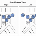
The natural history of testicular cancer provides the basis for its evaluation and management, and in turn is favorably influenced by effective therapy. Lymphatic spread is common to all forms of GCT, although in the case of choriocarcinoma, vascular dissemination is a common clinical feature. Early anatomic studies described that the primary drainage of the testis was to the area of its embryologic origin, the reptroperitoneum (11,12). Multiple surgical mapping studies have demonstrated that the primary landing zone for right-sided testicular tumors is the interaortocaval lymph nodes followed by the precaval and preaortic nodes (13,14). For left-sided testicular tumors, the primary landing zone is the paraaortic and preaortic lymph nodes followed by the interaortocaval nodes. Contralateral spread has been commonly reported for both right- and left-sided primary tumors, although it may occur more frequently from the right side (15).

The detailed mapping studies referred to above have significantly increased our understanding of the lymphatic spread of testicular cancer and have sharpened the focus of clinical staging, particularly radiographically. CT imaging is the most effective radiographic technique for identifying metastatic disease both above and below the diaphragm. The abdominal CT scan is normal in approximately 70% of patients with seminoma and 30% of patients with NSGCT. Lymph nodes in the primary landing zone measuring 10 to 20 mm are positive for GCT approximately 70% of the time, and nodes measuring 5 to 10 mm are involved approximately 50% of the time (16,17). The sensitivity and specificity for CT imaging are approximately 70% to 80% and 80% to 85%, respectively, for patients with clinical stage I and IIa testicular cancer (2).

MRI, like CT scan, is capable of identifying lymphadenopathy with similar sensitivity and specificity (18). MRI may provide additional information preoperatively regarding the vascular anatomy and patency of the great vessels in patients with bulky retroperitoneal disease, but overall, it contributes little to the management of most patients with GCT who have already undergone CT imaging. One potential advantage of MRI, however, is that it exposes patients to less radiation than a standard CT scan. However, this must be balanced against the higher cost, longer time to complete the examination, and inferior tolerability (especially for claustrophobic patients) of MRI compared with CT imaging. A randomized trial comparing CT and MRI in the follow-up setting (after completion of treatment) is currently ongoing in Europe. If positive, this trial could also lead to increased substitution of MRI for CT in the initial staging evaluation.

While PET imaging has shown promise in the postchemotherapy evaluation of patients with metastatic seminoma, no current role exists for PET imaging as an initial evaluation of GCT of either histology (19). Similarly, there is no role for PET scan in the routine postchemotherapy evaluation of patients with metastatic NSGCT (20,21). A particular disadvantage of PET in this situation is inability to differentiate between teratoma and normal tissue, both of which show little to no FDG-avidity.


EXTRARETROPERITONEAL STAGING

Accurate staging of extraretroperitoneal sites is essential in testis cancer guiding clinical management and IGCCCG risk stratification. The chest is the most common site of metastatic disease after the retroperitoneum. Approximately, 5% of patients with a radiographically normal retroperitoneum will be found to have pulmonary metastases, and this incidence increases to 40% for patients with retroperitoneal metastatic disease (22,23). CT imaging of the chest offers a higher sensitivity than chest roentogram and should be used as the initial staging modality. Additionally, CT imaging of the chest allows for evaluation of the supraclavicular and mediastinal lymph nodes. Imaging of the brain with either CT or MRI is not part of the routine staging of testicular cancer patients but should be included in patients with central neurological symptoms and should be considered for patients with pure choriocarcinoma, markedly elevated HCG (>100,000), or widespread pulmonary metastases since these features predict for a higher incidence of brain metastases. The constellation of these findings has been referred to as the “choriocarcinoma syndrome,” and such patients are at particular risk for hemorrhage at all sites of metastases, including within the brain. Bone scan is also not routinely recommended for staging but may be indicated in patients with symptoms suggestive of bone metastases or an unexplained elevation of the alkaline phosphatase. As previously mentioned, PET scan is not recommended for the standard staging evaluation of patients with testicular cancer.


CONCLUSION

The clinical presentation and staging and prognostic assessments discussed in this chapter are integral to the care of the testicular cancer patient. Together, AJCC stage and IGCCCG risk classification constitute the two major factors which influence the selection of treatment and likelihood of cure for any individual patient. Proper staging (namely STM measurement and CT imaging of the chest, abdomen, and pelvis) is important because both incomplete and excess evaluations can have negative consequences. Incomplete assessments can lead to undertreatment with the potential for particularly grave consequences, considering the high curability of this population. Similarly, unnecessary evaluations, such as bone scan, brain MRI, or PET scan can result in overtreatment, subjecting patients to unnecessary toxicities. Additionally, superfluous imaging exposes patients to excess radiation, results in unnecessary biopsies with potential morbidity, and increases healthcare costs. Establishing an accurate stage and risk classification is necessary so that patients have the highest cure rate with the least toxicity Therefore, understanding the principles guiding the diagnostic and staging evaluation is necessary to ensure that testicular cancer patients have the highest chance of cure with the least possible toxicity.


References

1. American Cancer Society. Cancer facts and figures 2009 Atlanta: American Cancer Society;2009.

2. Stephenson AJ, Bosl GJ, Motzer RJ, et al. Retroperitoneal lymph node dissection for nonseminomatous germ cell testicular cancer: impact of patient selection factors on outcome. J Clin Oncol 2005;23:2781-2788.

3. Carver BS, Serio AM, Bajorin D, et al. Improved clinical outcome in recent years for men with metastatic nonseminomatous germ cell tumors. J Clin Oncol 2007;25:5603-5608.

4. Moul J, Paulson D, Dodge R, et al. Delay in diagnosis and survival in testicular cancer: impact of effective therapy and changes during 18 years. J Urol 1990;143:520-523.

5. Bosl G, Goldman A, Lange P, et al. Impact of delay in diagnosis on clinical stage of testicular cancer. Lancet 1981;2:970-973.

6. Stephenson AJ, Russo P, Kaplinsky R, et al. Impact of unnecessary exploratory laparotomy on the treatment of patients with metastatic germ cell tumor. J Urol 2004;171:1474-1477.

7. Moul J, Moellman J. Unnecessary mastectomy for gynecomastia in a testicular cancer patient. Mil Med 1992;157:433-434.

8. Fleming I, ed. AJCC cancer staging handbook. Philidelphia: Lippincott-Raven, 1998.

9. Dash A, Carver BS, Stasi J, et al. The indication for postchemotherapy lymph node dissection in clinical stage IS nonseminomatous germ cell tumor. Cancer 2008;112:800-805.

10. IGCCCG. International Germ Cell Consensus Classification: a prognostic factor-based staging system for metastatic germ cell cancers. J Clin Oncol 1997;15:594-603.

11. Jamieson J, Dobsin J. Lymphatics of the testicle. Lancet 1910;1:493-495.

12. Skinner D, Leadbetter W. The surgical management of testis tumors. J Urol 1971;106: 84-93.

13. Donohue J, Zachary J, Maynard B. Distribution of nodal metastases in nonseminomatous testis cancer. J Urol 1982;128:315-320.

14. Weiscbach L, Boedefeld E. Localization of solitary and multiple metastases in stage II nonseminomatous testis tumor as a basis for a modified staging lymph node dissection in stage I. J Urol 1987;138:77-82.

15. Sogani P. Evolution of the management of stage I nonseminomatous germ cell tumors of the testis. Urol Clin North Am 1991;18:561-573.

16. Leibovitch I, Foster R, Kopecky K, et al. Improved accuracy of computerized tomography based clinical staging in low stage nonseminomatous germ cell cancer using size criteria of retroperitoneal lymph nodes. J Urol 1995;154:1759-1763.

17. Hilton S, Herr H, Teitcher J, et al. CT detection of retroperitoneal lymph node metastases in patients with clinical stage I testicular nonseminomatous germ cell cancer: assessment of size and distribution criteria. Am J Roentgenol 1997;169:521-525.

18. Sohaib SA, Koh DM, Barbachano Y, et al. Prospective assessment of MRI for imaging retroperitoneal metastases from testicular germ cell tumours. Clin Radiol 2009;64:362-367.

19. De Santis M, Becherer A, Bokemeyer C, et al. 2-18fluro-deoxy-D-glucose positron emission tomography is a reliable predictor for viable tumor in postchemotherapy seminoma: an update of the prospective multicentric SEMPET trial. J Clin Oncol 2004;22: 1034-1039.

20. Albers P, Bender H, Yilmaz H, et al. Positron emission tomography in the clinical staging of patients with Stage I and II testicular germ cell tumors. Urology 1999;53:808-811.

21. de Wit M, Brenner W, Hartmann M, et al. [18F]-FDG-PET in clinical stage I/II nonseminomatous germ cell tumours: results of the German multicentre trial. Ann Oncol 2008;19:1619-1623.

22. Tiffany P, Morse MJ, Bosl G, et al. Sequential excision of residual thoracic and retroperitoneal masses after chemotherapy for stage III germ cell tumors. Cancer 1986;57: 978-983.

23. Gels ME, Hoekstra HJ, Sleijfer DT, et al. Thoracotomy for postchemotherapy resection of pulmonary residual tumor mass in patients with nonseminomatous testicular germ cell tumors: aggressive surgical resection is justified. Chest 1997;112:967-973.


▪ 32B The Role of Serum Tumor Markers in the Diagnosis and Management of Testicular Cancer

Lamont J. Barlow

James M. Mckiernan


INTRODUCTION

Testicular germ cell tumors (GCTs) are one of the few malignancies for which specific biochemical markers have been identified that are simple to measure in serum and useful in the diagnosis and management of the disease. The prognostic utility of serum tumor markers in GCTs is reflected by the AJCC staging system that includes a separate category to account for elevation in three of these markers (Table 32B.1): alphafetoprotein (AFP), human chorionic gonadotropin (hCG), and lactate dehydrogenase (LDH; see separate chapter on staging for complete overview) (1). At least one of these markers is elevated in 85% of nonseminomatous GCTs, and a significant number of seminomas and nonseminomatous GCTs are associated with elevated markers prior to the radiographic or
clinical manifestations of disease (2,3,4). The production of the oncofetal substances AFP and hCG by trophoblastic and syncytiotrophoblastic cells within GCT is likely due to the expression of fetal genes that were deactivated during development or the malignant transformation of a pluripotential germinal cell that has retained the capacity to differentiate and produce oncofetal proteins (5).


TABLE 32B.1 COMMON SERUM TUMOR MARKERS IN TESTICULAR GERM CELL TUMORS


Marker


Normal Rangea


Half-Life in Serum


AFP


<40 ng/mL


5-7 d


hCG


<5 mIU/mL


24-36 hr


LDH


90-190 IU/L


Varies


a Normal ranges vary by institution or laboratory.


Alpha-fetoprotein

AFP is an oncofetal substance first described by Bergstrand and Czar in the 1950s (6). It is produced by fetal yolk sac, liver, and gastrointestinal tract and is found in high concentrations in human fetal serum, peaking by the 14th week of gestation (7). AFP may be produced in GCTs that contain components of embryonal carcinoma, yolk sac tumor, or teratoma. By definition, elevations in AFP do not occur in pure seminoma or pure choriocarcinomas, although molecular studies have demonstrated minute quantities of AFP mRNA in tumors previously believed to be pure seminomas (8). In general, any patient diagnosed with a seminoma who has elevated AFP should be assumed to have nonseminomatous components and treated accordingly (unless an alternate source of AFP elevation is identified as discussed later in this chapter). The half-life of AFP in human serum is 5 to 7 days (7).


Human Chorionic Gonadotropin

hCG is a glycoprotein discovered to be elevated in GCTs in 1930 by Zondek (9). It is produced by the placenta to maintain the corpus luteum during normal pregnancy, and a number of other malignancies are associated with elevations of this substance, including liver, lung, pancreas, and stomach cancers (7). In GCTs, syncytiotrophoblastic cells produce hCG and cause detectable elevations in a significant portion of both seminomas and nonseminomatous GCTs. Although these tumors produce intact hCG consisting of alpha and beta polypeptides, most commercially available assays detect only the beta subunit. The half-life of hCG in serum is 24 to 36 hours, although the beta subunit is cleared more rapidly (10).


Lactic Acid Dehydrogenase

The third marker most commonly relied upon in the management of GCTs is LDH. It is a cellular enzyme found in every tissue in the body with the highest concentrations in muscle (including smooth, cardiac, and striated), liver, and brain tissues. LDH was first shown to have a direct relationship to tumor burden in nonseminomatous GCTs by Boyle and Samuels in 1977 (11) and has also been associated with advanced pure seminoma. Although it is considerably less specific than AFP or beta-hCG, it is often the only elevated marker in some GCTs. There are at least four isoenzymes of LDH that may be elevated, and the half-lives of these isoenzymes range from 10 hours to 3 days.


Other Markers

There are several other measurable substances in serum that have been associated with GCTs. Gamma-glutamyl-transpeptidase (GGT) is an enzyme found primarily in the liver and is most commonly used as a marker of disease of the liver, biliary system, and pancreas. Up to one third of patients with seminomas have elevated GGT (12). Another marker, placental-like alkaline phosphatase (PLAP), is often elevated in seminomas, but like GGT, increased PLAP concentrations are also associated with a number of other malignancies as well as smoking (13). The lack of sensitivity and specificity for these and other investigated substances limit their clinical utility in comparison to the previously discussed markers.


TABLE 32B.2 CONDITIONS ASSOCIATED WITH ELEVATED SERUM AFP AND HCG


Condition


AFP


hCG


Genetic


Ataxia-telangiectasia


+


Hereditary tyrosinemia


+


Hereditary persistence of AFP


+


Metabolic/toxic


Primary hypogonadism


+a


Substance-induced liver damage


+


Marijuana use


±


Infectious


Viral hepatitis


+


Neoplastic


Nonseminomatous germ cell tumor


±


±


Pure seminoma


±


Gestational trophoblastic disease


±


±


Hepatocellular carcinoma


+


Gonadotroph adenoma


±a


Poorly differentiated adenocarcinoma


±


±


a False positive from increased LH cross-reacting with beta-hCG immunoassay.

Only gold members can continue reading. Log In or Register to continue

Stay updated, free articles. Join our Telegram channel

Jul 15, 2016 | Posted by in ONCOLOGY | Comments Off on Staging of Testicular Cancer

Full access? Get Clinical Tree

Get Clinical Tree app for offline access