Introduction
- ■
Radiation is an integral component of many patients’ treatment regimens. The goal of radiation therapy is to deliver a dose to the target while sparing the normal tissue so that the treatment can be well tolerated. Treatment toxicity is caused by dose to the normal organs that receive radiation as bystanders. All lung and mediastinal tumors have an adjacent boundary with the normal lung and thus pulmonary toxicity is fairly common. The lung is one of the most sensitive organs to ionizing radiation and thus pulmonary toxicity from radiation is seen in 5% to 50% of patients receiving radiation for lung cancer.
- ■
Radiation is used as a single modality treatment for early-stage medically inoperable lung cancer in the form of stereotactic body ablative radiotherapy (SBRT) mainly because these patients are unable to tolerate surgery secondary to poor pulmonary lung function and low pulmonary reserve. SBRT has recently become increasingly utilized due to excellent local control rates, improved survival, and its noninvasive nature. It is used in conjunction with chemotherapy for locally advanced lung cancer, especially when nodal metastasis is present. Concurrent chemotherapy and radiation have shown improved survival for both non–small-cell lung cancer (NSCLC) as well as small-cell lung cancer (SCLC), thus supporting its use; however, it comes at the price of increased rates of toxicities with concurrent treatment compared to sequential chemotherapy and radiation. Radiation has also been utilized for palliation of symptoms from lung tumors in the advanced disease setting for airway obstruction, dysphagia, hemoptysis, and superior vena cava syndrome. Both brachytherapy and external beam radiation have been utilized for palliation of symptoms.
- ■
Even with conformal techniques, such as three-dimensional (3-D) conformal radiation and the more modern intensity modulated radiation therapy (IMRT), planned with accurate assessment of target and organ at risk (OAR) motion with four-dimensional (4-D) computed tomography (CT) scans and delivered with image-guided radiation therapy (IGRT) and gating techniques, pulmonary toxicity still continues to be the main dose-limiting toxicity for thoracic radiation therapy both for primary cancers like NSCLC and SCLC as well as sarcomas, lymphomas, and other mediastinal tumors such as thymomas.
- ■
With the popularization of SBRT for primary as well as secondary malignancies of the lung and varying fractionation and dose schedules being utilized, the understanding of radiation-induced lung diseases is a focus of research. Side effects that are usually not noted with conventional fractionation are now being noted with SBRT. Some notable examples are bronchial necrosis and fistula formation.
- ■
This chapter focuses on radiation-induced pneumonitis and the changing patterns of toxicity with advanced radiation modalities like SBRT, IMRT, and brachytherapy. The mechanisms of action, various factors affecting the development of pneumonitis, and strategies for reducing the incidence of pneumonitis, and the pharmacological and nonpharmacological interventions to treat pneumonitis are discussed in this chapter.
Characteristics of Radiation-induced Lung Injury Radiation-induced lung injury was first described in 1898. It was then subdivided in 1925 into radiation pneumonitis and radiation fibrosis. Both types of lung injury are observed in patients who have undergone thoracic radiation treatment for lung, breast, or hematological malignancies. Radiation-induced damage to lung parenchyma remains a dose-limiting factor in chest radiotherapy.
- ■
Timing: Symptoms caused by acute radiation pneumonitis usually develop approximately 4 to 12 weeks following radiation; various symptoms of fibrotic radiation pneumonitis usually develop after 6 to 12 months.
- ■
Clinical presentation: The signs and symptoms of the two phases of radiation pneumonitis (acute and fibrotic) are similar, although fever is less likely to occur in the fibrotic phase. The following signs and symptoms have been described:
Symptoms
- ■
A nonproductive cough which may occur during therapy as a manifestation of bronchial mucosal injury or later as a manifestation of fibrosis.
- ■
Dyspnea may occur only with exertion or may be described as an inability to take a deep breath.
- ■
Fever is usually low grade but can be more pronounced in severe cases.
- ■
Chest pain may be pleuritic or substernal and can mimic cardiac chest pain.
- ■
Malaise and weight loss may be observed.
- ■
- ■
Physical signs include the following:
- ■
A pleural rub may be heard.
- ■
- ■
Dullness to percussion may be detected as a result of a pleural effusion that is seen in about 10% of patients.
- ■
Grading and severity: Tables 28.1 and 28.2 describe the grading and severity of radiation pneumonitis.
TABLE 28.1■
Commonly Utilized Acute Pneumonitis Grading Criteria
Scale
Grade 1
Grade 2
Grade 3
Grade 4
Grade 5
RTOG
Mild symptoms of dry cough or dyspnea on exertion
Persistent cough requiring narcotic, antitussive agents/dyspnea with minimal effort but not at rest
Severe cough unresponsive to narcotic antitussive agent or dyspnea at rest/clinical or radiological evidence of acute pneumonitis/intermittent oxygen or steroids may be required
Severe respiratory insufficiency/continuous oxygen or assisted ventilation
Death
CTCAE
Asymptomatic; clinical or diagnostic observations only; intervention not indicated
Symptomatic; medical intervention indicated; limiting instrumental ADL
Severe symptoms; limiting self-care ADL; oxygen indicated
Life-threatening respiratory compromise; urgent intervention indicated (e.g., tracheotomy or intubation)
Death
ADL, Activities of daily living; CTCAE, Common Terminology Criteria for Adverse Events; ROTG, Radiation Therapy Oncology Group.
TABLE 28.2■
Commonly Utilized Chronic Pulmonary Fibrosis Grading Criteria
Scale
Grade 1
Grade 2
Grade 3
Grade 4
Grade 5
RTOG
Asymptomatic or mild symptoms (dry cough); slight radiographic appearances
Moderate symptomatic fibrosis or pneumonitis (severe cough); low-grade fever; patchy radiographic appearances
Severe symptomatic fibrosis or pneumonitis; dense radiographic changes
Severe respiratory insufficiency/continuous oxygen/assisted ventilation
Death
CTCAE
Mild hypoxemia; radiological pulmonary fibrosis
Moderate hypoxemia; evidence of pulmonary hypertension; radiographic pulmonary fibrosis 25%–50%
Severe hypoxemia; evidence of right-sided heart failure; radiographic pulmonary fibrosis >50%–75%
Life-threatening consequences (e.g., hemodynamic/pulmonary complications); intubation with ventilatory support indicated; radiographic pulmonary fibrosis >75% with severe honeycombing
Death
CTCAE, Common Terminology Criteria for Adverse Events; ROTG, Radiation Therapy Oncology Group.
- ■
Diagnostic evaluation: Radiation pneumonitis is usually a diagnosis of exclusion. It should be suspected when a patient who has undergone thoracic radiation develops symptoms or signs such as shortness of breath, cough, fever, or malaise in weeks to months after radiation therapy. Appropriate evaluation is designed to assess the severity of respiratory impairment, to determine the correspondence of the radiographic changes with the radiation therapy portal, and to exclude other possible etiologies, such as infection, thromboembolic disease, drug-induced pneumonitis, spread of the underlying malignancy, tracheoesophageal fistula, exacerbation of underlying chronic obstructive pulmonary disease (COPD), interstitial lung disease, or heart failure.
- ■
Chest radiography: Perivascular haziness is an early radiation-induced abnormality, often progressing to patchy alveolar filling densities. Radiographs taken during the chronic phase may show volume loss with coarse reticular or dense opacities. A straight-line effect which does not conform to anatomical units but rather to the confines of the radiation field is virtually diagnostic of radiation-induced lung injury. However, conformal and stereotactic treatment strategies, such as 3-D conformal radiation therapy, SBRT, and tomography, do not cause this straight-line radiographic finding due to the complex distribution of the radiation dose. With these techniques, a focal area of opacity with ill-defined margins is seen in the radiated region.
- ■
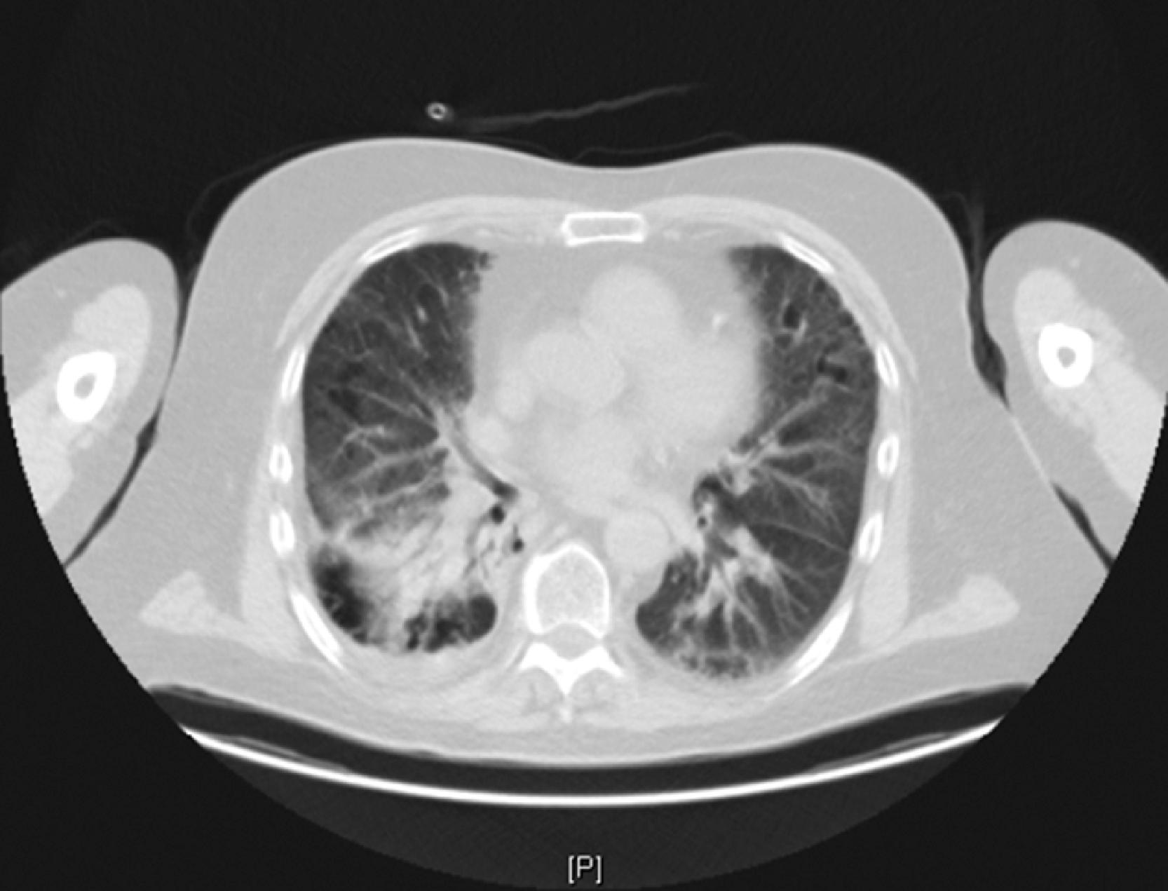
Chest CT: Chest CT ( Fig. 28.1 ) is more sensitive than chest radiography for detecting lung injury following radiation treatment and is often obtained in the evaluation of the patient with increased dyspnea or cough following radiation therapy, especially when other causes such as infective pneumonia or pulmonary thromboembolism have been ruled out. The key step in the evaluation of radiation pneumonitis is the comparison of the pretreatment CT images, containing rate deviation dosimetric information, with diagnostic CT images obtained at the time of symptom presentation by the radiation oncologist. Lung involvement in CT images of radiation pneumonitis typically aligns closely with the radiated area.