Introduction
- ■
Radiation dermatitis is one of the earliest known side effects of radiation. Almost all patients receiving radiation therapy have some changes in the skin. Acute and/or chronic skin changes may occur, which may have implications for quality of life during and after completion of radiation. These dermatological reactions may lead to delay in treatment or diminished cosmesis and functional deficits.
- ■
Radiation is used as adjuvant therapy after surgery in many situations such as after breast conserving therapy or mastectomy to reduce the risk of local recurrence for breast cancer, or as definitive treatment for early stage medically inoperable lung cancer concurrent with chemotherapy for locally advanced lung cancer, as well as with or without chemotherapy in the treatment of sarcomas, thymomas, and lymphomas. Radiation is also used as external beam radiation therapy or brachytherapy for the treatment of skin cancers. These broad indications increase the sheer numbers of potential patients, many of whom will develop some form of radiation dermatitis.
- ■
Radiation effects to the skin are usually unavoidable, as the radiation must enter, exit, or be deposited near the skin to reach the target volume. Skin cells, because they originate from a rapidly reproducing differentiated stem cell, are relatively radiosensitive. The side effects can sometimes lead to interruption of therapy and be dose-limiting for tumor control and thus prevention and treatment strategies become extremely important.
- ■
In this chapter, we will discuss the clinical manifestations, mechanism of action, treatment, and prevention of radiation dermatitis.
Description
The earliest reports of radiation-induced skin changes date back to 1896, nine months after Roentgen’s discovery, when Clarence E. Dally, a colleague of Thomas Edison participating in fluorescent lamp construction, developed epilation, skin ulceration of the hands and arms, carcinoma, and ultimately died in 1904 due to metastasis. This phenomenon of radiation dermatitis may be produced by both diagnostic and therapeutic radiation equipment.
TIMING
Radiation dermatitis typically occurs within 90 days of starting treatment. The specific skin changes depend upon the radiation dose and include erythema, edema, pigment changes, hair loss, and dry or moist desquamation. Late stage or chronic radiation dermatitis typically presents months to years after radiation exposure. It is characterized by dermal fibrosis and poikilodermatous changes, including hyper- and hypopigmentation, atrophy, and telangiectasias.
Clinical Presentation
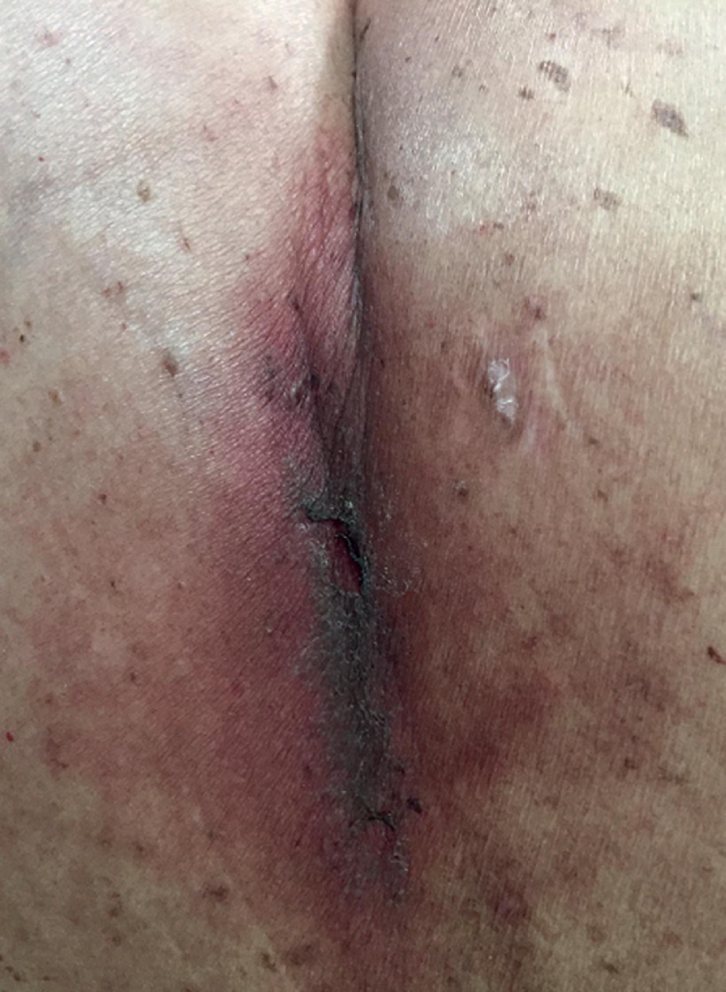
In order to be defined as radiation dermatitis, the skin reaction must occur within or at the margin of the radiotherapy field. Features of dermatitis are dose dependent. Early changes noted in the skin are: erythema at doses of 2 Gy or higher, dry desquamation at doses of 12–20 Gy, moist desquamation at greater than 20 Gy, and necrosis at 35 Gy or higher. All patients do not experience all acute skin reactions; however, there may be a combination of reactions occurring simultaneously in the radiation field.
Late radiation changes are progressive and may begin to appear 10 weeks after the radiation. Reactions may progress slowly and subclinically, beginning months to years following treatment. Changes may include photosensitivity, hyper- or hypopigmentation, atrophy, fibrosis, telangiectasia, ulceration, and necrosis.
GRADING AND SEVERITY
The grading and severity of radiation dermatitis is reflected in Table 27.1 .
| Grade 1 | Grade 2 | Grade 3 | Grade 4 | Grade 5 | |
|---|---|---|---|---|---|
| Skin and subcutaneous tissue disorders – Other, specify | Asymptomatic or mild symptoms; clinical or diagnostic observations only; intervention not indicated | Moderate; minimal, local, or noninvasive intervention indicated; limiting age-appropriate instrumental ADL | Severe or medically significant but not immediately life-threatening; hospitalization or prolongation of existing hospitalization indicated; disabling; limiting self-care ADL | Life-threatening consequences; urgent intervention indicated | Death |
| Skin atrophy | Covering <10% BSA; associated with telangiectasias or changes in skin color | Covering 10%–30% BSA; associated with striae or adnexal structure loss | Covering >30% BSA; associated with ulceration | ||
| Skin hyperpigmentation | Hyperpigmentation covering <10% BSA; no psychosocial impact | Hyperpigmentation covering >10% BSA; associated psychosocial impact | – | ||
| Skin hypopigmentation | Hypopigmentation or depigmentation covering <10% BSA; no psychosocial impact | Hypopigmentation or depigmentation covering >10% BSA; associated psychosocial impact | |||
| Skin infection | Localized, local intervention indicated | Oral intervention indicated (e.g., antibiotic, antifungal, antiviral) | IV antibiotic, antifungal, or antiviral intervention indicated; radiological or operative intervention indicated | Death | |
| Skin ulceration | Combined area of ulcers <1 cm; nonblanchable erythema of intact skin with associated warmth or edema | Combined area of ulcers 1–2 cm; partial thickness skin loss involving skin or subcutaneous fat | Combined area of ulcers >2 cm; full-thickness skin loss involving damage to or necrosis of subcutaneous tissue that may extend down to fascia | Death | |
| Dry skin | Covering <10% BSA and no associated erythema or pruritus | Covering 10%–30% BSA and associated with erythema or pruritus; limiting instrumental ADL | Covering >30% BSA and associated with pruritus; limiting self-care ADL | – |
DIAGNOSTIC EVALUATION
The diagnosis of acute radiation dermatitis is clinical, based upon the findings of the skin changes.
- 1.
Mild dermatitis (Common Terminology Criteria for Adverse Events [CTCAE] grade 1) is characterized by mild blanchable erythema or dry desquamation. The onset is typically within days to weeks of initiating therapy and symptoms generally dissipate within a month. Pruritus and hair loss are commonly associated symptoms.
- 2.
Moderate dermatitis (CTCAE grade 2) is characterized by painful erythema and edema that may progress to focal loss of epidermis and moist desquamation, usually limited to skin folds. Moist desquamation is characterized by epidermal necrosis, fibrinous exudates, and often considerable pain. Bullae, if present, may rupture or become infected. This reaction usually peaks 1–2 weeks after the end of treatment.
- 3.
Severe dermatitis (CTCAE grades 3 and 4) is characterized by confluent moist lesions which can become secondarily infected. Pain is usually severe.
Chronic radiation dermatitis is characterized by sustained loss of certain skin structures such as sebaceous glands, hair follicles, and nails; as well as textural changes to the skin. , Atrophy of the epidermis and dermis may be observed, although some patients may develop induration and thickening of the dermis. Telangiectasia may occur as a result of blood vessel dilatation, while damage to blood vessels may also result in tissue hypoxia, predisposing the patient to development of skin ulceration and/or chronic wounds. Radiation induced fibrosis is a potentially serious consequence of radiation that may cause poor cosmesis, lymphedema, skin retraction, persistent hyperpigmentation, and joint immobility.
Pathogenesis
The pathogenesis of radiation dermatitis is complex but is often characterized by the following findings :
- ■
Erythema: increased blood supply via the dermal vascular plexus
- ■
Dry desquamation: inability of basal cells to replicate quickly enough to replenish the damaged squamous epithelium
- ■
Moist desquamation: focal loss of epidermis with a serous drainage and exudate
- ■
Alopecia: loss of hair follicles
- ■
Telangiectasias: dilated superficial blood vessels
- ■
Fibrosis and contraction: tightening of skin secondary to injury to fibroblasts
- ■
Radiation necrosis: dermal ischemia and impairment of the reparative process
Radiation dermatitis is the overall result of injury to multiple skin structures including epidermal keratinocytes, dermal fibroblasts, cutaneous vasculature, and hair follicles. It interferes with maturation, reproduction, and the repopulation of these cells and is the result of direct tissue injury, as well as inflammation recruited to the injured skin. DNA damage is mediated by the production of free radicals. Leukocytes and other immune cells migrate from the circulation to the skin. Increased release of cytokines and chemokines has been associated with acute radiation dermatitis. Production of reactive oxygen species cause further damage to the skin. This process triggers vasodilatation, increased vascular permeability, and recruitment of inflammatory cells. The immune cells secrete proinflammatory cytokines such as transforming growth factor beta which cause fibroblast differentiation into myofibroblasts, and subsequently produce collagenous and smooth muscle actin, resulting in much of the observed findings in fibrosis.
Factors Influencing the Onset, Duration, and Intensity of the Radiation Dermatitis
- ■
Patient-related factors


