Fig. 5.1
A chest CT showing a 10.2 × 5.0 × 4.8 cm anterior mediastinal mass with mild narrowing of the trachea, compression of the upper esophagus, and moderate narrowing of the right jugular vein and superior vena cava
Primary mediastinal (thymic) large B-cell lymphoma (PMBL) accounts for approximately 5 % of all cases of non-Hodgkin lymphoma. The median age at presentation is in the third to fourth decades, with a female predominance [1, 2]. The majority of cases present with bulky disease localized to the anterior mediastinum, often with associated pleural and pericardial effusions. Superior vena cava syndrome is a common clinical presentation. Although PMCL are largely localized at diagnosis, relapses tend to occur in extranodal organs such as the gastrointestinal tract, kidneys, adrenals, liver, and ovaries.
The postulated cell of origin of PMBL is thymic B cell. Although previously classified as a subtype of diffuse large B-cell lymphoma, in the World Health Organization (WHO) classification, PMBL is considered a distinct entity [3]. Morphologically, immunophenotypically, and according to gene expression profiling studies, PMBL is thought to be more closely related to classical Hodgkin lymphoma [4, 5]. The malignant cells of PMBL express B-cell markers such as CD19 and CD20 but lack CD5 and CD10 expression. Expression of CD30 is often present but is weak. More recently, the pathogenesis of PMBL has been associated with the Janus Kinase-Signal Transducer and Activator of Transcription (JAK-STAT) and nuclear factor kappa-b (NF-kb) signaling pathways, and overexpression of PD-1 ligands has been identified, findings that may guide the development of new therapeutic targets for PMBL [6–8].
Staging and Prognostic Factors
Imaging
Case 1
Positron emission tomography–computed tomography (PET–CT) was performed, showing a large superior anterior mediastinal mass with intense fluorodeoxyglucose (FDG) uptake with a standardized uptake value (SUV) max of 15.4 (Fig. 5.2).
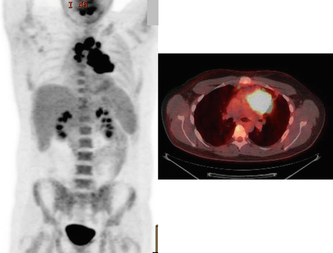
Fig. 5.2
Positron emission tomography-computed tomography (PET-CT) showing a large superior anterior mediastinal mass with intense fluorodeoxyglucose (FDG) uptake with a standardized uptake value (SUV) max of 15.4
Case 2
PET–CT was performed, showing FDG–avid disease involving the left supraclavicular region (SUV max 8.2) and the anterior mediastinum extending from the thoracic inlet to the level of the aortic root with an SUV max of 14 (Fig. 5.3).

Fig. 5.3
PET-CT showing FDG-avid disease involving the left supraclavicular region (SUV max 8.2) and the anterior mediastinum extending from the thoracic inlet to the level of the aortic root with an SUV max of 14
Blood work in both cases included complete blood count (CBC) with differential, serum lactate dehydrogenase (LDH), blood urea nitrogen (BUN), and creatinine; electrolytes were all within normal limits. Human immunodeficiency (HIV) and hepatitis B and C serologies were negative. Bone marrow biopsies were performed in both cases, both showing no evidence of marrow involvement.
The staging workup for PMBL is similar to that for diffuse large B-cell lymphoma. Relevant history and physical examination include documentation of signs and symptoms that may be related to the mediastinal disease including chest pain, shortness of breath, cough, voice changes, facial or neck swelling, and chest wall masses. Imaging studies include PET-CT and optional chest CT with contrast which may aide subsequent radiation treatment planning. Although bone marrow involvement is rare in PMBL, bone marrow biopsy should be performed especially in the absence of evidence of bone/bone marrow involvement on PET-CT [9].
Modifications to the Ann Arbor staging system were recently made in the Lugano Classification for the staging of lymphoma [9]. The designation of “X” for bulky disease was eliminated, and instead, it was recommended that the largest tumor diameter be recorded. This may be especially pertinent to PMBL, which tend to present with bulky disease. In addition, specifically for non-Hodgkin’s lymphoma, the designation of “B” symptoms was eliminated.
The overall prognosis of patients with PMBL has been shown to be more favorable than patients with diffuse large cell lymphoma [10]. The international prognostic index (IPI) and the subsequent revised IPI [11], developed for patients with aggressive non-Hodgkin lymphoma treated with anthracycline-based chemotherapy, can be extrapolated for use in patients with PMBL, although available data suggest that age-adjusted IPI may not be predictive of overall survival in patients with PMBL [10, 12]. In a recent study focusing on 123 patients with PMBL treated with rituximab, cyclophosphamide, doxorubicin, vincristine, and prednisone (R-CHOP) chemotherapy, the presence of pleural or pericardial effusion at diagnosis was the only factor that independently predicted for primary refractory disease or early relapse within 12 months [13].
Treatment
Case 1
The patient was initiated on R–CHOP chemotherapy. Restaging PET–CT after 3 cycles of chemotherapy showed a partial response with a Deauville score of 4 (Fig. 5.4a). The patient completed 3 additional cycles of chemotherapy, and restaging PET–CT performed 4 weeks after the chemotherapy showed further response with a Deauville score of 2 (Fig. 5.4b). The patient was then referred for radiation therapy.

Fig. 5.4
(a) Restaging PET-CT after 3 cycles of chemotherapy showing a partial response with a Deauville score of 4 (a). (b) After 3 additional cycles of chemotherapy, restaging PET-CT performed 4 weeks after the chemotherapy showing further response with a Deauville score of 2
Radiation Treatment
The patient was simulated in an arm–down position with a Vac–LoK bag for immobilization and a face mask to keep the neck in an extended position. CT simulation was performed using a three–dimensional scan and deep–inspiration breath-hold technique. The planning CT was fused with the pre–chemotherapy PET–CT, and the clinical target volume (CTV) was determined according to the International Lymphoma Radiation Oncology Group and involved site radiation therapy (ISRT) guidelines [14, 15]. The CTV included the initially involved disease extent but excluded the previously displaced, uninvolved normal structures (lungs, major vessels, heart, bones, muscles) (Fig. 5.5). An expansion to internal target volume (ITV) was not performed because breath–hold technique was used. A 1–cm CTV to planning target volume (PTV) expansion was used to account for daily setup errors. A primarily anterior–posterior/posterior–anterior beam arrangement intensity-modulated radiation therapy (IMRT) approach was used (Fig. 5.6a), in order to reduce low doses to large volume of lungs. Since no lateral beams were used, the patient was simulated and treated in an arm–down position (which is especially important in cases of female patients in which an arm–up position will bring more breast tissue into the radiation field). This IMRT technique is referred to as the butterfly technique [16]. A total dose of 30 Gy in 15 fractions was prescribed. The dose volume histogram (DVH) is shown in Fig. 5.6b. The mean heart dose (MHD) was 11.5 Gy, and the mean lung dose (MLD) was 9.1 Gy. The lung V5 and V20 were 42 % and 20 %, respectively.


Fig. 5.5
CTV including the initially involved disease extent but excluding the previously displaced, uninvolved normal structures (lungs, major vessels, heart, bones, muscles).
Fig. 5.6
(a) Axial view showing IMRT planning with anteroposterior beam arrangement used. (b) Dose volume histogram
Case 2
The patient was started on dose–adjusted etoposide, doxorubicin, and cyclophosphamide with vincristine, prednisone, and rituximab (DA–EPOCH–R) with prompt resolution of the facial and neck swelling. Restaging PET–CT after 3 cycles of chemotherapy showed a complete metabolic response (Deauville 2) (Fig. 5.7). The patient received a total of 6 cycles of DA–EPOCH–R, and restaging PET–CT 1 month post–chemotherapy showed continued complete response. Radiation oncology was not consulted as complete remission was achieved and there were also concerns with late toxicity of mediastinal irradiation in a young woman.


