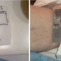
Fig. 6.1
(a–c) Axial CT scan slices showing a mixed sclerotic lytic lesion in the right scapula. There was associated soft tissue swelling in the surrounding musculature
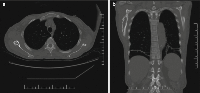
Fig. 6.2
Axial (a) and coronal (right panel) CT scan with bone windows, showing the partly sclerotic and partly lytic nature of the lesion. Note splenomegaly in the coronal CT (b)
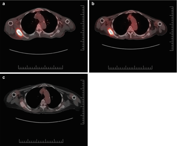
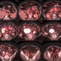
Fig. 6.3
PET/CT scan (axial slices) at initial diagnosis (a, left), 6 months (b, middle) and 16 months (c, right) after radiation treatment (35 Gy). The right scapula osteosclerotic solitary plasmacytoma is avid for FDG uptake with SUVmax of 24.3 prior to RT, decreasing to 10.7 at 6 months, and became normal (1.4) at 16 months, and metabolic local control was maintained at 4.5 years follow-up. However some sclerosis of the bone persisted
The Diagnosis of POEMS Syndrome and Solitary Plasmacytoma
POEMS syndrome is a rare condition with the acronym representing some (but not all) key paraneoplastic features of the syndrome: polyneuropathy, organomegaly, endocrinopathy, monoclonal plasma cell disorder, and skin abnormalities. There are established diagnostic criteria with the mandatory ones being polyneuropathy and a monoclonal plasma cell disorder [1]. In addition, one of three major criteria and one of six minor criteria are required to secure the diagnosis [1]. In this case, the solitary plasmacytoma serves as one of the mandatory criteria, despite the absence of systemic multiple myeloma [1]. The co-diagnosis of Castleman’s disease and a sclerotic bone lesion serves as major criteria. Bone lesion, if present in POEMS syndrome, is characteristically sclerotic [2], in contrast with solitary plasmacytoma without POEMS that is typically a purely lytic lesion. She also has a plethora of minor criteria conditions, characterized by splenomegaly (organomegaly), diabetes (endocrinopathy), thrombocytosis with prior thrombotic disease, papilledema, and edema. However she did not have characteristic features of skin changes such as hyperpigmentation or acrocyanosis [3] nor pulmonary manifestations [4]. Patients with POEMS syndrome have these disparate symptoms and signs, and it is quite common to have some typical features but not others, which is the reason why it is difficult to diagnose, in addition to its rarity (prevalence <0.5 per 100,000). Delays in establishing a diagnosis is unfortunately common with median time from onset of symptoms to diagnosis of 19 months in a series of 38 patients [5], similar to that observed in this case. The pathogenesis of POEMS is poorly understood, but it is known that treating the underlying monoclonal plasma cell disorder can result in improvement of the syndrome [1, 6, 7]. There is no documentation of IgA nephropathy to be associated with development of POEMS or plasma cell diseases, although one can speculate whether the immunosuppression might have a role in her developing the plasma cell neoplasia. It appears that elevated VEGF levels reflect the disease activity, but anti-VEGF therapy alone has not been successful suggesting that it is another manifestation of the disease rather than having a causative role [1].
The workup of a suspected diagnosis of solitary plasmacytoma is directed to exclude multiple myeloma. The following criteria must be satisfied: a biopsy-confirmed single lesion with negative skeletal imaging elsewhere, normal bone marrow biopsy (<10 % clonal plasma cells), and no myeloma-related organ dysfunction (normal blood counts, calcium and renal function) [8, 9]. A monoclonal protein can be present in blood or urine, but it is usually only minimally elevated. Solitary plasmacytomas present more commonly in the bone, and usual symptoms are pain, neurologic compromise (e.g., from vertebral lesion causing nerve or spinal cord compression), and sometimes pathologic fracture. It is unusual to be locally asymptomatic as in this case with the scapula lesion. Solitary plasmacytoma uncommonly presents in an extramedullary site (20 %), usually as a soft tissue mass in the head and neck areas [10]. After adequate local treatment, it is known that an extramedullary presentation is associated with a lesser probability of progression to multiple myeloma [10], in contrast with a bone presentation where the disease recurs as multiple myeloma with a high likelihood, usually within 5–10 years [9, 11].
Treatment Management
When the patient was assessed for radiation therapy, she was severely disabled and totally bedridden (ECOG performance status 4) due to her neuropathy and generalized edema. She required frequent care in hospital, and have not experienced any improvement following multiple pulse treatments with high-dose glucocorticoids and intravenous immunoglobulin treatment. It was elected to treat her with radiation therapy to the right scapula lesion, her only documented plasmacytoma. She received 35 Gy in 15 fractions (2.33 Gy fractions) over 3 weeks. This shorter regimen was chosen as she required inter-hospital transfer daily for her RT. Other acceptable regimens in common use are 40–45 Gy in conventional fractionation. However one of the largest multi-institutional reviews showed no evidence of dose response beyond 30 Gy in terms of local control, regardless of size of the tumor [11]. A 3D conformal treatment plan was used choosing beam angles and segmental fields to minimize lung exposure (Figs. 6.4 and 6.5). She tolerated RT well and was transferred to a rehabilitation facility following completion of radiation. At 6 months follow-up, she improved neurologically with recovery in her arms, but still unable to walk. Her edema resolved. Her serum VEGF level returned to normal. Repeat FDG PET/CT scan showed improvement in the scapula lesion, SUVmax decreased to 10.7 and no new lesions detected. Repeat bone marrow biopsy remained negative. One year following RT, she started to ambulate and was able to be discharged home. Repeat FDG PET/CT scans 16 months and 21 months post-RT showed complete metabolic response (no visual uptake in the right scapula), although CT scan continues to show some residual sclerosis at the local site. The patient functioned well apart from residual mild leg weakness.
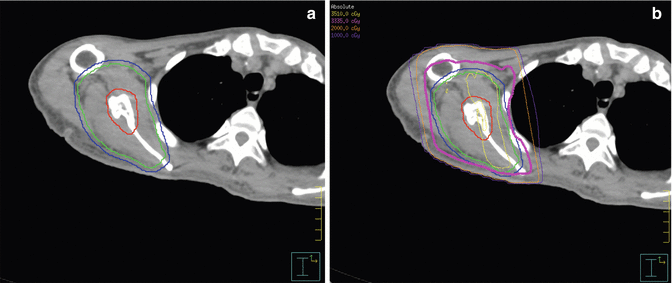
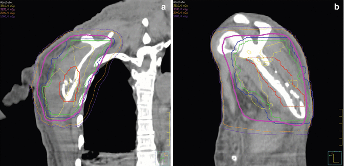

Fig. 6.4
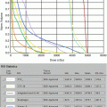
Radiation treatment of right scapula plasmacytoma. (a) Left panel: Contours of the bony GTV (red line), CTV (green line), and PTV (blue line). (b) Right panel: Isodose distribution, with prescribed dose 35 Gy and the objective of PTV covered by the 95 % isodose line 33.25 Gy

Fig. 6.5
RT treatment of right scapula plasmacytoma. Isodose distributions on CT coronal perspective (a) and sagittal perspective (b) and bone windows. The sclerotic lytic nature of the lesion is best appreciated on the sagittal image
Four years after RT, she experienced some worsening of her motor strength, and investigations revealed a recurrence of her POEMS syndrome with multiple FDG PET-avid spinal sclerotic lesions (Fig. 6.6) which were new, although small and asymptomatic. The scapula lesion remained controlled with no FDG uptake. She had elevation of serum VEGF (875 pg/ml). However her bone marrow biopsy remained negative for multiple myeloma, and she maintained absence of M-protein in blood and urine. After a brief course of chemotherapy with oral cyclophosphamide and prednisone, she received an autologous peripheral blood stem cell transplant with conditioning regimen consisting of melphalan 200 mg/M2 and tolerated it well. Her further follow-up is awaited.
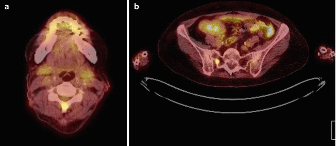

Fig. 6.6
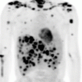
PET/CT scan (axial slices) at time of relapse of disease, 4 years after radiation therapy to the plasmacytoma of the right scapula, showing multiple bone lesions in the spine, with FDG uptake at C5 spinous process (a) and right sacroiliac joint (b)
Discussion
Radiation therapy is the standard treatment for solitary plasmacytoma. In the presence of POEMS syndrome, provided that there is no evidence of a bone marrow plasma cell clone, radiation therapy is the best initial treatment, even for up to three isolated bone lesions. In one of the largest series of POEMS patients from the Mayo Clinic (n = 146), 38 patients (26 %) satisfied this criteria and hence was found suitable for radiation therapy (RT) [5]. After median RT doses of 35–54 Gy (median 45 Gy), up to half of patients had clinical improvement or stability of POEMS-related symptoms, but most have some residual disability as in this case. With median follow-up of 43 months, the 4-year overall survival was 97 %, although eventually 48 % of patient required subsequent salvage therapy due to neurologic deterioration or worsened bone disease [5]. Most were treated with autologous stem cell transplantation, as in this case [12]. The expected 5-year progression-free survival is 75 % with transplant [12]. The pretreatment disease features associated with a poor prognosis were pulmonary impairment with a DCLO <75 % and elevated urinary total protein [5]. The case described in this report did not have these adverse features. Response to RT is also prognostic, not surprisingly, but as the case illustrates, residual sclerosis persist for many months to years after treatment, not indicative of disease, and this makes routine radiographs or CT scans difficult as a means to assess for residual disease. FDG PET is very helpful in this regard, but the response is very gradual as observed in this case, with a slow decrease in metabolic activity over a period of a year, with disappearance of metabolic activity only after 16 months from completion of RT. Local control has been maintained even when the patient progressed with multiple sclerotic lesions in her thoracic spine, as demonstrated by lack of FDG uptake in the scapula prior to her receiving the autologous stem cell transplant.
Although it is common practice to give a higher dose than 30–35 Gy for solitary plasmacytoma, with some historical data showing a lower local relapse rate of 6 % with doses of ≥40 Gy, compared with 31 % for lower doses [13], a large series from multiple institutions showed no evidence of improved local control with doses ranging 30–50 Gy, even for tumors >4 cm in maximum diameter [11]. In the case under discussion, a dose of 35 Gy in a slightly hypofractionated regimen have resulted in local control with a 4.5 years follow-up duration from RT. In general, a local control rate of 85–90 % is expected from RT for solitary plasmacytoma. Gross target volume and clinical target volume require careful definition of the bone disease and soft tissue infiltration, if present, taking care to encompass adjacent bone which may contain microscopic disease; this can be done using an MRI and following the abnormal signal seen in the bone/bone marrow to include it in the CTV; this is based on the fact that myeloma is a marrow disease, and initially it starts by forming a tumor collection before it gets to produce the lytic lesion that can be seen on CT or to much lesser degree on a simple X-ray. As a general rule CTV is made by adding information from PET scan (avid sites) + MRI abnormal signal in the marrow + CT scan (typically lytic abnormality). In general PTV would vary depending on the site to be treated taking into consideration internal organ motion as well as daily setup, but generally no more than 2 cm is to be added if bone site is considered. Coverage of the whole bone is not required. In general regional nodes are not required to be covered as nodal failures are rare, with the exception that if the plasmacytoma was extramedullary and involve a lymphatic structure (e.g., Waldeyer’s ring), the drainage lymph node region deemed at high risk of subclinical disease may be treated.
Stay updated, free articles. Join our Telegram channel

Full access? Get Clinical Tree


