Pathology and Cytology of Tumors of the Urinary Tract


Pathology and Cytology of Tumors of the Urinary Tract


Hikmat Al-Ahmadie

Oscar Lin

Victor E. Reuter


NORMAL ANATOMY AND HISTOLOGY

The mucosal surface of the renal pelvis, ureters, urinary bladder, and urethra is lined by a multilayered epithelium, the most superficial of which consists of “umbrella cells.” Although this epithelial lining has historically been called “transitional epithelium,” it is currently preferentially referred to as urothelium. The endoderm of the urogenital sinus gives rise to the urothelium of the urinary bladder and urethra, whereas the lamina propria, muscularis propria, and adventitia develop from the surrounding splanchnic mesenchyme. The urothelium of the renal pelvis and ureters is mesodermally derived (1).

The wall of the urinary bladder is formed of four layers: (a) epithelium (urothelium), (b) lamina propria, (c) muscularis propria, and (d) adventitia or serosa (2,3). Depending on the location, these layers may be surrounded by perivesical fat. The thickness of the urothelium varies, as does the shape of the epithelial cells, depending on the degree of bladder distension.

The lamina propria consists of abundant connective tissue containing a rich vascular network, lymphatic channels, sensory nerve endings, and a few elastic fibers. Its thickness varies between the empty versus the distended bladder but is generally thinner in the areas of the trigone and bladder neck. It is important to note that the superficial lamina propria may contain wisps of smooth muscle, either isolated or forming complete or incomplete muscularis mucosae (3,4). These superficial muscle fascicles must not be confused with the smooth muscle bundles of the muscularis propria because this might result in errors of tumor staging and treatment. The muscularis propria has loosely anastomosing, ill-defined internal and external longitudinal layers and a more prominent middle circular layer of muscle. In the bladder neck of the male, the fascicles of the muscularis propria are continuous with the fibromuscular tissue of the prostate, whereas in the female, they are continuous with the muscle fibers in the wall of the urethra (2). The outermost layer of the viscus is an adventitia of connective tissue, and only the superior surface is covered by serosa of the pelvic peritoneum. Fat may be present in the lamina propria and between the muscle bundles of the muscularis propria. The renal pelvis and ureters have a similar structure of urothelium, lamina propria, and, in the ureter, a relatively thick muscularis that increases gradually from proximal to distal ureter.


VARIANTS OF NORMAL HISTOLOGY

The urothelium has the ability to transform its morphologic appearance in response to a local stimulus, resulting in morphologic features that are so common that they are considered variants of normal histology. The most common reactive proliferative change within the urothelium is the formation of Von Brunn nests, which represent invaginations of the surface urothelium into the underlying lamina propria that may lose continuity with the surface, becoming isolated within the superficial lamina propria (5,6,7,8). The term cystitis cystica has been coined to describe when the nests become cystically dilated and when the lining undergoes glandular metaplasia; this will give rise to what is called cystitis glandularis (9). Brunn nests, cystitis cystica, and cystitis glandularis represent a continuum of proliferative or reactive changes seen along the entire urothelial tract, and it is common to see all three in the same specimen. One or all of these changes are commonly present in biopsy specimens containing bladder cancer, but the coexistence may be coincidental rather than representing a causal relationship; or the cancer itself may be producing the local inflammatory insult that causes them and not vice versa (5,6,7,8,9,10,11,12).

Squamous metaplasia, particularly in the area of the trigone, is a common finding in women. It is responsive to estrogen production and is likely to represent a normal histologic variant unassociated to local injury (13).


UROTHELIAL NEOPLASIA

Approximately 98% of malignant tumors arising in the urinary bladder are of epithelial origin, and of these, 90% are “usual” urothelial carcinomas (transitional cell carcinoma [TCC]) (13). Most urothelial carcinomas at initial diagnosis are papillary and superficial, and as many as 70% are characterized by a prolonged clinical course characterized by multiple recurrences following local resection without tumor progression (14). There is, however, a smaller but significant percentage of patients whose tumors present with an aggressive clinical course over a short period of time. A number of pathologic features have been identified that are accurate predictors of the clinical course of bladder cancer, the most important of which are depth of invasion, if any, at presentation, tumor multifocality, a history of prior urothelial tumors, tumor size, and grade (14). As a rule, only high-grade tumors develop regional lymph node metastasis. Cytologically benign papillary tumors may and do recur, but do not invade. Tumors are as likely to recur at a different site in the bladder, indicating multifocal occurrence rather than recurrence of the original tumor.


CLASSIFICATION/GRADING OF UROTHELIAL TUMORS

Classification of urothelial tumors has undergone many revisions in the past, but for practical purposes what is referred to here is the latest classification of World Health Organization (WHO) of 2003 (Table 16.1). This classification was the result
of WHO guided and sponsored consensus meeting in 2002 in Lyon, France that involved a group of urologic pathologists who met to discuss and finalize the new “Blue Book” on the pathology and genetics of tumors of the urinary system. The 1998 classification of WHO and the International Society of Urological Pathology (ISUP) (15) was accepted with few changes and few dissenting votes and now has become the 2003 WHO classification of urothelial tumors (16).


TABLE 16.1 THE WORLD HEALTH ORGANIZATION/INTERNATIONAL SOCIETY OF UROLOGICAL PATHOLOGY CONSENSUS CLASSIFICATION OF UROTHELIAL (TRANSITIONAL CELL) NEOPLASMS OF THE URINARY BLADDER


Normal


Flat lesions:


Flat hyperplasia


Flat lesions with atypia


Reactive (inflammatory) atypia


Atypia of unknown significance


Dysplasia (low-grade intraurothelial neoplasia)


Carcinoma in situ (high-grade intraurothelial neoplasia)


Papillary lesions:


Papillary hyperplasia


Papillary neoplasms


Papilloma


Papillary urothelial neoplasm of low malignant potential


Papillary carcinoma, low grade


Papillary carcinoma, high grade


Adapted from Epstein JI, Amin MB, Reuter VR, et al. The World Health Organization/International Society of Urological Pathology consensus classification of urothelial (transitional cell) neoplasms of the urinary bladder. Bladder Consensus Conference Committee. Am J Surg Pathol 1998;22:1435-1448.


HYPERPLASIA

Flat Urothelial Hyperplasia consists of a markedly thickened mucosa without cytological atypia, whereas Papillary Urothelial Hyperplasia is characterized by undulating or papillary growth lined by urothelium of varying thickness, and lacking atypia and generally lacking a well-developed fibrovascular core. This lesion is usually asymptomatic and generally found on routine follow-up cystoscopy for papillary urothelial neoplasms.


FLAT UROTHELIAL LESIONS


Reactive Atypia

Reactive (inflammatory) atypia consists of nuclear abnormalities occurring in acutely or chronically inflamed urothelium.


Atypia of Unknown Significance

This term is reserved for situations when it is difficult to differentiate between reactive and neoplastic atypia. There may be a greater degree of pleomorphism and/or hyperchromatism out of proportion to the extent of the inflammation. This designation allows patients to be followed more closely for reevaluation.


Dysplasia (Low-Grade Intraurothelial Neoplasia)

Dysplastic urothelium has appreciable cytologic and architectural changes felt to be preneoplastic, yet falling short of the diagnostic threshold for urothelial carcinoma in situ. This term is controversial due to problems with interobserver reproducibility, lack of a uniform definition, and confusing reports in the literature.


Carcinoma In Situ (High-Grade Intraurothelial Neoplasia)

Urothelial carcinoma in situ (CIS) is a flat lesion of the urothelium that is a documented precursor of invasive cancer in many cases. It is characterized by the presence of cells with large, irregular, hyperchromatic nuclei that may be present throughout either the entire thickness of the epithelium or only part of it (Fig. 16.1). Mitoses are commonly seen in the mid to upper urothelium. CIS cells may be found scattered within the urothelium, have a pagetoid spread, or present as isolated cells in a partially denuded surface. CIS is a high-grade lesion and is most often found in association with
papillary or invasive carcinoma, where it has been reported in as many as 90% of cases (17,18,19,20,21); or in patients with a history of prior tumors. A substantial proportion of patients with CIS alone are asymptomatic. Symptoms, when they appear, suggest cystitis: dysuria, frequency, and nocturia (19,20,22,23,24,25,26,27). Gross hematuria, usually associated with tumor, is rare; however, most patients with CIS do have microscopic hematuria. Cystoscopically, the mucosa appears to be inflamed, with an erythematous, velvety, or cobblestone appearance. It differs from banal cystitis only by its discrete gross boundaries compared with the diffuseness of cystitis. The epithelium in areas of CIS is only loosely cohesive, cells readily desquamate, and the epithelium has a rapid turnover rate. Thus, there is extensive exfoliation from even limited areas of CIS and it is common for biopsied areas of CIS to exhibit a predominantly denuded bladder wall (so-called erosive cystitis), making urine cytology a very important and sensitive means to establish the correct diagnosis. Although some patients may have only a limited focus of CIS, the majority will have multifocal or diffuse involvement.


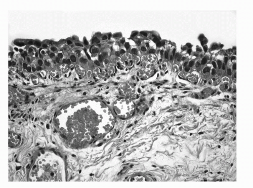
FIGURE 16.1. Urothelial Carcinoma in situ. Markedly hyperchromatic and pleomorphic cells lacking polarity in relation to the basement membrane. (See color insert.)

If left untreated, most cases of CIS will progress to invasive carcinoma, and in patients who underwent cystectomy for CIS, invasion was detected in approximately one third of them (27,28,29). Nevertheless, not all patients with CIS develop subsequent invasive disease (30).


PAPILLARY UROTHELIAL LESIONS


Grading of Papillary Urothelial Lesions

The most common tumors of the urinary tract (excluding prostate) are papillary tumors of urothelial origin. They frequently are multiple, and often recur after being removed, yet most of these tumors are easily managed by local resection. Patients who have had papillary tumors of the bladder are at risk of developing invasive carcinoma, and some very experienced pathologists regard nearly all papillary tumors as carcinoma (31,32). The issue of grading papillary urothelial tumors has been vigorously debated among urologic pathologists, but in this chapter, we follow the classification adopted by the WHO/ISUP (Table 16.1). This classification system relies on changes in the architectural and cytologic features within the urothelium.


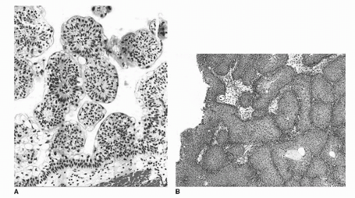
FIGURE 16.2. Urothelial papilloma (A) discrete, exophytic papillary growth. The lining urothelium is of normal thickness and cytology. Inverted urothelial papilloma (B). Invaginated fronds of histologically benign urothelium occupy the lamina propria, giving the overlying mucosa a polypoid or nodular appearance. (See color insert.)


Urothelial Papilloma

This is a rare benign condition typically occurring as a small, isolated growth seen primarily, but not exclusively, in younger patients. Morphologically it is a discrete, exophytic papillary growth with a central fibrovascular core lined by urothelium of normal thickness and cytology (Fig. 16.2A). Recurrences are rare and progression to carcinoma is exceedingly rare (33,34). Inverted Urothelial Papilloma is, by definition, an endophytic mass that shares several features with exophytic urothelial papilloma (Fig. 16.2B). Inverted papillomas have a low risk of recurrence when completely excised. Rarely, hybrid cases exist with portions of the lesion being endophytic and others exophytic. They may be seen in patients with a history of urothelial carcinoma, and rarely, urothelial carcinoma may arise within inverted urothelial papilloma.


Papillary Urothelial Neoplasm of Low Malignant Potential

This term describes a papillary urothelial neoplasm with an orderly arrangement of cells within papillae with minimal architectural abnormalities and minimal nuclear atypia,
irrespective of cell thickness (Fig. 16.3). Mitoses are infrequent and usually limited to the basal layer. When strictly defined, PUNLMP is not associated with invasion or metastasis. Nevertheless, these patients are at risk of developing new bladder tumors (recurrence), usually of similar histology. Occasionally (7%-10%), these subsequent lesions manifest as urothelial carcinoma, such that follow-up of the patient is warranted.


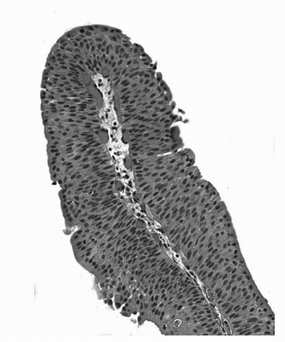
FIGURE 16.3. Papillary urothelial neoplasm of low malignant potential (PUNLMP). Papillary fronds lined by variably thickened urothelium surrounding a fibrovascular core. Cytologically the cells are arranged in an orderly fashion and lack cytologic atypia or pleomorphism. The chromatin pattern is homogeneous. (See color insert.)


Low-Grade Noninvasive Papillary Urothelial Carcinoma

Low-grade carcinomas maintain an overall orderly appearance but exhibit architectural or cytologic atypia that is easily recognizable at scanning magnification (Fig. 16.4). Variability in polarity, nuclear size, shape, and chromatin texture are minimal but comprise the hallmarks of the cytologic atypia seen in low-grade papillary urothelial carcinoma. Mitotic figures are infrequent. Although mitoses may be seen at any level of the urothelium, they are not atypical and usually are limited to the lower half. These tumors frequently recur and have a low (less than 5%) risk of progression.


High-Grade Noninvasive Papillary Urothelial Carcinoma

High-grade carcinomas are characterized by a disorderly appearance because of marked architectural and cytologic abnormalities, recognizable at low magnification (Fig. 16.5). Cellular pleomorphism ranges from moderate to marked, and mitotic figures, including atypical forms, frequently are seen at all levels of the urothelium. They are commonly associated with invasive carcinoma at the time of initial presentation and the adjacent mucosa may show evidence of carcinoma in situ. Progression rate varies from 15% to 40%, much higher than low-grade lesions.


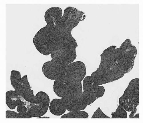
FIGURE 16.4. Noninvasive papillary urothelial carcinoma, low grade. Tumor cells exhibit mild pleomorphism and architectural disorder. (See color insert.)

In a papillary urothelial neoplasm with variable histology, the tumor should be graded according to the highest grade, although it is not uncommon for minuscule areas of higher grade morphology to be ignored in a tumor with overwhelming low-grade component. It is yet to be determined how significant a minor component must be in order to have an impact on prognosis.


INVASIVE UROTHELIAL CARCINOMA

The histology of invasive UC is variable and assessing the depth of invasion is of paramount importance. The histology of invasive UC has no specific features and shows cohesive
nests of cells with moderate to abundant cytoplasm and large hyperchromatic nuclei. Nuclear pleomorphism is common with irregular nuclear contours, occasionally prominent nucleoli, and readily identifiable mitotic figures. There is, however, some confusion related to the use of some terminology such as applying the term “muscle invasion” without further qualification. This term does not distinguish between invasion of the muscularis mucosae or the muscularis propria. Also, the term “superficial bladder cancer” should not be used as it groups three biologically different lesions: CIS, noninvasive papillary neoplasia, and carcinoma with lamina propria invasion.


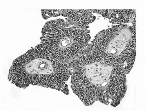
FIGURE 16.5. Noninvasive papillary urothelial carcinoma, high grade. Neoplastic cells with marked pleomorphism, hyperchromasia, and disorder. (See color insert.)


TABLE 16.2 MORPHOLOGIC CRITERIA TO DETERMINE INVASION INTO LAMINA PROPRIA


Histologic grade


Invasion more common in high-grade lesion


Characteristics of the invading epithelium


Irregular nests or single infiltrating cells


Irregular or absent basement membrane


Irregular invasive nests with more cytoplasm than the overlying noninvasive component (paradoxic differentiation)


Characteristics of stromal-host reaction


Presence of retraction artifact


Presence of stromal desmoplasia or myxoid stroma


Inflammatory reaction


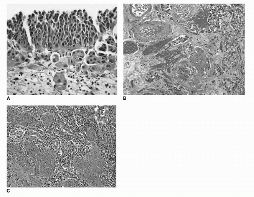
FIGURE 16.6. Invasive urothelial carcinoma. Invasion into lamina propria (A). Irregular clusters of tumor cells with abundant eosinophilic cytoplasm. Tissue retraction causing peritumoral clearing is evident. Invasion into deep lamina propria (B). Irregular tumor nests surrounding muscularis mucosae and large vessels of the deep lamina propria. Invasion into muscularis propria (C). Tumor nests and cells infiltrating thick bundles of smooth muscle of the muscularis propria. (See color insert.)

There are several morphologic criteria useful in the determination of lamina propria invasion (Table 16.2). The presence of urothelial nests, clusters, or single cells within the lamina propria, sometimes with prominent retraction artifact is characteristic. The invasive tumor cells may show abundant eosinophilic cytoplasm at the advancing edge of the infiltrating nests (Fig. 16.6A). Another feature of invasive tumor is the presence of associated desmoplastic or inflammatory stromal response.

Attempts to subclassify T1 tumors based on their depth of invasion have been successful only in some cases in which a
well-delineated muscularis mucosae or large vessels are readily identified in the deep lamina propria and which serve as anatomical landmarks to assess the depth of invasion (Fig. 16.6B). However, more work is necessary to arrive at criteria that can be universally adopted and applied to all transurethral resection and biopsy specimens.

Invasion into the muscularis propria should be diagnosed when tumor is seen infiltrating thick smooth muscle bundles (Fig. 16.6B) with all effort applied to distinguish that from muscle fibers of the muscularis mucosae.


Carcinomas with Divergent (Aberrant) Differentiation

Urothelial carcinomas, particularly high-grade tumors, have a propensity for divergent differentiation and may contain variable proportions of different histologic patterns, including a number of variants of urothelial carcinoma that have been recognized by the WHO (Table 16.3). This feature is seen primarily in invasive tumors but may occasionally be present in in situ disease (35). In a series of 300 consecutive cystectomies performed at Memorial Sloan-Kettering Cancer Center, 27% of cancer-bearing specimens contained some form of divergent differentiation. This incidence, however, was lower in transurethral resection specimens at approximately 7%. When divergent differentiation is seen together with usual urothelial carcinoma, the pathologist should use the terminology urothelial carcinoma with ____ differentiation, inserting the type of differentiation observed.


Squamous Cell Carcinoma

The diagnosis of squamous cell carcinoma (SCC) should be reserved for urothelial tumors that exhibit exclusive or predominant squamous differentiation (Fig. 16.7). For urothelial tumors with variable amounts of squamous differentiation, the term urothelial carcinoma with squamous differentiation should be used. Overall, squamous cell carcinomas constitute 2% to 7% of urothelial cancers except in West African regions and along the Nile Valley where, as a consequence of the endemic schistosomal infections, they are the most common form of cancer (36,37,38). Risk factors associated with the development of bladder squamous cell carcinoma include long-term indwelling catheterization, a nonfunctioning bladder, bladder calculi, urinary obstruction, and chronic infection with Schistosoma hematobium (39,40). Squamous carcinoma of the urothelial tract is thought to arise through a process of metaplasia of the urothelium. A large percentage of patients with SCC have squamous metaplasia of the adjacent urothelium. Many have a history of severe, long-term chronic inflammation associated with stones, chronic infection, bilharziasis and, in a few examples, prior systemic chemotherapy with cyclophosphamide (41). A number of morphologic alteration in the urothelial lining have been associated with the development of bladder squamous cell carcinoma including keratinizing squamous metaplasia, verrucous squamous hyperplasia, condyloma acuminatum, and squamous cell carcinoma in situ (42,43).


TABLE 16.3 CLASSIFICATION OF UROTHELIAL CARCINOMA AND VARIANTS


Urothelial (transitional cell) carcinoma


Urothelial carcinoma with mixed epithelial features (divergent differentiation)


Squamous cell carcinoma


Adenocarcinoma


Enteric type


Clear cell (“mesonephric”) type


Signet ring cell type


Urachal carcinoma


Small cell-neuroendocrine carcinoma


Lymphoepithelioma-like carcinoma


Nested variant of urothelial carcinoma


Micropapillary carcinoma


Sarcomatoid carcinoma/Carcinosarcoma


Microcystic carcinoma


Urothelial carcinoma with trophoblastic differentiation


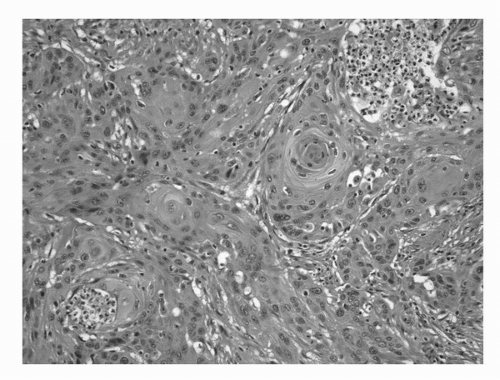
FIGURE 16.7. Squamous cell carcinoma. An invasive high-grade carcinoma exhibiting predominant squamous differentiation. (See color insert.)

Squamous cell carcinoma of the bladder tends to be sessile, ulcerated, and infiltrative at the time of diagnosis. The histologic hallmarks of pearl formation, intercellular bridges, and keratotic cellular debris are those of squamous carcinoma at any site. With the exception of the verrucous variant, most of these carcinomas are moderately or poorly differentiated and more deeply invasive at the time of diagnosis than the majority of transitional cell carcinomas, which may contribute to the generally poor prognosis of these tumors. When these tumors are evaluated stage for stage, however, the prognosis is similar to that of “usual” urothelial carcinoma (38,44,45).


Adenocarcinoma

Primary pure adenocarcinomas of the bladder are rare, representing no more than 2.5% of all malignant vesical neoplasms (46,47). By definition, the tumor should be composed entirely, or virtually entirely, of glandular elements. As with squamous carcinoma, they arise through a process of metaplasia of the urothelium and frequently are associated with long-standing local irritation, nonfunctioning bladder, and obstruction (48,49,50,51,52,53,54). Up to 90% of carcinomas associated with bladder extrophy are of adenocarcinoma type and this tumor is more frequently encountered in a setting of schistosomiasis (55,56). They can arise anywhere on the bladder surface, although a large percentage originates from the trigone and posterior wall. A major clinical difference as compared to usual urothelial carcinoma is that two thirds of adenocarcinomas are single discrete lesions, whereas TCC tends to be multifocal (48,49). Grossly the tumors can be papillary, nodular, or flat and ulcerated. Microscopically, the tumor is most often composed of colonic-type glandular epithelium (Fig. 16.8) and may contain abundant extracellular mucin. Regardless of histologic pattern, cystitis cystica et glandularis or surface glandular metaplasia is commonly present in the adjacent benign urothelium. At the time of initial diagnosis, most adenocarcinomas are locally advanced and infiltrated deeply, most likely accounting for their poor prognosis. Stage for stage, however, they appear to have similar survival to TCC (46,52,53,54,57,58).

It is important to consider the possibility of metastasis or direct invasion from another primary site in the differential diagnosis of an adenocarcinoma involving the bladder. Tumors that directly invade the bladder and mimic primary vesical adenocarcinoma include those arising in rectum, prostate, appendix, and endometrium (59,60,61). For this reason, we have routinely added the following disclaimer to our pathology report of the biopsy specimen: We would accept as primary at this site if a metastasis or direct extension from an adjacent organ can be ruled out clinically. The treating clinician is in the best position to evaluate the patient and consider other primary sites.


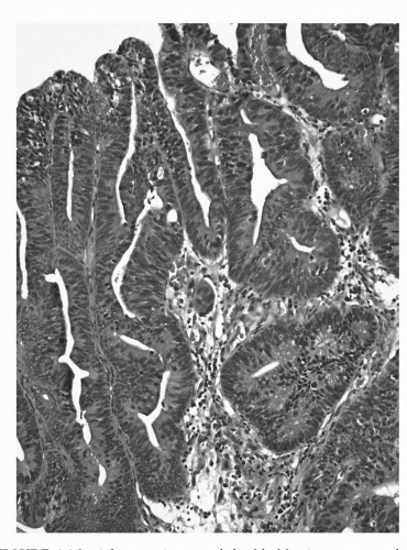
FIGURE 16.8. Adenocarcinoma of the bladder in a transurethral resection specimen. The morphologic features of this lesion overlap with those seen in colorectal adenocarcinoma. (See color insert.)

Signet ring cell carcinoma of the bladder is a rare variant of adenocarcinoma with fewer than 70 cases reported in the English literature, which tends to infiltrate the bladder wall diffusely, giving it an indurated and thickened quality similar to the “linitis plastica” seen in gastric signet ring cell carcinomas (Fig. 16.9). Most of these tumors additionally exhibit morphology in which tumor cells have eccentrically situated nuclei and abundant eosinophilic cytoplasm giving them the appearance of plasma cells and hence the term “plasmacytoid” urothelial carcinoma. The tumor also commonly infiltrates extensively throughout adjacent soft tissue, making primary resection for cure virtually impossible. In the differential diagnosis, one must rule out direct extension, usually from a rectal or prostatic carcinoma, or metastasis from stomach or lobular carcinoma of the breast, or other organs (62,63,64,65,66,67,68,69).

Clear cell adenocarcinoma is another rare variant that is characterized by cuboidal tumor cells forming duct or tubulelike structures that resemble its müllerian counterpart in the female genital tract. These tumors were initially thought to arise from mesonephric nests in the trigone area, but are now considered to arise through a process of metaplasia of the surface urothelium or from müllerian rest (69,70,71,72,73). This tumor has a strong female predominance, which is unusual for urothelial tumors and suggests that müllerian origin may account for an even larger percent of these cases. Immunohistochemical studies are not useful in establishing histogenesis (72). Histologically, these tumors are characterized by tubular and papillary structures that are generally lined by flat, cuboidal, or rarely columnar cells; with hobnail cells present in most tumors (Fig. 16.10). Clear cells are abundant in most tumors, but cells with abundant eosinophilic cytoplasm are also common.


Nephrogenic Adenoma (Metaplasia)

This is a distinct metaplastic lesion that may mimic adenocarcinoma and is characterized by aggregates of cuboidal or hobnail cells with clear or eosinophilic cytoplasm and small discrete nuclei without prominent nucleoli (74). The architectural patterns include thin papillary fronds on the surface and tubular structures within the lamina propria of the bladder
(Fig. 16.11). This lesion is usually fairly well circumscribed and does not extend into the muscularis propria and the tubules are often surrounded by a thickened and hyalinized basement membrane. Usually, there is no stromal response to the lesion, but variable amount of acute and chronic inflammatory cells is commonly encountered. Nephrogenic adenoma is thought to be due to an inflammatory insult or local injury (75,76,77). It is most commonly seen in the bladder but may occur in the prostatic urethra as well (74,77).


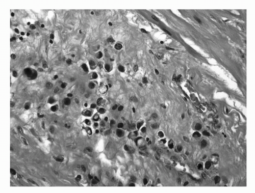
FIGURE 16.9. Signet ring cell carcinoma of the bladder. The tumor is deeply infiltrative into perivesical tissue in a diffuse pattern. Some tumor cells exhibit plasmacytoid morphology. (See color insert.)


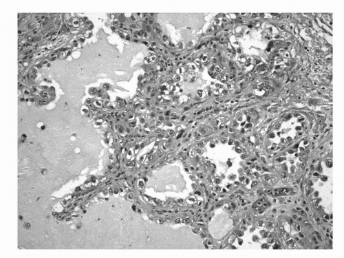
FIGURE 16.10. Clear cell adenocarcinoma. Characteristic tumor cells forming ductular, tubular, or papillary structures. Tumor cells may have clear or eosinophilic cytoplasm and hobnail appearance. (See color insert.)


Urachal Carcinoma

Urachal carcinoma is a primary carcinoma of the bladder arising from urachal remnants, the vast majority of which is adenocarcinoma. Urachal remnants have been found in up to 35% of bladders at the time of autopsy; usually in the dome, but also along the anterior and rarely along the posterior bladder wall (78,79). Although their epithelial lining usually is transitional in type, it can be glandular in 33% of cases. Urachal carcinoma is rare and comprises 0.35% to 0.7% of all bladder cancers and 22% to 35% of vesical adenocarcinomas (79,80,81). Most of these carcinomas have enteric features and may be mucinous (Fig. 16.12); however, rare examples containing signet ring cell, micropapillary, squamous cell, small cell/neuroendocrine, lymphoepithelioma-like, usual urothelial and anaplastic carcinoma components have been reported (79,82,83,84). Adenomatous changes have been identified in the adjacent urachal remnant in 37% of cases in one study (79).


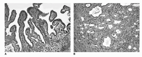
FIGURE 16.11. Nephrogenic adenoma (metaplasia). The lesion consists of bland epithelial cells in a simple papillary formation on the surface (A) and tubular structures in the submucosa (B). In the background, vascular congestion and mixed inflammatory infiltrate is evident. (See color insert.)

Although the criteria for a diagnosis of urachal carcinoma are somewhat controversial, most investigators agree with those set forth by Sheldon et al. (85) and Mostofi et al. (49). These include (a) tumor in the dome of the bladder; (b) absence of cystitis cystica and cystitis glandularis; (c) predominant invasion of the muscularis or deeper tissues with a sharp demarcation between the tumor and surface bladder urothelium that is free of glandular or polypoid proliferation; (d) presence of urachal remnants within the tumor; (e) extension of tumor into the bladder wall with involvement of the space of Retzius, anterior abdominal wall, or umbilicus; and (f) no evidence of a primary neoplasm elsewhere.

A few staging systems have recently been proposed (84,86), but commonly followed is that proposed by Sheldon et al. (85): pT1—no invasion beyond the urachal mucosa; pT2—invasion confined to the urachus; pT3—local extension to the (a) bladder, (b) abdominal wall, and (c) viscera other than the bladder; and pT4—metastasis to (a) regional lymph nodes and (b) distant sites.

Because the urachus usually is found along the free surface of the bladder, urachal carcinomas frequently are amenable to partial cystectomy. The entire length of the median umbilical ligament may harbor urachal remnants that may develop carcinoma synchronously or metachronously (87). For this reason, the surgery of choice should include en bloc resection of the entire length of the ligament, including the umbilicus (84,88).

The differential diagnosis includes metastatic adenocarcinoma and adenocarcinoma arising in the bladder surface. The latter usually is associated with an intraluminal mass and the diagnosis is established by finding CIS or extensive glandular metaplasia of the adjacent urothelium. Because these features are rarely seen on a TUR specimen of an ulcerated lesion, we include the following note in the pathology report of any solitary glandular lesion involving the dome of the bladder: We would accept as primary at this site, including the possibility of urachal origin, if a metastasis or
direct extension from an adjacent organ has been ruled out. Once again, the urologist is in the best position to make this determination.


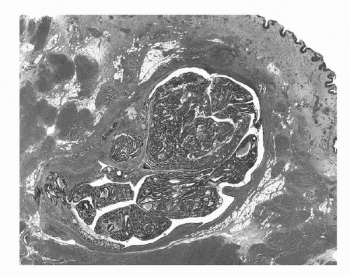
FIGURE 16.12. Urachal carcinoma. Tumor arising within and distending a urachal remnant. The tumor is an adenocarcinoma, enteric type with adjacent adenomatous change. (See color insert.)


Small Cell-Neuroendocrine Carcinoma

Small cell carcinoma is a malignant neuroendocrine neoplasm derived from the urothelium that histologically resembles its pulmonary counterpart (16,89,90,91,92). It consists of small cells with nuclear molding, scant cytoplasm, and dark nuclei containing finely stippled chromatin and inconspicuous nucleoli (Fig. 16.13). By immunohistochemical studies, most tumors express chromogranin and synaptophysin. In most cases, they are associated with CIS of the urothelium and show microscopically foci of other divergent histologic patterns, usually transitional cell carcinoma, but also adenocarcinoma, squamous cell carcinoma, or spindle cell carcinoma (89,93,94). Small cell or neuroendocrine carcinomas should be considered high-grade tumors. They usually present at an advanced stage; up to one third of patients have metastases at the time of diagnosis.


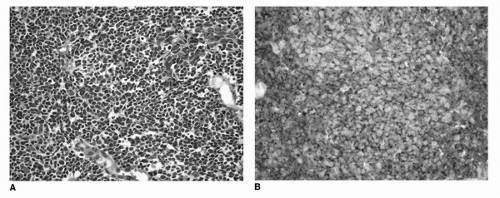
FIGURE 16.13. Small cell/neuroendocrine carcinoma of the urinary bladder. Histologic features are similar to small cell carcinoma of the lung and other sites. Tumor cells are with nuclear molding, scant cytoplasm, and dark nuclei (A). Expression of neuroendocrine markers synaptophysin (B) and chromogranin by immunohistochemistry is a consistent finding and can be helpful in establishing the diagnosis. (See color insert.)

Only gold members can continue reading. Log In or Register to continue

Stay updated, free articles. Join our Telegram channel

Jul 15, 2016 | Posted by in ONCOLOGY | Comments Off on Pathology and Cytology of Tumors of the Urinary Tract

Full access? Get Clinical Tree

Get Clinical Tree app for offline access