(1)
State University of New York at Buffalo Kaleida Health, Buffalo, NY, USA
The removal of the duodenum and the head of the pancreas is performed in patients with a malignant tumor in the head of the pancreas, the ampulla of Vater, or the distal choledochus. Because of its gravity, this operation is applied primarily with curative intent in the absence of demonstrable metastatic disease. Rarely, it can be used to remove a solitary metastatic lesion in this area, if the biology of the disease and the specifics of the case favor the removal of such a tumor according to the literature, and if an uncomplicated resection is anticipated.
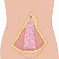
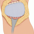
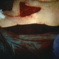
A midline incision from the xiphoid to a few centimeters below the umbilicus provides expeditious entry and closure and excellent exposure in most cases. For patients with a wide abdominal girth, a tri-star (Mercedes) incision can be used, consisting of bilateral subcostal incisions with an upward extension to the lower sternum at the angular joining of the two subcostal incisions.
With either incision, the xiphoid may be resected for additional proximal exposure. A thorough exploration is first performed to rule out the presence of metastatic deposits on the omentum, the parietal or visceral peritoneum, and the pelvis. The right and left lobes of the liver are examined carefully. The falciform ligament is incised all the way to the diaphragm following ligation and division of the round ligament. The porta hepatis is examined by inspection and palpation as it is encircled by the examining hand with the index and middle fingers in the foramen of Winslow and the thumb in front of the hepatoduodenal ligament. The goal is to rule out the presence of abnormal lymph nodes at some distance from the pancreas. Lymph nodes close to the pancreas that are expected to be encompassed in the specimen are not contraindications to resection. Lymph nodes beyond the field encompassed in the procedure require biopsy and frozen section. The gastrohepatic ligament is incised in an avascular area (close to the liver), and the right gastric vessels are ligated and divided close to the lesser curvature of the stomach. (For a pylorus-preserving operation, the right gastric vessels are preserved, the duodenum is divided 2 cm below the pylorus, and the gastroduodenal artery is ligated and divided close to its origin.) The superior border of the pancreas is visualized and examined for any adjacent adenopathy. The common hepatic, gastroduodenal, and proper hepatic arteries are easily palpable. The gastrocolic ligament is divided, and the inferior border of the pancreas is visualized and palpated.
The duodenum is extensively kocherized, exposing the inferior vena cava and aorta. Palpation is carefully carried out. A search for replaced or accessory hepatic arteries in the hepatoduodenal ligament is made; if they are present, they should be protected. The base of the mesocolon at the location of the superior mesenteric vessels and the uncinate process is visualized and palpated. The middle colic or an adjacent vein is gently traced along its superior aspect, leading to the anterior surface of the superior mesenteric vein (SMV). Gentle, blunt finger or clamp dissection is then performed between the SMV and the neck of the pancreas. At the superior border of the pancreas, the gastroduodenal artery is ligated and divided, if not done already, exposing the portal vein. Gentle finger dissection is carried out from above the superior border of the pancreatic neck and from below the inferior border until the two dissecting fingers meet, establishing freedom from invasion of the anterior surface of the SMV and the beginning of the portal vein. If the uncinate process is also separable from the superior mesenteric vessels, the tumor is resectable.
Two sutures are placed at the superior pancreatic border and two at the inferior border, on each side of the proposed plane of pancreatic transection. These sutures are tied, to control blood vessels running along these borders, and the neck of the pancreas is then divided. With a support such as a clamp or forceps behind it, bleeders from the cut parenchyma are controlled, and as the pancreatic head is laterally retracted, tributaries to the SMV and the portal vein are ligated and divided, as are branches of the superior mesenteric artery more posteriorly, toward the head. The choledochus is divided well above the duodenum. The stomach is divided a few centimeters above the pylorus, and the first and second portions of the duodenum are separated from the major structures of the hepatoduodenal ligament. The jejunum is divided with a stapling device just below the ligament of Treitz, and then, as the specimen is retracted inferolaterally, small branches of superior mesenteric vessels between their posterior surface and the specimen are ligated and divided, completing the resection.
Stay updated, free articles. Join our Telegram channel

Full access? Get Clinical Tree


