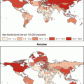
Fig. 1
Prevalence of current cigarette smoking, by race/ethnicity and sex, 2010, National Health Interview Survey, United States [2]
While personal cigarette smoking has been causally linked with lung cancer, second-hand cigarette smoking, also called involuntary smoking or environmental tobacco smoke (ETS) , has been associated with lung cancer risk in exposed nonsmoking individuals. In 1986, the Surgeon General released a report detailing the chemical composition of sidestream smoke, noting it is qualitatively similar to the mainstream smoke inhaled by the smoker and that both mainstream and sidestream smoke act as carcinogens [3]. This report also concluded ETS exposure is associated with lung cancer in nonsmoking individuals. Various epidemiologic studies have confirmed the association with lung cancer, although the findings for ETS are neither as strong nor as consistent as the risk estimates reported for current smoking. This is not unexpected, as measuring ETS exposure is less standardized than estimating years smoked, or number of cigarettes used per day. Despite difficulties with assessing ETS exposure, most epidemiologic evidence supports a modest association between ETS exposure and lung cancer. A meta-analysis of 35 case–control and 5 cohort studies suggested nonsmoking women exposed to ETS from their spouse’s smoking had a 1.2-fold increase in risk compared to women who were not exposed (OR = 1.20, 95 % CI: 1.10–1.29) [4]. Since personal smoking is more prevalent among men, similar studies estimating risk of lung cancer among men from exposure to ETS through their smoking spouse are not available. For workplace ETS, another meta-analysis of 22 workplace studies found a 24 % increase in risk of lung cancer among workers exposed to ETS (RR = 1.24, 95 % CI: 1.18, 1.29) [5]. Longer durations of exposure, whether in the workplace or at home, have been associated with even greater increases in risk.
Other methods to smoke tobacco, such as pipe and cigar smoking, are also associated with increased risk of lung cancer. Two studies from the American Cancer Society’s Cancer Prevention Study cohort provide estimates of risk for men; however, data for women are not available because of low usage of these products among women. Men who reported current or former exclusive pipe smoking (i.e., did not also smoke cigarettes) had a 5-fold increased risk of death from lung cancer compared to men who reported never using tobacco (including cigarettes) (RR = 5.00, 95 % CI 4.16, 6.01) [6]. In this same cohort, men who reported current exclusive cigar smoking at baseline had an nearly identical risk of dying from lung cancer compared to those who never used tobacco (RR = 5.1, 95 % CI: 4.0–6.6) [7]. A cohort study from Europe of 102,395 men reported slightly lower estimates for exclusive users of pipes (HR = 3.0, 95 % CI: 2.1, 4.5) or cigars (HR = 2.2, 95 % CI: 1.3, 3.8) compared to men who did not use any tobacco [8]. While these estimates are lower than for cigarette smoking, it should be noted that pipe or cigar smoking is not a safer alternative to cigarette smoking, but the lower risk is likely explained by lower smoking intensity and perhaps lesser degrees of inhalation of these products.
Other products are also potential risk factors for lung cancer that will likely generate more research in the upcoming decades. First, marijuana (cannabis) use is reported to be the most widely consumed illicit drug worldwide, and the smoke contains many of the same carcinogens found in tobacco. In the U.S., marijuana use has been decriminalized for medical purposes in some regions and to a lesser extent, for personal use. The long-term effects of marijuana use on lung cancer are largely unknown. A 40-year cohort study of 49,321 Swedish men enrolled between the ages of 18 and 20 suggests that “heavy” use of marijuana smoking (defined as having used more than 50 times prior to enrollment) was associated with a 2-fold increase in risk of lung cancer (HR = 2.12, 95 % CI: 1.08, 4.14) after adjustment for tobacco use at baseline and other potential confounders [9]. Unfortunately, these data were only collected at baseline, so updated exposure status could not be incorporated into the models. Marijuana use is prevalent among youth in the U.S., with data from 2011 suggesting that 39.9 % of high school students (9th–12th grades) had tried marijuana at least once, and 23.1 % had used in the 30 days prior to the survey [10]. Thus, marijuana is poised to become an increasingly important risk factor for lung cancer.
Another potential factor for lung cancer is hookah use (water pipe tobacco smoking). Research suggests that a single session of hookah use results in similar mean peak plasma nicotine concentration levels compared to smoking a cigarette, but has 3.75-fold greater carboxyhemoglobin (COHb) levels, and 56-fold greater inhaled smoke volume [11]. The tobacco products used in hookah pipes are often enhanced with various flavorings, and the potential health effects of these chemicals have not been studied. The Monitoring the Future survey found that in 2011, 18.5 % of 12th grade students in the United States had used hookahs in the past year [12]. Other studies indicate that hookah smoking is more prevalent among university students in the United States, with past-year use ranging from 22 % to 40 %, and note that hookah users are more likely to use cigarettes and marijuana [13, 14]. Lastly, electronic cigarettes (e-cigs) or “vapors” are also emerging as an alternative way to inhale nicotine, although little research has been done on these products. It will be necessary for future studies of lung cancer risk to include comprehensive exposure questionnaires to account for various routes of inhaled nicotine, tobacco, and marijuana exposure.
Environmental Exposures
Compared to cigarette smoking, the proportion of lung cancers associated with environmental or occupational exposures is relatively low in the United States, but of significant concern to the 10–15 % of never smokers who develop lung cancer, and also because many environmental exposures may act synergistically with cigarette smoking. Many agents have been examined as potential risk factors associated with lung cancer but are often difficult to quantify and thus the evidence is unclear. Below we describe exposures which have been linked to lung cancer and are of particular interest due to their ubiquitous nature: radon, air pollution, asbestos, diesel exhaust, and ionizing radiation.
Radon is widely accepted as the first identified environmental cause of lung cancer in studies of underground miners (1920s). An inert gas, it is naturally produced from radium in the decay series of uranium (found in rocks and soil) and is a ubiquitous contaminant of indoor air. A meta-analysis of 13 European case–control studies suggests risk of lung cancer is increased by 8.4 % (95 % CI: 3.0–15.8) per 100 Becquerels/m3 increase in measured radon (p-value = 0.0007) and they noted a linear dose–response relationship. There was a synergistic effect with current cigarette smoking, with absolute risk at least 25 times greater for smokers [15, 16]. A pooled analysis of over 4000 cases and 5000 controls from 7 North American case–control studies of lung cancer reported similar findings [17]. Estimates suggest that 20,000 lung cancers diagnosed annually in the United States are attributed to radon exposure [18]. As radon is odorless and colorless, most people are unaware of this potential household hazard. Thus, in 2011, 10 United States federal agencies, led by the Environmental Protection Agency, developed a plan to increase awareness and to reduce the risk from radon exposure [19].
Radon can be considered indoor air pollution, as can ETS, but there are additional indoor contaminates that may increase lung cancer risk. In particular, the use of soft coal for cooking and heating has been associated with lung cancer. A meta-analysis of 25 case–control studies with over 10,000 cases and 13,000 controls noted that household coal use was associated with lung cancer in all studies (meta-OR = 2.15, 95 % CI: 1.61–2.89), and stronger associations were seen in studies from China [20]. Several studies of Chinese nonsmoking women report that heating cooking oils to high temperatures is associated with increased risk of lung cancer [21, 22]. The combination of burning coal, cooking fumes, and ETS exposure play an important role in the development of lung cancer for this population. Outdoors, long-term ambient fine particulate matter (PM2.5) air pollution has been studied as a potential risk factor for lung cancer, usually through cohort studies that link with air monitoring networks. These studies have shown increased risk of lung cancer mortality as PM2.5 levels rise, among individuals living in these areas longer-term [23, 24]. These associations are small, and may be confounded by other exposures, such as cigarette smoking. Regardless of the source, air pollution is a source of concern and continual study for lung cancer and other pulmonary conditions is needed.
A well-established occupational risk factor for lung cancer is asbestos. Asbestos refers to naturally occurring silicate mineral fibers, which have been widely used in industry. Asbestos exposure is related to both mesothelioma and lung cancer, responsible for a combined 10,000 deaths annually in the United States [25]. Asbestos-induced effects in the lungs appear to be dose-dependent and related to the size and composition of the fiber inhaled, with effect sizes ranging from OR = 2.0 to 6.0, depending on the fiber type [25, 26]. There also appears to be a synergistic relationship between cigarette smoking and asbestos exposure, highlighting the need for smoking prevention and cessation for workers in this industry [27].
Evidence supporting increased lung cancer risk with occupational diesel exposure is less established, but a pooled analysis of 11 case–control studies suggested about a 30 % increase in risk among the exposed (OR = 1.3, 95 % CI: 1.2, 1.4) and a significant dose–response trend [28]. A recent review of various studies and exposure assessment methods argues that evidence is still insufficient to make this claim, although the particles found in diesel exhaust contain known carcinogens [29]. In addition, millions of people living in urban areas are exposed to various levels of diesel across their lifespans, and little is known about lung cancer risk associated with low level, chronic exposure.
Lastly, another common exposure that may increase risk of lung cancer is ionizing radiation. Studies of the Hiroshima and Nagasaki atomic bomb survivors suggest increased lung cancer incidence (as well as other solid tumors) among those exposed, with risk increasing in a linear dose response pattern [30]. This single, high dose exposure differs from the smaller doses the general population may receive during X-ray or computed tomography (CT) screenings [31]. Risks associated with repeated CT screenings, while relatively low for an individual, have been considered when making recommendations for implementing population-based lung cancer screening, so that the increased risk of screening does not outweigh the potential benefit [32].
Family History of Lung Cancer
Epidemiologic evidence demonstrates familial aggregation of lung cancer after adjusting for familial clustering of cigarette smoking and other risk factors. Familial aggregation of lung cancer was first noted 50 years ago by Tokuhata and Lilienfeld [33, 34]. In a study of 270 lung cancer patients and 270 matched controls, and their relatives, they found 2.0 to 2.5-fold increased lung cancer mortality in smoking relatives of cases as compared with smoking relatives of controls. A similar finding was noted in nonsmoking relatives. There was an interaction between family history and smoking, with smoking relatives of lung cancer patients having a higher risk of lung cancer than either nonsmoking relatives of lung cancer patients or smoking relatives of controls. This was the first study to account for age and smoking status in a study of familial aggregation of lung cancer, however, smoking intensity or duration was not available.
Several other studies have since reported familial aggregation of lung cancer [35, 36], with the best studies taking into account the number of relatives in the families and the risk factor profiles for each relative to ensure that clustering of smoking habits is not driving aggregation of lung cancer. Studies in southern Louisiana, Houston, Detroit and Iceland reported an increased familial risk of lung cancer among relatives of lung cancer probands (the index case leading the family to be studied) after accounting for the effects of age, sex, and smoking history, and occupation or history of COPD [37–41]. These studies suggested a 2 to 4-fold increased risk associated with having a first degree relative with lung cancer after accounting for risk factors, including smoking amount and duration, among the relatives, with variation in risk estimates by age of the proband, smoking status and race.
While the studies described above included risk factor data among relatives, pooled and meta-analyses have been conducted that include a broader range of studies. A meta-analysis of 28 case–control studies and 17 cohort studies demonstrated fairly consistent findings of an approximately 2-fold increased risk of lung cancer associated with family history [35]. Risk was generally higher in relatives of cases diagnosed at a young age and when multiple family members were affected. The International Lung Cancer Consortium study included data from approximately 24,000 lung cancer cases and 23,000 controls and reported a significant 1.5-fold increased risk of lung cancer associated with family history after adjustment for smoking and other potential confounders in cases and controls, and a significant 1.3-fold increased risk for lung cancer among never smokers [36]. Risk estimates were similar when evaluating only those studies with risk factor data for each family member; relative risks for lung cancer among relatives with a family history were 1.6 overall, 1.5 for white, 2.1 for African American, and 2.0 for early-onset (<age 50) case relatives. These studies provide substantial evidence for familial aggregation of lung cancer that remains after adjustment for clustering of cigarette smoking within family members.
Genetic Susceptibility
Evidence of familial aggregation of lung cancer suggests that there is a genetic contribution to lung cancer susceptibility, and typically suggests a rare, highly penetrant inherited mutation. In addition, smokers have differential susceptibility to lung carcinogens; only 15 % of smokers develop lung cancer and 10–15 % of lung cancers develop in never smokers. It is possible that variation in genetic profiles contributes to this differential susceptibility, most likely in the form of a more common, low penetrant genetic alteration.
Rare, High Penetrance Genes
Only one large, family-based lung cancer study has been conducted providing the first evidence of a lung cancer susceptibility locus on chromosome 6 [42]. In this consortium study, multipoint parametric linkage under the simple dominant low-penetrance affected only model yielded a maximum heterogeneity LOD (HLOD) score of 2.79 at 155 cM (marker D6S2436) on chromosome 6q23-25, with 67 % of the families estimated to be linked. Higher HLODs at this location were reported for more highly affected families: families with four affected relatives gave an HLOD of 3.47, families with five or more affected members in two or more generations, gave an HLOD was 4.26, with 94 % of the families estimated to be linked to this region. In expanded analyses with additional families, the region on 6q was again identified [43]. In addition, lung cancer risk among putative carriers was estimated and found to be higher than among noncarriers, even among never smokers. The usual dose response curves of increasing lung cancer risk with increasing amount smoked was demonstrated among smoking noncarriers. Among smoking carriers, risk was higher than among noncarriers, but a dose response relationship was not apparent suggesting that any level of tobacco exposure increases risk among those with inherited lung cancer susceptibility. Additional evidence suggestive for linkage was also found for regions on chromosomes 1q, 8q, 9p, 12q, 5q, 14q and 16q [43, 44].
Common, Low Penetrance Genes
Initial studies designed to identify more common, low penetrance genes with more moderate effects evaluated small numbers of genetic polymorphisms in biologically plausible pathways including metabolic genes, growth factors, growth factor receptors, DNA damage and repair genes, oncogenes and tumor suppressor genes [45, 46]. More recently, genome-wide association studies (GWAS) have been conducted that rely on very large samples and more than 300,000 markers across the genome. Unlike the candidate gene studies, the GWAS have provided highly significant and reproducible results.
The first three publications of lung cancer GWAS findings identified the same region of chromosome 15q as significantly associated with lung cancer risk [47–49]. This region includes a neuronal nicotinic acetylcholine receptor gene cluster comprising CHRNA3, CHRNA5 and CHRNA4 subunits. Genetic variation in this 15q25 region was associated with an approximately 1.3-fold increased risk of lung cancer among individuals carrying a heterozygous mutation (44.2 % of controls for marker rs8034191) and about a 1.8-fold increase for individuals homozygous for the mutation (10.7 % of controls). This region has also been associated with smoking behavior. One study suggested that the region affected smoking behavior [48], another found stronger effects on lung cancer risk that remained after adjusting for smoking behavior [49], while the third study did not find any association with smoking behaviors [47]. A meta-analysis of smokers, lung cancer cases and lung cancer-free controls, and chronic obstructive pulmonary disease (COPD) cases and COPD-free controls reported that multiple loci within this region are associated with cigarettes smoked per day and at least one locus associated with lung cancer independent of amount smoked [50].
Two other regions, on chromosomes 6p21 and 5p15, identified from GWAS have been consistently associated with lung cancer risk [47–49, 51, 52]. BAT3 and MSH5 are located in the 6p21 region, while TERT and CLPTM1L are located in the 5p15 region. In a large meta-analysis of 14,900 lung cancer cases and 29,485 controls from 16 GWAS, all of European ancestry (as were the initial GWAS), additional support was provided for loci associated with increased lung cancer risk at 5p15, 6p21, and 15q25 [53]. Lung cancer GWAS have also been conducted in the Han Chinese population where evidence was found for lung cancer risk associations at 5p15, 3q28 (TP63), 13q12 (MIPEP-TNFRSF19), and 22q12 (MTMR3-HORMAD2-LIF) [54], and at 10p14, 5q32 and 20q13 [55]. In the Japanese population, the findings on 5p15, 3q28, and 6p21 were replicated [56].
Genetic susceptibility for lung cancer in never smokers is less well studied due to the smaller number of never smokers with lung cancer. In a GWAS of never smoking women in Asia, the 6p21, 5p15 and 3q28 findings were replicated and new regions on 10q25 and 6q22 were identified as being associated with lung cancer [57]. A large GWAS in European American never smokers is underway. In addition, a lung cancer GWAS in African Americans is being conducted. While the findings from the GWAS in African Americans have yet to be published, associations between lung cancer risk and SNPs on 15q25, 5p15 and 6p21 have been replicated in African Americans [58, 59]. A GWAS in lung cancer cases with a strong family history of lung cancer has also been conducted, but results have yet to be published. GWAS in various population subsets who have different genetic backgrounds and smoking behaviors will provide important information for the eventual identification of lung cancer susceptibility genes.
Chronic Obstructive Pulmonary Disease (COPD)
COPD and lung cancer share a common risk factor, cigarette smoking, but studies also suggest that COPD itself is a risk factor for lung cancer independent of smoking habits. A COPD diagnosis has been consistently reported to be associated with a 2- to 3-fold risk of developing lung cancer [60–66], even among never smokers [67]. Lung cancer risk varies with specific COPD phenotypes, i.e., emphysema and chronic bronchitis [62, 66, 68–72]. In a meta-analysis, lung cancer was associated with a previous history of COPD (OR = 2.2, 95 % CI 1.7–3.0), chronic bronchitis (OR = 1.5, 95 % CI 1.3–1.8), and emphysema (OR = 2.0, 95 % CI 1.7–2.4) [65]. In a large, population-based case–control study in women in Detroit, non-small cell lung cancer cases with a joint chronic obstructive lung disease phenotype were more likely to be white, heavy smokers, be exposed to environmental tobacco smoke, have childhood asthma, and have a history of asbestos exposure than lung cancer cases without a history of COPD [64]. Most epidemiologic studies of COPD, however, rely on self-report of COPD phenotype and are subject to both recall bias and misclassification.
Prospective studies have evaluated the association between computed tomography (CT) evidence of emphysema and/or spirometry-defined measures of airflow obstruction and risk of lung cancer, reducing the potential for disease misclassification. These studies report a 2- to 4-fold increased risk of lung cancer in the presence of CT evidence of emphysema, with no or lower risks associated with airflow obstruction [64, 73–75]. In studies using quantitative image analysis of CTs, no increased lung cancer risk among patients with emphysema was reported [76, 77]. Risk of lung cancer has also been shown to increase with decreasing forced expiratory volume in 1 second (FEV1) even in smokers with only minimal declines in FEV1 [72]. For these studies to move forward, consistently defined COPD will need to be evaluated in individuals with the joint COPD-lung cancer phenotype.
The lung cancer-COPD connection also is evidenced in family and genetic studies. First degree relatives of lung cancer patients show impaired FEV1 [78] and a family history of COPD increases risk of lung cancer development [79], suggesting a common underlying genetic contribution to these diseases. In family-based genetic studies for COPD, a region on 6q, just beyond the lung cancer linkage region and extending to the end of the chromosome, was linked to FEV1 [80, 81]. There was also evidence for linkage of lung function to moderate obstructive lung disease in smokers on chromosome 12p [82, 83]. These data provide some regions of potential overlap in areas linked to lung function, COPD and lung cancer on chromosomes 6q and 12p.
Candidate gene studies in COPD and lung cancer have focused on inflammation, extracellular matrix proteolysis, and oxidative stress pathways [84–86], with some consistent findings for SNPs in epoxide hydrolase 1 (EPHX1), matrix metalloproteinases, and interleukin 1β (IL1B) [87–91]. Inflammatory pathway genes have been targeted for study because of the chronic inflammation caused by cigarette smoke. Van Dyke et al. showed that SNPs in IL7R, IL15, TNF, TNFRSF10A, IL1RN, and IL1A were associated with lung cancer risk in women with self-reported COPD, but not among women without COPD [92]. SNPs in IL1A have also been reported to be more strongly associated with lung cancer risk in those with emphysema [89]. GWAS for COPD-related phenotypes have identified some of the same regions identified in studies of lung cancer, namely 15q25.1 [50, 93–96]. Few studies, however, have evaluated a joint lung cancer and COPD phenotype. Young et al. summarize findings and report that the 15q25 locus is associated with risk of both diseases, genetic variation on 4q31 and 4q22 are associated with reduced risk of both diseases, loci on 6p21 are most strongly associated with lung cancer risk in smokers with COPD, and variants on 5p15 and 1q23 alter lung cancer risk when COPD is not present [97]. Taken together, these findings suggest that lung cancer occurrence is linked to COPD and more detailed studies of the joint phenotype using clearly defined COPD traits are needed to better untangle the relationship [98].
Infectious Agents
The role of infectious agents in lung cancer risk has had a varied focus over time with the changes in prevalent exposures. An association between TB and lung cancer has been reported for many years. In a meta-analysis of 37 case–control studies and 4 cohort studies, Liang et al. report significant associations between TB and subsequent lung cancer diagnoses, with risk estimates of 1.7 (95 % CI 1.5–2.0) adjusting for smoking history [99]. Similar risk estimates were reported in never smokers and highest risk was seen within 5 years of the TB diagnosis. In a more recent, population-based cohort study in Taiwan, a 1.8-fold increased risk of lung cancer was reported after a diagnosis of TB [100]. Human papillomavirus (HPV) infections have also been studied with regard to lung cancer risk. The prevalence of HPV in lung tumor tissue ranges from 0 % to 100 %, with great heterogeneity of findings across geographic regions, histology type of the lung cancer, sex, and HPV type [101, 102]. This is an area which will require additional study in high risk populations.
Another focus of current research is on lung cancer risk among individuals with HIV infection. With more effective treatments, HIV-infected patients are living longer and lung cancer now ranks as one of the most frequently diagnosed non-AIDS-defining malignancies. Studies comparing lung cancer incidence in HIV-infected individuals to the general population have shown a 1.5 to 5.0-fold increased risk in infected individuals. In the review by Hou et al., standardized incidence ratios (SIRs) and incidence rate ratios (IRRs) adjusted for age, gender, race/ethnicity, smoking and route of infection are presented for 65 publications [103]. Lung cancer risk in HIV-positive populations varied with geographic region; SIRs or IRRs were 1.5–3.4 in Europe, 0.7–6.9 in the United States and 5.0 in Africa. Risk estimates were 5.4 in Europe and 2.8–3.0 in the United States for individuals with AIDS. Most studies showed little difference in lung cancer risk among HIV-infected patients receiving highly active antiretroviral therapy (HAART) and those not [104–106]. There are a number of limitations to the current body of literature, and continued follow-up of HIV-positive individuals will be needed to fully evaluate lung cancer risk that considers smoking more fully and focuses on race/ethnicity in populations at high risk of infection.
Risk Models
With the anticipated launch of population-based lung cancer screening on the horizon, identification of the group who would most benefit from early detection (i.e., those at highest risk of lung cancer) is critical to the success of a screening program. Over the last decade, various models have been proposed, as shown in Table 1. The eight proposed models are from both cohort (n = 4) [107–110] and case–control studies (n = 4) [111–114]. The majority of these studies are for current or former (usually defined as quit within one year of diagnosis or study entry) smokers, and use a combination of demographic characteristics (e.g., age and sex), intensity and/or duration of cigarette smoking, medical history, and occupational exposures as variables in the predictive model.
Table 1
Risk models for lung cancer in the general population
First author, (year) | Population type | Population details | Variables | Model discrimination | Other information |
|---|---|---|---|---|---|
Bach et al. [107] | N = 18,172 Carotene and Retinol Efficacy Trial participants | All participants were either 20 pack year smokers (current or quit within 6 years) or asbestos-exposed men (current smokers or quit within 15 years) | -Age -Sex -Asbestos exposure -Duration of smoking -Cigarettes per day -Time since smoking cessation | One year risk: c-statistic = 0.72 Also presented a 10-year risk table | Within study validation and the Mayo Clinic CT study |
Spitz et al. [112] | 1851 lung cancer patients and 2001 control subjects (matched on age (±5 years), sex, ethnicity, and smoking status (never, former, or current)) | Cases recruited from the Thoracic Center at The University of Texas M. D. Anderson Cancer Center Controls recruited from Kelsey – Seybold Clinics in metropolitan Houston, TX | Never smokers: -ETS -Family history of any cancer Former smokers: -Emphysema -Dust exposure -Hay fever -Family history of any cancer -Age stopped smoking Current smokers: -Emphysema -Pack-years -Dust exposure -Hay fever -Asbestos exposure -Family history of smoking-related cancer | One year risk : c-statistic: -Never smokers: 0.59 -Former smokers: 0.63 -Current smokers: 0.65 | 25 % of population used for validation |
Spitz et al. [114] | 725 lung cancer patients and 615 control subjects, (matched on age (±5 years), sex, ethnicity, and smoking status (current or former smokers only)) | Cases recruited from the Thoracic Center at The University of Texas M. D. Anderson Cancer Center Controls recruited from Kelsey – Seybold Clinics in metropolitan Houston, TX | In addition to the variables listed above in Spitz (2007), data from two assays were included: a DNA repair capacity measure and a bleomycin sensitivity measure | Former smokers: AUC = 0.70 Current smokers: AUC = 0.73 | Within study 3-fold cross-validation |
Etzel et al. [113] | 491 African American lung cancer patients and 497 African American control subjects (matched on age ±5 years), sex, ethnicity, and smoking status
Stay updated, free articles. Join our Telegram channel
Full access? Get Clinical Tree
 Get Clinical Tree app for offline access

|


