Non-Muscle-Invasive Bladder Cancer


Non-Muscle-Invasive Bladder Cancer


▪ 22A Low-Risk Noninvasive Bladder Cancer

J. Alfred Witjes


WHAT IS LOW-RISK NON-MUSCLE-INVASIVE BLADDER CANCER?

Non-muscle-invasive urothelial cell carcinoma (UCC) of the bladder is a spectrum of fairly benign bladder tumors with a low recurrence rate and an extremely low rate of progression in patients classified as low risk and higher recurrence and progression in patients classified as high risk. Recurrences occur in the vast majority of high-risk patients, and a substantial percentage of these patients will experience progression to muscle-invasive bladder cancer during the course of their disease. It seems obvious that the differentiation between the risk groups is not black and white, and subsequently, the definition of the low-risk group varies over time and between different definitions and guidelines. For example, pTa, grade 1, solitary and primary bladder tumors, was an often-used definition of low-risk non-muscle-invasive bladder cancer (NMIBC) in the 1990s that is now no longer used.

Currently, definitions and guidelines per continent or country differ slightly. The American Urologic Association (AUA) guidelines classify patients using a system of index patients ranging from 1 to 5, where index patient 1 is “a patient who presents with an abnormal growth on the urothelium but who has not yet been diagnosed with bladder cancer” and index patient 5 is “a patient with high-grade Ta, T1, and/or carcinoma in situ bladder cancer which has recurred after prior intravesical therapy.” Index patient 2 is described as “a patient with small volume, low-grade Ta bladder cancer” (1). Multiplicity and the difference between primary and recurrent tumors are not specified. Using similar definitions, the First International Consultation on Bladder Tumors (FIBCT) defines low risk as low-grade Ta, and the National Comprehensive Cancer Network (NCCN) defines low risk as G1-2Ta (2,3). The current European Association of Urology (EAU) guidelines for classifying risk are based on the previously published European Organization for Research and Treatment of Cancer (EORTC) risk tables (4). For these risk tables, the individual data of 2,596 patients with NMIBC from 7 EORTC trials were used to calculate the chance of recurrence and progression following 1 to 5 years of initial treatment with transurethral resection (TUR) in all patients and intravesical therapy in the majority of patients. With this dataset, six readily available clinical and histological factors could be identified to predict disease outcome. Three of these factors are valuable in predicting recurrence including the number of tumors, tumor size, and prior recurrence rate; the remaining three factors are valuable in predicting progression including T category, carcinoma in situ (CIS), and grade.

For each of the six factors, a number of points are given for both recurrence and progression. The total score gives an indication for the probability of recurrence and progression. The risk for recurrence and progression at 1 year for low-risk patients is 15% and <1%, respectively. For high-risk patients, these figures are 61% and 17%, respectively. At 5 years, the probability for recurrence and progression is 31% and still <1% for low-risk patients, and 78% and 45% for high-risk patients, respectively. The total score also refers to the risk group a patient belongs to. When there are no points scored, a patient is in the low-risk group for both recurrence and progression. By the EORTC definition, a low-risk patient has a single, <3 cm, primary pTaG1 tumor. Between 1 and 9 points for recurrence means the intermediate-risk group for recurrence, and between 2 and 6 points for progression means the intermediate-risk group for progression. Both high-risk groups are defined as 10 to 17 points for recurrence and 7 to 23 points for progression. Practical use of these tables is relatively easy, and with the electronic calculator, available through the internet (http://www.eortc.be/tools/bladdercalculator/); its use has become common practice in many countries.

Several remarks need to be made regarding the use of this bladder calculator. It is important to note that of the trials analyzed, only 171 patients were treated with bacille Calmette-Gurein (BCG), and none of the patients received BCG maintenance therapy, resections for tumors, or any immediate intravesical instillation after TUR. This means that the predicted recurrence rates and progression rates are rather close to the natural history of NMIBC. However, with current treatments, these recurrence and progression numbers might be lower. This is nicely illustrated by the Spanish Club Urológico Español de Tratamiento Oncológico (CUETO) group. Data from 1,062 patients treated in four trials with BCG were used to develop a risk stratification model with estimates of the probability of recurrence and progression after BCG treatment (5). Patients were divided into four risk groups for recurrence and progression. Factors for predicting recurrence were gender, age, grade, tumor status, multiplicity, and associated CIS; factors for predicting progression were age, grade, tumor status, T category, multiplicity, and associated CIS. As expected, the calculated risk for recurrence and progression for high-risk patients was lower when using the CUETO tables as compared to the EORTC tables.

Another important limitation in the EORTC tables is the use of the 1973 World Health Organization’s (WHO) grading system (grades 1, 2, and 3). In 2004, a new grading system was published by the WHO (WHO Classification of Tumours. Pathology and Genetics, WHO 2004) based on the consensus text published by Epstein et al. (6) in 1998, to allow for broader accepted terminology, better definitions, and less interobserver variability. One goal of the WHO’s new grading system was to prevent use of the term “cancer”
with patients at very low risk of progression. Therefore, classification of tumors changed considerably, especially in the lower-risk range. In this new classification system, a new category was defined: papillary urothelial neoplasm of low malignant potential (PUNLMP). However, this has led to considerable discussion (7,8) and a lack of general acceptance since reproducibility of PUNLMP is low (9). The distinction between low- and high-grade tumors, after exclusion of PUNLMP, fortunately did improve (10).

An additional concern for the EORTC tables relates to staging, as it is not 100% accurate. In a 1990s randomized controlled trial, the concordance of pTa classification was 81.5% (11). pTa tumors were initially diagnosed in 254 patients via local pathology and confirmed in 207 of these patients after review pathology. For all stages, the conformity between local and review pathology results was 79.3%, and overstaging appeared to be more common than understaging. It should to be mentioned that local pathologists were instructed before the trial about criteria for staging and grading. Similar results were found for pTa tumors in a recent review of three fluorescence cystoscopy studies (12). Comparison of the TUR specimens between local and review pathology revealed an overall conformity for staging in 56.6% of the patients; this was much higher in Ta disease, 80.4%, as compared to T1 disease, 47.6%. Less favorable results were described by van der Meijden et al. (13), who reported on the impact of pathological review in five EORTC trials. The conformity between local and review pathology for pTa tumors was 68.7%, which was lower than pT1 tumors, 77.1%. In all, histological classification of stage Ta seems fairly reproducible, but staging may be incorrect in approximately one out of five patients.

Unfortunately, current research efforts are focused on specific markers used to identify high-risk tumors, especially those with high chance of recurrence and progression. As a result, research on markers to identify low-risk NMIBC is limited. Additionally, little emphasis is placed on the clinical relevance of low-risk NMIBC. One of the few markers that specifically identifies low-risk NMIBC seems to be mutations of the Fibroblast Growth Factor Receptor 3 gene (FGFR3).

In a multicenter study of 286 patients with primary UCC, Van Rhijn et al. (14) found FGFR3 mutations in 60% of the cases. An FGFR3 mutation was detected in 88% of grade 1 tumors and only 16% in grade 3 tumors, with the mutation being linked to a favorable disease course. Gomez et al. (15) also found a marked overexpression of both FGFR3 mRNA and protein in UCC patients with a greater expression in pTa and pT1 bladder cancer compared to invasive bladder cancer. Van Oers et al. (16) studied 255 unselected patients with primary UCC. Mutations in the FGFR3 gene were detected in 47% of the 208 bladder tumors of which 61% occurred in pTa and pT1 tumors. The highest frequency of FGFR3 mutations was found in stage pTa (73%) and grade 1 (82%) tumors. While these authors concluded that an FGFR3 mutation is a useful marker in identifying superficial tumors with low malignant potential, grade and stage were still better predictors their series.

Most recently, Kompier et al. (17) studied FGFR3 mutations in primary and recurrent bladder tumors using urinary samples of 118 patients. They confirmed that these mutations are indeed common in NMIBC (63%) with no difference between primary and recurrent tumors. Recurrences in patients with an FGFR3 mutation were of lower stage and grade than those of patients with a wild-type primary tumor (p < 0.001). In conclusion, FGFR3 seems to play an important role in the development of NMIBC in general and low-grade disease in particular. Used as a molecular marker, it is indicative for low-grade/low-stage NMIBC, and FGFR3 mutations seem to be associated with favorable tumor characteristics. Apart from clinical prognosis, FGFR3 gene status provides insight into the molecular pathways that are involved in the development of low-risk UCC.

In summary, most clinical or guideline definitions of low risk are comparable and refer to a patient with a small Ta, low-grade UCC, usually primary and solitary. There is virtually no risk of progression and the risk of recurrence is approximately 15% in the first year. Changes in the definition of low-grade UCC have yet to lead to a better identification of this group. Currently, markers are not used in clinical practice, but FGFR3 mutation appears to be able to further identify the best possible outcome of the low-risk category.


INITIAL DIAGNOSIS OF LOW-RISK NMIBC

The diagnosis of any bladder tumor, including low-risk NMIBC, is based on the recognition of complaints (predominantly hematuria), the combination of two diagnostic golden standards (cystoscopy and urinary cytology), and subsequent pathologic confirmation of the tumor by TUR and histological examination. The degree of hematuria, typically macroscopic and painless, is not an indicator for the risk category of the tumor. The role of imaging in patients suspected to have a bladder tumor is limited. Routine intravenous urography (IVU) does not show small (low-risk) bladder tumors, and the value of upper urinary tract imaging in low-risk tumors is at best questionable. The incidence of upper urinary tract tumors is low (1.8%) but increases to 7.5% in tumors located in the trigone (18). Ultrasound is increasingly being used to study the bladder when a bladder tumor is suspected; ultrasound is noninvasive and no contrast agents are necessary. Computed tomography (CT) and magnetic resonance imaging (MRI) can also be used to detect bladder abnormalities but with limited sensitivity.

Moreover, staging still is insufficient with both modalities. Additionally, CT scanning is associated with radiation, and the most important limitation of MRI is its availability, cost, and duration of the procedure. Overall, local imaging has a limited value, certainly in low-risk tumors. However, according to the EAU guidelines, in cases where a bladder tumor is visualized during an imaging study, a separate diagnostic cystoscopy can be omitted before TUR (19). Imaging for potential associated upper urinary tract abnormalities is only recommended based on specific indications that are seldom identified in low-risk tumors.

A cornerstone of diagnosis is urinary cytology. The most important advantage of cytology is that it is highly specific (>90%) (20). Sensitivity remains problematic especially in low-grade, low-risk tumors. Bladder wash cytology may play a role in improved diagnostics as it provides a better diagnostic yield than voided urine cytology (21). Therefore, negative urinary cytology certainly does not exclude the presence of low-grade disease (19). On the other hand, positive cytology in a patient with a low-risk tumor should trigger upper urinary tract investigation as the cytology suggests the presence of a higher grade lesion. Both AUA and EAU guidelines have indicated that cytology is best applied for diagnosis and follow-up of high-grade, high-risk tumors. An alternative for urinary cytology might be the use of diagnostic urinary markers. Many tumor markers have been studied and tested in patient cohorts with bladder cancer, usually in diagnostic studies that include patients with non-muscle-invasive and invasive bladder cancer. In general, most markers have a lower specificity but an increased sensitivity than urinary cytology (20). This increased sensitivity is mainly due to better detection of low-grade, low-risk tumors. Therefore, these tumor urinary markers have some additional value over cytology. However, the costs of these tests may be
considerable (19,21). Additionally, to date, no single marker has been identified that can guide in the screening, diagnosis, or surveillance of bladder cancer and lower the frequency of cystoscopy (20).

The gold standard for diagnosing bladder cancer is cystoscopy. As indicated above, this may be omitted when imaging studies clearly show a bladder tumor. Cystoscopy will give information on tumor size, number, and location. However, cystoscopy is not perfect. In a combined analysis of seven EORTC trials, Brausi et al. (22) looked at the variability between institutions and surgeons in the rate of recurrences at initial follow-up cystoscopy after TUR in papillary NMIBC. The idea behind this analysis was quality control of the TUR performed, since a tumor that is seen 3 months after a TUR is much more likely to be a missed lesion during the initial TUR than a new occurrence of a bladder tumor, especially in low-risk patients. Data of 2,410 patients were analyzed, including tumor multiplicity and adjuvant intravesical therapy. In higher-risk patients, those with multiple tumors, the 3-month recurrence rate varied between 7.4% and 45.8% in spite of additional therapy. In lower-risk patients, those with single tumors, the 3-month recurrence rate also varied widely between institutions from 3.4% to 20.6% for patients without adjuvant therapy, and from 0% to 15.4% in those receiving it. The authors suggested that the quality of the TUR and the skill level of the individual surgeons may be responsible for these high recurrence rates and differences between institutions. Although a TUR may appear to be a straightforward operation, especially in a small solitary low-risk tumor, this apparently is not the case. A recent development that has improved detection of bladder tumors during cystoscopy is fluorescence cystoscopy. With the help of a photosensitizing agent, bladder tumors are more visible. This technique is predominantly used during TUR, which is discussed in the following section.

In summary, cystoscopy is the most important tool for diagnosing bladder cancer, particularly low-risk bladder cancer. If a tumor is found during imaging, outpatient cystoscopy can be omitted. Imaging of the upper urinary tract should only be done in selected patients, rarely in low-risk patients. Sensitivity of urinary cytology in low-grade tumors is low. Markers improve sensitivity but are costly, and a marker certainly cannot replace cystoscopy.


TREATMENT OF LOW-RISK BLADDER TUMORS

After a bladder tumor is detected on cystoscopy or imaging, patients will undergo TUR to confirm the presence of a bladder tumor, irrespective of the risk category. Guidelines indicate that a complete resection of all visible lesions should be the goal (1,19), because a complete and well-executed TUR is important for the prognosis of the patient (22). Small tumors like those in low-risk patients can be resected en bloc. The use of a bladder diagram is advised. Random mucosal biopsies, meaning biopsies from normal-looking mucosa, are not routinely recommended. The chance to detect CIS is <2%, especially in low-risk tumors, and the choice for adjuvant intravesical therapy is not influenced by the biopsy result (23). In case of mucosal abnormalities or abnormal cytology with an apparent low-grade lesion, random mucosal biopsies are indicated.

As indicated above, fluorescence cystoscopy makes bladder tumors better visible. This is especially the case for CIS, where detection rate is increased by approximately 20% as compared to white light cystoscopy (24,25). Also, the detection rate is higher in papillary tumors and both T1 and Ta tumors; resections are more complete and the subsequent recurrence rate is lower. For example, in one of the first studies published on the use of hexaminolevulinate fluorescence cystoscopy, 376 pTa tumors were found, of which 328 were found with white light and 365 with blue light (26).

A second resection is advised in high grade and/or pT1 NMIBC, or in cases where an initial resection may not have been complete, such as multiple or very large tumors. Since none of these criteria are applicable for low-risk tumors, a second resection is not routinely indicated for these patients unless a complete initial resection could not be performed.

Intravesical instillation therapy in low-risk patients after resection is a single instillation of chemotherapy. The effect might be twofold, destruction of circulating tumor cells immediately after TUR, or an ablative effect (chemoresection) of residual tumor cells at the resection site. The AUA guidelines advise administering an initial single dose of intravesical chemotherapy immediately postoperatively for patients who are categorized as index patient 2 (4). This advice is based on the panel’s own data collection and meta-analysis of randomized controlled trials. The AUA guidelines panel indicates that the data specifically referring to patients with low-grade, Ta bladder cancer are limited. However, when looking at all patient risk groups, they calculated that a single dose of mitomycin C after TUR reduced the recurrence rate by 17% (95%, CI: 8-28) as compared to TUR alone. They also did not find any additional value of multiple adjuvant instillations of BCG or chemotherapy in patients initially diagnosed with a Ta grade 1 bladder tumor. The EAU guidelines give similar advice (19). In patients at low risk for tumor recurrence and progression, one immediate instillation of chemotherapy is strongly recommended as the complete adjuvant treatment. The grade of this recommendation is A, which is the highest possible grade of recommendation, based on a meta-analysis published by Sylvester et al. (27) in 2004. In this analysis of seven randomized trials, data from 1,476 patients with a median follow-up of 3.4 years were analyzed. Half of these patients had one immediate instillation of chemotherapy after TUR, which resulted in a decreased recurrence rate of 12% (from 48.4%-36.7%, relative reduction of 39%). This benefit was seen both in patients with single and multiple tumors. Looking at the number of patients requiring treatment, the EAU guidelines consider this approach cost effective. A recent study questioned the value of an immediate instillation in patients with multiple tumors. Gudjonsson et al. studied the effect of an early instillation within 24 hours after TUR with 80 mg of epirubicin versus placebo in 209 evaluable low- and intermediate-risk patients (28). After a median follow-up of 3.9 years, recurrences were seen in 62% of the epirubicin group versus 77% of the control group (p = 0.016). However, this difference was only seen in patients with primary solitary tumors, whereas there was no benefit in patients with recurrent or multiple tumors. Finally, a recent meta-analysis of eight randomized trials with 1,776 patients confirmed the significant reduction in recurrence rate after one immediate instillation (24%-62% vs. 48%-77% after TUR only) (29). Again, this difference was not present in patients with multiple tumors. In all the studies reviewed above, the instillation was given within 24 hours. However, the exact timing of instillation remains unknown. A study by the Finnbladder group, for example, suggested that an immediate instillation should be given the day of surgery (30). In patients receiving the instillation the next day, the recurrence rate in this study was twice as high. What is clear from the meta-analysis published by Sylvester et al. is that there is no superior chemotherapeutic drug for this indication. Although one immediate instillation is generally safe, an important contraindication is a suspected bladder wall perforation during extensive resection.

While it seems clear that one immediate instillation after resection remains the recommended therapy for low-risk patients, it has been known for a long time that the impact of
this one immediate instillation is only seen in the first 2 years after treatment (27,31). However, some recent reports imply that this advice should be balanced. Berrum-Svennung et al. (32) studied 404 patients who had an instillation of 50 mg of epirubicin or placebo within 6 hours after TUR. The recurrence rate in the epirubicin group was 51% as compared to 62.5% in the placebo group (p = 0.04). However, predominantly small recurrences were prevented that could easily have been fulgurated using local anesthesia at follow-up cystoscopy. Therefore, the authors questioned the benefit of single instillations. This was also questioned by Hendricksen et al. (33) who compared the additional effect of an early instillation and maintenance instillations of epirubicin to a standard 6-month epirubicin schedule in a randomized controlled trial with 731 patients. Neither of the additions to the standard schedule reduced the recurrence rates. Of note, this study included intermediate- and high-risk patients, not low-risk patients. Moreover, an early instillation was advised to be given within 48 hours, not 24 hours, and only 68% of patients received an early instillation within 24 hours. Finally, Bohle et al. (34) recently compared an early instillation with 2,000 mg of gemcitabin to placebo in 248 NMIBC patients. After a median follow-up of 12 months, 22.3% of the patients in the gemcitabine group had a recurrence as compared to 24.7% in the placebo group (p = 0.78). Although gemcitabine is a relatively new drug, it is effective and probably not the reason for the lack of difference. Since both groups had additional continuous bladder irrigation for at least 20 hours, this could have led to the good results in both study groups. The authors also suggested that current TUR/cystoscopy techniques may have contributed to the high recurrence-free survival in both groups.

An alternative therapy for low-risk patients who are not willing or not fit to undergo resection is chemoresection. The first and most important problem with this approach is that no will be removed for pathologic study. For patients with a recurrent tumor after a previous low-risk tumor, the majority of the recurrences again will be low risk, which can be predicted by an experienced urologist during outpatient cystoscopy (35). For these patients, resection can be postponed safely (36). When chemoresection is considered, most of the information on the ablative effect of intravesical chemotherapy is from marker lesion studies where usually all but one lesion is resected. Since these patients by definition have multiple lesions to start with, their initial classification is always at least intermediate risk. However, the remaining solitary lesion usually is of low grade and stage, and this lesion is subsequently treated with a course of instillations. Although BCG has been used in marker lesion studies, it may be overtreatment in low-risk patients. Recognizing this, the EORTC studied the ablative effect of a ¼ dose of BCG in a small study with 44 low- and intermediate-risk patients (37). Although the complete response rate was promising (27/44, 61%), 54% of patients had dysuria and 39% had macroscopic hematuria. Gemcitabine is another drug that has been used in a marker lesion setting. The complete response rates were variable. Gardmark et al. (38) studied 32 intermediate-risk patients. They found a 10% complete response rate in patients treated with one instillation of 2,000 mg gemcitabine in 100 mL of unbuffered saline as compared to 44% of patients who had a course of gemcitabine with two instillations weekly. Gontero et al. (39) treated 39 intermediate-risk patients with weekly instillations of 2,000 mg gemcitabine in 50 mL of saline for 6 weeks. Their complete marker lesion response was 22/39 (56%) with very low toxicity. Last but not least, apaziquone, a new drug for intravesical use, was tested in 46 low- and intermediate-risk patients with marker lesions (40). The complete response rate was 67%, and side effects were low. This study is worth mentioning, especially since both the United States and the Canada have two large randomized controlled trials using one immediate instillation of apaziquone in low- and intermediate-risk patients after TUR. These studies should lead to the Federal Drug Administration’s (FDA) registration of apaziquone as the first drug available for immediate intravesical use. Although currently there are more drugs under investigation, some are still in preclinical use, some are in initial clinical trials, and most are studied for other indications such as BCG refractory high-risk disease. The same is true for deviceassisted instillation therapy, such as chemothermotherapy.

In summary, therapy should start with a well-executed and complete TUR, possibly with the help of fluorescence techniques. After resection, low-risk patients should receive one instillation of a chemotherapeutic drug on the day of surgery, although more recent studies seem to question this. In any patient with more than a solitary low-grade and low-stage tumor, the value of one immediate instillation seems at least debatable. Chemoresection does not seem applicable to low-risk patients, and the concept is still relatively investigational.


FOLLOW UP

As mentioned above, the risk of progression in low-risk NMIBC is very low (<1%, even after 5 years), but the risk of recurrence, even in this low-risk group, is considerable (15% after 1 year to 31% after 5 years). Therefore, it seems logical that follow-up is not necessary to detect life-threatening disease, but follow-up should be directed at inconvenient recurrences, which are predominantly of the same low stage and grade. In this respect, the first check-up cystoscopy is probably the most important one to predict subsequent recurrences (19,41,42). Thus, EAU guidelines are a logical conclusion (19): low-risk patients should have a cystoscopy at 3 months. If negative, the following cystoscopy is advised at 9 months and consequently yearly for 5 years (grade of recommendation: B). As discussed earlier, the roles of cytology and markers are very limited in the diagnosis of low-risk disease. Similarly, these diagnostic tools seem to play no role in follow-up of low-risk patients.

A marker that might be helpful in the follow-up of these patients is the FGFR3 mutation (17). In a study of 118 patients who underwent 2,133 cystoscopies with a median follow-up of 8.8 years, patients with FGFR3 mutations appeared to have continued recurrences after 10 years, which was not seen in patients with wild-type FGFR3. The authors suggested that a reduced cystoscopy frequency based on the FGFR3 mutation is worth further study.

Findings by Herr are also interesting for this risk group (35). An experienced urologist is able to predict histology when he sees a recurrence on cystoscopy during patient follow-up for NMIBC. Herr reported on 150 patients with recurrent bladder tumors. His objective was recognition of low-risk tumors, suitable for office fulguration. Outpatient cystoscopy classified 84 of 150 patients as TaG1. This appeared correct in 78 cases (93%). With knowledge that these patients are not at risk for life-threatening events, it seems feasible not only to do office fulguration but also to postpone resection. This has also been reported by several groups, most recently by Hernandez et al. (36) They followed a selected group of 64 patients with recurrent, predominantly low-risk, NMIBC. After a mean observation time of more than 10 months, the stage and grade of these tumors appeared comparable to the initial histology in approximately 90% of cases. Moreover, no patients progressed to muscle-invasive disease.

The main objective for follow-up in patients with low-risk NMIBC is to detect for tumor recurrence using cystoscopy. Follow-up is yearly, except for the first cystoscopy that is performed 3 months after resection. Even when a tumor is detected in follow-up, these patients do not need aggressive treatment or follow-up.


References

1. Hall MC, Chang SS, Dalbagni G, et al. Guideline for the management of nonmuscle invasive bladder cancer (stages Ta, T1, and Tis): 2007 update. J Urol 2007;178(6):2314-2330.

2. Nieder AM, Brausi M, Lamm D, et al. Management of stage T1 tumors of the bladder: International Consensus Panel. Urology 2005;66(6 Suppl 1):108-125.

3. National Comprehensive Cancer Network. Clinical practice guidelines in oncology: bladder cancer including upper tract tumours and urothelial carcinoma of the prostate. Version 1. NCCN 2007.

4. Sylvester RJ, van der Meijden AP, Oosterlinck W, et al. Predicting recurrence and progression in individual patients with stage Ta T1 bladder cancer using EORTC risk tables: a combined analysis of 2596 patients from seven EORTC trials. Eur Urol 2006;49(3):466-465; discussion 475-467.

5. Fernandez-Gomez J, Madero R, Solsona E, et al. Predicting nonmuscle invasive bladder cancer recurrence and progression in patients treated with bacillus Calmette-Guerin: the CUETO scoring model. J Urol 2009;182(5): 2195-2203.

6. Epstein JI, Amin MB, Reuter VR, et al. The World Health Organization/International Society of Urological Pathology consensus classification of urothelial (transitional cell) neoplasms of the urinary bladder. Bladder Consensus Conference Committee. Am J Surg Pathol 1998;22(12):1435-1448.

7. MacLennan GT, Kirkali Z, Cheng L. Histologic grading of noninvasive papillary urothelial neoplasms. Eur Urol 2007;51(4):889-897; discussion 897-888.

8. Bostwick DG, Mikuz G. Urothelial papillary (exophytic) neoplasms. Virchows Arch 2002;441(2):109-116.

9. Murphy WM, Takezawa K, Maruniak NA. Interobserver discrepancy using the 1998 World Health Organization/International Society of Urologic Pathology classification of urothelial neoplasms: practical choices for patient care. J Urol 2002;168(3):968-972.

10. Mamoon N, Iqbal MA, Jamal S, et al. Urothelial neoplasia of the urinary bladder—comparison of interobserver variability for WHO Classification 1972 with WHO/ISUP Consensus Classification 1998. J Ayub Med Coll Abbottabad 2006;18(2):4-8.

11. Witjes JA, Kiemeney LA, Schaafsma HE, et al. The influence of review pathology on study outcome of a randomized multicentre superficial bladder cancer trial. Members of the Dutch South East Cooperative Urological Group. Br J Urol 1994;73(2):172-176.

12. Witjes JA, Moonen PM, van der Heijden AG. Review pathology in a diagnostic bladder cancer trial: effect of patient risk category. Urology 2006;67(4):751-755.

13. Van Der Meijden A, Sylvester R, Collette L, et al. The role and impact of pathology review on stage and grade assessment of stages Ta and T1 bladder tumors: a combined analysis of 5 European Organization for Research and Treatment of Cancer Trials. J Urol 2000;164(5):1533-1537.

14. van Rhijn BW, Vis AN, van der Kwast TH, et al. Molecular grading of urothelial cell carcinoma with fibroblast growth factor receptor 3 and MIB-1 is superior to pathologic grade for the prediction of clinical outcome. J Clin Oncol 2003;21(10):1912-1921.

15. Gomez-Roman JJ, Saenz P, Molina M, et al. Fibroblast growth factor receptor 3 is overexpressed in urinary tract carcinomas and modulates the neoplastic cell growth. Clin Cancer Res 2005;11(2 Pt 1):459-465.

16. van Oers JM, Wild PJ, Burger M, et al. FGFR3 mutations and a normal CK20 staining pattern define low-grade noninvasive urothelial bladder tumours. Eur Urol 2007;52(3):760-768.

17. Kompier LC, van der Aa MN, Lurkin I, et al. The development of multiple bladder tumour recurrences in relation to the FGFR3 mutation status of the primary tumour. J Pathol 2009;218(1):104-112.

18. Palou J, Rodriguez-Rubio F, Huguet J, et al. Multivariate analysis of clinical parameters of synchronous primary superficial bladder cancer and upper urinary tract tumor. J Urol 2005;174(3):859-861; discussion 861.

19. Babjuk M, Oosterlinck W, Sylvester R, et al. EAU guidelines on non-muscle-invasive urothelial carcinoma of the bladder. Eur Urol 2008;54(2):303-314.

20. Vrooman OP, Witjes JA. Urinary markers in bladder cancer. Eur Urol 2008;53(5):909-916.

21. Kirkali Z, Chan T, Manoharan M, et al. Bladder cancer: epidemiology, staging and grading, and diagnosis. Urology 2005;66(6 Suppl 1):4-34.

22. Brausi M, Collette L, Kurth K, et al. Variability in the recurrence rate at first follow-up cystoscopy after TUR in stage Ta T1 transitional cell carcinoma of the bladder: a combined analysis of seven EORTC studies. Eur Urol 2002;41(5):523-531.

23. van der Meijden A, Oosterlinck W, Brausi M, et al. Significance of bladder biopsies in Ta,T1 bladder tumors: a report from the EORTC Genito-Urinary Tract Cancer Cooperative Group. EORTC-GU Group Superficial Bladder Committee. Eur Urol 1999;35(4):267-271.

24. Babjuk M, Soukup V, Petrik R, et al. 5-aminolaevulinic acid-induced fluorescence cystoscopy during transurethral resection reduces the risk of recurrence in stage Ta/T1 bladder cancer. BJU Int 2005;96(6):798-802.

25. Jichlinski P, Jacqmin D. Photodynamic diagnosis in non-muscle-invasive bladder cancer. Eur Urol 2008;7:529-535.

26. Schmidbauer J, Witjes F, Schmeller N, et al. Improved detection of urothelial carcinoma in situ with hexaminolevulinate fluorescence cystoscopy. J Urol 2004;171(1):135-138.

27. Sylvester RJ, Oosterlinck W, van der Meijden AP. A single immediate postoperative instillation of chemotherapy decreases the risk of recurrence in patients with stage Ta T1 bladder cancer: a meta-analysis of published results of randomized clinical trials. J Urol 2004;171(6 Pt 1):2186-2190; quiz 2435.

28. Gudjonsson S, Adell L, Merdasa F, et al. Should all patients with non-muscle-invasive bladder cancer receive early intravesical chemotherapy after transurethral resection? The results of a prospective randomised multicentre study. Eur Urol 2009;55(4):773-780.

29. Dobruch J, Herr H. Should all patients receive single chemotherapeutic agent instillation after bladder tumour resection? BJU Int 2009;104(2):170-174.

30. Kaasinen E, Rintala E, Hellstrom P, et al. Factors explaining recurrence in patients undergoing chemoimmunotherapy regimens for frequently recurring superficial bladder carcinoma. Eur Urol 2002;42(2):167-174.

31. Solsona E, Iborra I, Ricos JV, et al. Effectiveness of a single immediate mitomycin C instillation in patients with low risk superficial bladder cancer: short and long-term followup. J Urol 1999;161(4):1120-1123.

32. Berrum-Svennung I, Granfors T, Jahnson S, et al. A single instillation of epirubicin after transurethral resection of bladder tumors prevents only small recurrences. J Urol 2008;179(1):101-105; discussion 105-106.

33. Hendricksen K, Witjes WP, Idema JG, et al. Comparison of three schedules of intravesical epirubicin in patients with non-muscle-invasive bladder cancer. Eur Urol 2008;53(5):984-991.

34. Bohle A, Leyh H, Frei C, et al. Single postoperative instillation of gemcitabine in patients with non-muscle-invasive transitional cell carcinoma of the bladder: a randomised, doubleblind, placebo-controlled phase III multicentre study. Eur Urol 2009;56(3):495-503.

35. Herr HW. Does cystoscopy correlate with the histology of recurrent papillary tumours of the bladder? BJU Int 2001;88(7):683-685.

36. Hernandez V, Alvarez M, de la Pena E, et al. Safety of active surveillance program for recurrent nonmuscle-invasive bladder carcinoma. Urology 2009;73(6):1306-1310.

37. Mack D, Holtl W, Bassi P, et al. The ablative effect of quarter dose bacillus Calmette-Guerin on a papillary marker lesion of the bladder. J Urol 2001;165(2):401-403.

38. Gardmark T, Carringer M, Beckman E, et al. Randomized phase II marker lesion study evaluating effect of scheduling on response to intravesical gemcitabine in recurrent Stage Ta urothelial cell carcinoma of the bladder. Urology 2005;66(3):527-530.

39. Gontero P, Casetta G, Maso G, et al. Phase II study to investigate the ablative efficacy of intravesical administration of gemcitabine in intermediate-risk superficial bladder cancer (SBC). Eur Urol 2004;46(3):339-343.

40. van der Heijden AG, Moonen PM, Cornel EB, et al. Phase II marker lesion study with intravesical instillation of apaziquone for superficial bladder cancer: toxicity and marker response. J Urol 2006;176(4 Pt 1):1349-1353; discussion 1353.

41. Holmang S, Johansson SL. Stage Ta-T1 bladder cancer: the relationship between findings at first followup cystoscopy and subsequent recurrence and progression. J Urol 2002;167(4):1634-1637.

42. Mariappan P, Smith G. A surveillance schedule for G1Ta bladder cancer allowing efficient use of check cystoscopy and safe discharge at 5 years based on a 25-year prospective database. J Urol 2005;173(4):1108-1111.

▪ 22B High-Risk Non-Muscle-Invasive Bladder Cancer: Ta High Grade and Carcinoma In Situ

Andrew J. Lightfoot

Henry M. Rosevear

Michael A. O’Donnell


INTRODUCTION

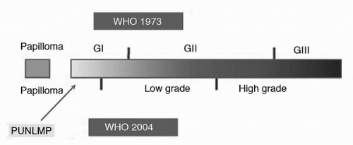
Over 90% of all newly diagnosed bladder cancers are urothelial in origin. At the time of presentation, 80% of patients are found to have non-muscle-invasive bladder cancer (NMIBC) (1,2). NMIBC is defined as tumor that does not invade the detrusor muscle. Accurate pathological staging and grading of cystoscopically obtained bladder tumor tissue is essential in determining clinical prognosis and directing therapy. In the tumor-node-metastasis (TNM) classification system, NMIBC can be one of three types: stage Ta (confined to the mucosa), stage T1 (invasive into the submucosa or lamina propria), and carcinoma in situ (CIS). Roughly, 70% of NMIBC at initial assessment is Ta, 20% is T1, and 10% is CIS (3,4). Since it is quite rare to have lymph node or distant metastasis with NMIBC, stage is usually akin to tumor depth. The intrinsic aggressive potential of this malignancy, on the other hand, is strongly associated with tumor grade. Currently, there is no uniformly accepted grading system for bladder cancer. The two systems in use include that first introduced by the World Health Organization (WHO) in 1973 that grades bladder cancer on a scale from 1 to 3, from well to moderately to poorly differentiated (4,5). The second classification system introduced in 1998 by the World Health Organization and International Society of Urological Pathology (WHO/ISUP) characterizes grade as papillary urothelial neoplasm of low malignant potential (PUNLMP), low-grade and high-grade carcinoma, from well differentiated to poorly differentiated (Fig. 22B.1) (5). By definition, CIS is high grade. Ta and T1 disease can be either low grade or high grade. For the purpose of discussion, reference to high grade in this chapter refers to the 1973 grade 3 definition or 1998 high-grade tumors, as different studies use different grading systems depending on the year they were written.

Considerable amount of work has been done in order to stratify the risk of recurrence, progression, and death associated with NMIBC. In 2000, Millan-Rodriquez et al. (6) sought to identify patients at risk for recurrence, progression, and death from NMIBC based on the stage and grade of cancer. Shortly thereafter, Sylvester et al. (7) proposed an eloquent classification system that characterized patients with NMIBC into low, intermediate, and high risk of recurrence and progression based on the number of tumors, tumor diameter, prior recurrence rate, stage category, concomitant CIS, and grade. More recently, Fernandez-Gomez et al. (8) proposed a classification system based on multiplicity, prior tumor recurrence, gender, and presence of CIS.


FIGURE 22B.1. Comparison of the World Health Organization (WHO) grading systems: 1973 versus 2004. (PUNLMP, papillary urothelial neoplasm of low malignant potential.)

Although there are multiple different definitions and formulas to determine patients with high-risk bladder cancer, most would agree that all patients with high-grade disease are at a higher risk of recurrence and a much higher risk of progression when compared to low-grade disease. In 2000, Herr (9) showed that the progression of bladder tumors is primarily dependent upon grade. For these reasons, both Ta high grade and CIS are considered high risk and are discussed in more detail throughout the chapter. Discussion of T1 high-grade disease will be specifically reviewed in another chapter.


Ta HIGH GRADE


Incidence

Stage Ta bladder cancer accounts for 51% to 73% of all Ta/T1 papillary lesions and 51% to 71% of all NMIBC (10,11,12). Sylvester et al. (13) reviewed 12 different series and found that the overall incidence of high-grade disease accounted for only 6.9% (range of 2.9%-18%) of all Ta tumors. Overall, Ta high-grade tumors accounted for 4.1% (range of 1.7%-9.3%) of all papillary tumors.


Pathology

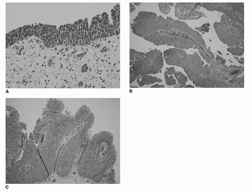
The TNM classification system defines stage Ta as papillary bladder cancer confined to the mucosa. Ta bladder cancer can be identified as low grade or high grade (Fig. 22B.2). Low-grade papillary urothelial carcinoma is associated with an overall orderly appearance. Minimal cytologic atypia is appreciated with respect to nuclear size, shape and chromatin texture. Mitotic activity is minimal but when present typically occurs in the lower half of the urothelium. In contrast, high-grade papillary urothelial carcinoma is represented by marked cytologic and architectural disorder. Nuclear pleomorphism ranges from moderate to severe. Mitotic activity is easily appreciated throughout the entire epithelium. The papillary urothelium closely resembles flat CIS (5,14).

The evaluation of genomic alterations associated with Ta NMIBC has provided further details into the nature of the disease. Cappellen et al. (15) were the first to describe the expression of fibroblast growth factor receptor 3 (FGFR3) in bladder cancer. Multiple subsequent studies confirmed the presence of FGFR3 expression in greater than 70% of patients with low-grade Ta disease (16,17,18). The earliest alterations to low-grade disease include deletions of chromosome 9 and activating mutations to FGFR3 (19,20). In contrast, high-grade Ta disease is associated with mutations to P53 or chromosome 17p (16,21,22). Other common changes in Ta disease include 20p, 20q, 18q, 11p, 10q and 1q (23,24,25,26). However, these findings are more common in high-grade disease (19).

Going a step further, many groups have explored the gene expression in bladder cancer through DNA microarray analysis (27,28). Aaboe et al. (28) first profiled the genetic expression of both high-grade and low-grade Ta urothelial cancers in 2005. Low-grade tumors were associated with gene alterations involved in cytoskeleton, protein folding, transferase activity,
and transcription. In contrast, the genes most commonly overexpressed in high-grade disease involved genes related to regulation of the cell cycle, cell growth and transcription. When correlated with clinical outcome, those tumors that had genetic expression consistent with high-grade disease were significantly more likely to have recurrence when compared to those tumors that had genetic expression consistent with PUNLMP.


FIGURE 22B.2. (A) Normal urothelium. (B) Ta low grade. (C) Ta high grade. (Images courtesy of Laila Dahmoush, MD, University of Iowa Department of Pathology.) (See color insert.)

Although the diagnosis and prognosis of bladder cancer may one day rely on molecular makeup of the tumor in question, current practice relies solely on the gross and histologic pathologic specimen. This is complicated by the fact that differences between grade and even stage can be subtle on histologic specimens. Three recent trials have pointed out the difficulty of well-trained pathologists to agree on stage and grade of bladder tumors. Witjes et al. (29) first recognized the discrepancy between local and review pathology when determining the grade and stage of bladder tumors. Local pathologists recognized 16 of 450 bladder cancers as Ta high grade. Review pathologists agreed with 5 of 16 (31%) being Ta high grade. Five of the original 16 diagnosed as Ta high grade were upgraded to T1 high grade by the review pathologist. The review pathologists classified a total of 13 Ta high-grade tumors. This means eight tumors classified by the review pathologist as Ta high grade were not classified as such by the local pathologist (initially described as 3 TaG2, 4 T1G2 and 1 T1G3). Similar findings were also noted in a review of 1,400 patients treated in five European Organization for Research and Treatment of Cancer (EORTC) trials (30). Likewise, Bol et al. (31) reviewed 130 consecutive stage Ta/T1 bladder cancers in Norway with similar findings using the 1998 WHO/IUSP grading system. It is estimated that 23% of cases of Ta High grade by local pathology were confirmed by review (13).

The importance of accurately assessing stage and grade cannot be overstated. Grade (primary) and stage (secondary) are the most important risk factors of progression in NMIBC (6,9).


Recurrence/Progression/Death

Assessment of recurrence, progression and disease-specific survival is difficult when comparing multiple studies. Limitations involved in comparing multiple studies include the use and variation of treatment following initial TURBT, presence of concomitant CIS, degree of progression, grading system used, change in follow-up regimens, and change in overall follow-up time, as well as the aforementioned accuracy of the pathology in question. With an understanding of the limitations of assessing recurrence, progression, and disease-specific survival, we compiled a review of eight relevant trials that evaluated Ta high-grade disease (Table 22B.1) (9,10,32,33,34,35,36,37).


TABLE 22B.1 RATE OF RECURRENCE, PROGRESSION, AND DEATH ASSOCIATED WITH TA HIGH-GRADE DISEASE


Study


Treatment after TURBT


Recurrence


Progression (≥T1)


Progression (≥T2)


Death from Disease


Follow-Up


Chen et al. (32)


Chemotherapy


8/14 (57%)


4/14 (29%)


3/14 (21%)


Mean 74.3 mo


Heney et al. (37)


None


1/4 (25%)


1/4 (25%)


Median 39 mo


Herr (9)


BCG


49/125 (39%)


32/125 (26%)


Minimum 15 yr


Holmang et al. (33)


None, Cystectomy, Radiotherapy


2/9 (22%)


1/9 (11%)


1/9 (11%)


Minimum 20 yr


Holmang et al. (34)


None, Chemotherapy, Radiotherapy


6/13 (46%)


Minimum 5 yr


Jakse et al. (36)


None


4/16 (25%)


1/16 (6%)


1/16 (6%)


Median 106 mo


Larsson et al. (10)


None, Chemotherapy, Cystectomy


9/15 (60%)


3/15 (20%)


1/15 (7%)


2/15 (13%)


Minimum 5 yr


Lebret et al. (35)


BCG


23/32 (72%)


15/32 (47%)


8/32 (25%)


4/32 (13%)


Median 58.4 mo


Adapted from Sylvester RJ, van der Meijden A, Witjes JA, et al. High-grade Ta urothelial carcinoma and carcinoma in situ of the bladder. Urology 2005;66(6 Suppl 1):90-107.


As is the case for most NMIBC, Ta high-grade cancers are at high risk for recurrence. Findings from four different series suggest an overall risk of recurrence of 68% with a range of 22% to 73% (Table 22B.1) (10,32,33,35). A retrospective review by Holmang et al. (33) in 1995 reviewed 176 patients with primary Ta/T1 bladder cancer evaluated between 1963 and 1972. A total of 77 patients had Ta disease, and 9 had high-grade disease. Of the nine patients with Ta high grade disease, two underwent cystectomy and one underwent radiation as primary therapy. Of the six remaining who underwent “partial bladder resection,” or TURBT, two developed recurrence. With the change in current therapy of NMIBC one would expect a much higher rate of recurrence. In a more recent study, Lebret et al. (35) followed 32 patients with Ta high-grade disease with a mean follow-up of 58.4 months. This group was found to have a 72% recurrence rate.

More concerning than the rate of recurrence of Ta high-grade disease is the rate of progression. A review of eight major studies assessing the rate of progression in Ta high-grade bladder cancer revealed a 29% risk with a range of 11% to 39% (Table 22B.1) (9,10,32,33,34,35,36,37). In 2000, Herr (9) reviewed his 15-year outcomes with Ta high-grade disease, following 125 patients, where 49 (39%) had progressed to invasive disease. These results were nearly equivalent to the patients with T1 high-grade disease. Lebret et al. (35) followed 32 patients with Ta high-grade disease, and with an average of 58-month follow-up, 8 (25%) had progression of disease. These findings have led to the conclusion that grade is the most important risk factor in the development of progression.

Along with an increased risk of progression comes an increased risk of death. In the four reviews that discussed death due to bladder cancer in patients initially treated for Ta high-grade disease, the overall rate of death due to disease was 22% (range of 13%-26%) (9,10,35,36). In other words, disease-specific survival was 78% with a range from 74% to 87% (Table 22B.1).


Prognosis

In 2006, Sylvester et al. (7) compiled data from 2,596 patients with a known history of NMIBC from seven randomized intravesical therapy EORTC trials in an attempt to accurately predict the risk of recurrence and progression in individual patients. Multiplicity, prior recurrence rate and tumor size were associated with the greatest risk of recurrence. The most important factors in determining the risk of progression were concomitant CIS, grade and stage of disease. Likewise, Fernandez-Gomez et al. (8) also reviewed factors that accurately predict the risk of recurrence and progression. Their study consisted of patient information involving 1,062 patients with Ta or T1 urothelial carcinoma from three randomized trials. Multiplicity, recurrent tumor, female gender and concomitant CIS increased the likelihood of recurrence. Stage, grade, recurrent tumor, and recurrence at first cystoscopy were all associated with increased risk of progression.


Treatment

There have been no randomized controlled trials to date analyzing different treatment options in patients with Ta high-grade disease. Based on risk of progression, Ta high-grade disease should be considered high risk and treated as such (9,13,38).

As in all papillary bladder cancers, the initial step in treatment is TURBT, followed by an immediate dose of intravesical chemotherapy (39,40). This recommendation is based on meta-analyses of seven randomized trials that showed a decrease in recurrence of 12% when compared to TURBT alone for all patients treated with one dose of chemotherapy immediately postoperatively (39). In a recent study from Sweden, 305 patients with NMIBC were randomized to receive either epirubicin or placebo following TURBT (41). There was no significant difference in recurrence rates for patients with intermediate to high risk disease when treated with epirubicin or placebo. This study initially intended to evaluate the recurrence in patients with low to intermediate risk NMIBC; therefore, concrete conclusions cannot be drawn regarding high-risk patients. Current recommendations from the European Association of Urology (EAU) include the use of one immediate dose of chemotherapy following TURBT for all patients (42). Following the initial resection and administration of postoperative chemotherapy, the EAU further recommends a second transurethral resection (TUR) for all high-grade papillary NMIBC (42,43). The American Urological Association (AUA) recommends re-resection of Ta high-grade lesions if muscularis propria is lacking from the initial specimen (43). Re-resection may allow for increased accuracy of clinical staging.

Only gold members can continue reading. Log In or Register to continue

Stay updated, free articles. Join our Telegram channel

Jul 15, 2016 | Posted by in ONCOLOGY | Comments Off on Non-Muscle-Invasive Bladder Cancer

Full access? Get Clinical Tree

Get Clinical Tree app for offline access