NEUROLOGIC MANIFESTATIONS OF VARICELLA AND HERPES ZOSTER
JOHN W. GNANN, JR. AND RICHARD J. WHITLEY
Varicella-zoster virus (VZV) causes two clinically distinct diseases. Varicella (chickenpox), which results from primary infection of a susceptible individual, is a common, extremely contagious, and usually benign acute illness that occurs in epidemics and is characterized by a generalized vesicular rash. Like all other herpesviruses, VZV establishes latency following primary infection. Reactivation of latent VZV results in a localized cutaneous eruption termed herpes zoster or shingles, a common disorder among the elderly. Both varicella and herpes zoster can be complicated by central nervous system (CNS) involvement (1).
EPIDEMIOLOGY
Varicella
Humans are the only known reservoir for VZV. Primary infection occurs when a susceptible individual is exposed to airborne virus by the respiratory route. Patients with chickenpox are contagious for about 48 hours before and 4 to 5 days after rash onset. Infection is usually acquired after exposure to another individual with varicella but can also result from exposure to a patient with herpes zoster. Varicella is highly infectious; attack rates of 61% to 87% have been observed following household exposure.
In the United States, varicella epidemics occur annually in the late winter and early spring, with peak numbers of cases reported in March. Before the availability of the varicella vaccine, about 3.8 million cases of chickenpox occurred each year in the United States (15 cases per 1,000 population), which approximately equaled the annual birth cohort (2). Varicella was predominantly a disease of school-aged children. About 50% to 60% of varicella cases occurred in children between 5 and 9 years of age, and 90% of cases occurred in patients younger than 15 years. Previous surveys indicated that more than 90% of the U.S. population was VZV seropositive by age 20 years (3). Mortality estimates for varicella in children (ages 1 to 14 years) are 1.4 per 100,000 cases and in adults 31 per 100,000 cases (4).
Introduction of the varicella vaccine in the United States in 1995 resulted in striking changes in the epidemiology of chickenpox. By monitoring vaccine and disease activity at three sentinel sites, the Centers for Disease Control and Prevention (CDC) showed that vaccine coverage among preschool-aged children increased from 40% in 1997 to 70% in 1999. Between 1995 and 1999, varicella incidence declined 80% in the surveillance areas, with the greatest decline seen in children aged 1 to 4 years (5).
Herpes Zoster
Herpes zoster occurs as a result of reactivation of latent VZV. The annualized incidence of herpes zoster is 1.5 to 3.0 cases per 1,000 persons in the population (6,7). An incidence figure of 2.0 cases per 1,000 persons would predict about 500,000 cases of zoster annually in the United States. Expressed another way, an individual has a 10% to 20% risk of developing herpes zoster at some point during his or her lifetime. Increasing age is the most important risk factor for the development of herpes zoster. There is an increase in the age-specific incidence of herpes zoster beginning at around age 55 years; individuals older than 75 years have a zoster incidence of more than 10 cases per 1,000 person-years (7). Zoster occurs with equal frequency in men and women, with no seasonal association. The other well-defined risk factor for herpes zoster is altered cell-mediated immunity. Patients with neoplastic diseases, especially lymphoproliferative malignancies and organ transplant recipients, are at very high risk for development of herpes zoster. Approximately 15% to 30% of patients with Hodgkin disease experience herpes zoster, compared with 2% of patients with various solid tumors (8). The disease occurs in 3% to 10% of renal transplant recipients, 20% to 25% of cardiac transplant recipients, and 20% to 50% of allogeneic hematopoietic stem cell transplant recipients (9). Herpes zoster is also seen with increased frequency in persons infected with human immunodeficiency virus (HIV) and often serves as an initial marker of disease in that population (10). A longitudinal study of a cohort of HIV-seropositive men in San Francisco demonstrated an incidence of zoster of 29.4 cases per 1,000 person-years, compared with 2.0 cases per 1,000 person-years in a matched population of HIV-seronegative controls (11).
INFECTIOUS AGENT
VZV shares structural characteristics with other members of the family Herpesviridae. The complete virion is approximately 180 to 200 nm in diameter. It is composed of an icosahedral nucleocapsid measuring 90 to 95 nm in diameter, an amorphous tegument, and a lipid-containing envelope with glycoprotein spikes (12). The VZV genome consists of a linear, double-stranded DNA molecule containing 125,000 base pairs with guanosine-plus-cytosine content of 46% (13). The genome is organized in a manner similar to that of other herpesviruses, having unique long (UL; 104.5 kb) and unique short (US; 5.2 kb) regions flanked by inverted repeats. VZV encodes approximately 69 polypeptides, including seven glycoproteins.
VZV can be propagated in vitro in a limited number of continuous and discontinuous cell culture monolayers, mostly of human or simian origin. In human embryonic lung fibroblasts, cytopathic effects begin as a focal process with subsequent cell-to-cell spread. Approximately 8 to 10 hours after infection, virus-specific immunofluorescence can be detected in cells adjacent to the initial focus of infection. VZV is highly cell associated, with very limited release of infectious virions into the media.
PATHOGENESIS AND PATHOLOGY
Pathogenesis of Varicella
Varicella is transmitted via the respiratory route. Virus in airborne droplets enters the susceptible host via mucosal surfaces of the conjunctiva, oropharynx, or upper respiratory tract. VZV undergoes an initial round of replication, presumably in cervical lymph nodes (14). When local immune responses are overcome, a primary viremia occurs, with widespread dissemination of VZV to the reticuloendothelial system and possibly to epithelial cells of other organs. VZV then undergoes multiple cycles of replication, resulting in a second viremic phase (about 1 week after the initial viremia) that is accompanied by the onset of clinical symptoms. VZV localizes to endothelial cells of cutaneous capillaries and then extends to epithelial cells of the epidermis, where replication results in formation of the characteristic vesicles. Viremia and new vesicle formation continue for 3 to 5 days and then terminate when humoral and cellular immune responses appear.
Pathology of Varicella
The cutaneous manifestations of varicella begin with hematogenous infection of the endothelial cells of cutaneous blood vessels. VZV then begins to replicate in the skin, leading to ballooning degeneration of epithelial cells in the prickle cell (malpighian) layer of the epidermis. Local collections of extracellular edema fluid result in acantholysis, with elevation of the stratum corneum to form a clear vesicle. Multinucleated giant cells are found at the base of the lesion. Infected cells contain eosinophilic intranuclear inclusion bodies (Cowdry type A inclusions) surrounded by a clear zone. A perivascular infiltration of mononuclear cells is seen around cutaneous vessels. The vesicular fluid becomes cloudy as it accumulates inflammatory cells and desquamated epidermal cells. The vesicular fluid is resorbed, resulting in drying and crusting of the lesion. Healing occurs with regeneration of the epithelial cell layers.
Pathogenesis of Herpes Zoster
As VZV replicates in the skin during acute varicella, some virions are transported via sensory nerves to the corresponding dorsal root ganglia (15). The virus establishes a latent infection within the ganglion, preferentially infecting satellite cells rather than neurons (16). An alternative explanation is that VZV may reach the ganglion by viremic spread. VZV may periodically reactivate and undergo limited replication, but replication is suppressed by the immune response before any clinical symptoms result (17,18). The specific immune responses that limit reactivation of VZV from the sensory ganglia are poorly understood. The most important factor that predisposes to the development of herpes zoster appears to be decline or suppression of VZV-specific cellular immunity. This may occur naturally with aging or be induced by immunosuppressive illness or therapy. Following reactivation and replication in the ganglion, virus travels along the sensory nerve to the skin, where it again replicates in epithelial cells, producing the characteristic dermatomal vesicular rash of shingles. Unlike the lesions of varicella, in which different stages are seen simultaneously, most zoster lesions are in the same stage of development. A limited viremia occurs during many episodes of herpes zoster, reflected by the appearance of a few extradermatomal vesicles.
Pathology of Herpes Zoster
Replication of VZV in the sensory ganglion results in intense inflammation, neuronal destruction, and focal hemorrhage. Less severe inflammatory changes often occur in the adjacent ganglia. Occasionally, inflammation and necrosis also extend to the anterior nerve root, resulting in localized motor neuropathy. These changes are accompanied by lymphocytic pleocytosis. Movement of virus from the ganglion down the sensory nerve to the skin produces acute inflammation of the nerve (19). Virus reaching the skin replicates in epithelial cells of the epidermis, producing pathologic changes identical to those described for varicella. Inflammatory changes in the sensory nerve persist for months and may result in demyelination, wallerian degeneration, and sclerosis.
CLINICAL MANIFESTATIONS
Natural History of Varicella
Varicella is generally a benign disease in healthy children, although symptoms are often more severe in adolescents and adults. Fewer than 5% of primary VZV infections are subclinical. Symptoms develop after an incubation period of about 15 days (range, 10 to 20 days). A prodrome of fever, malaise, headache, and anorexia is variably present, occurring more commonly among older children and adults and lasting 1 to 2 days. A transient scarlatina-like rash is occasionally noted just before or coincident with the appearance of the varicella lesions. The lesions first appear on the head, then the trunk, and finally the extremities. The greatest concentration of lesions is on the trunk and proximal extremities. The rash of varicella is characterized by rapid evolution of lesions over 8 to 12 hours and by successive crops of new lesions. Consequently, lesions of all stages are present simultaneously on involved skin surfaces. Lesions begin as pink macules that quickly become papular and evolve into fragile vesicles 1 to 4 mm in diameter surrounded by a zone of erythema. As inflammatory cells migrate into the vesicular fluid, the lesions become pustules; these are often centrally umbilicated. The pustules become crusted and the crusts detach after 1 to 3 weeks, usually healing without scarring. Vesicles also appear on mucosal surfaces and rapidly evolve into shallow ulcerations. New lesion formation continues for 2 to 4 days, accompanied by pruritus, fever (100° to 102°F), headache, malaise, and anorexia. The rash peaks on about the fifth day, with an average lesion count of 250 to 500; fewer lesions are seen in children younger than 5 years (20). Older children and secondary cases within a household tend to have higher lesion counts and higher fever.
The most common complication of varicella in otherwise normal children is bacterial superinfection (usually staphylococcal or streptococcal infection) that can occasionally progress to serious necrotizing cellulitis (1). Varicella pneumonia is rare in children but occurs more often in adults (21). Pregnant women with chickenpox may be at especially high risk for severe varicella pneumonia. Neonatal varicella can occur when the mother develops chickenpox within a period of 5 days before to 2 days after delivery. The infected neonate can develop severe disseminated varicella with a mortality of 20% to 30%. Maternal varicella occurring during the first trimester of pregnancy has been associated with congenital abnormalities, but the risk appears low (about 1% to 2%) (1).
In immunocompromised children, varicella is a serious and potentially fatal infection. Children at highest risk are those with acute leukemia, although children with other malignancies and those on cytotoxic or immunosuppressive medications (including high-dose corticosteroids) can also develop complications. Immunocompromised children may develop severe hemorrhagic or necrotic skin lesions (purpura fulminans or hemorrhagic varicella) or severe bacterial superinfection. Persistent viremia can result in dissemination of VZV, producing pneumonitis, hepatitis, or encephalitis. A chronic form of varicella has been reported in HIV-infected children.
Neurologic Complications of Varicella
The incidence of CNS complications with varicella is reported to be 1 to 3 per 10,000 cases (3,21). Because many uncomplicated cases of varicella do not come to medical attention, any calculation of the frequency of complications is likely to be an overestimation. The CNS manifestations most frequently associated with chickenpox are cerebellar ataxia and encephalitis (22,23). Uncommon neurologic complications include transverse myelitis, aseptic meningitis, strokes, and Guillain-Barré syndrome (23). Optic neuritis has been reported as a rare complication of varicella in both pediatric and adult patients, with good visual recovery expected in most cases (24). Reye syndrome, a triad of acute hepatic failure, encephalopathy, and hypoglycemia, was previously associated with varicella (and with other viral infections) but is now known to be more specifically related to salicylate therapy in febrile children. However, many cases of Reye syndrome are included in older reviews of varicella encephalitis, resulting in misleading estimates of incidence and mortality.
Cerebellar Ataxia
Cerebellar ataxia, the most common neurologic abnormality associated with varicella, is diagnosed in approximately 1 per 4,000 cases of chickenpox (21). Children can develop ataxia from several days before to 2 weeks after the onset of the rash, although neurologic symptoms most often occur simultaneously with the rash (23). Symptoms include vomiting, headache, and lethargy accompanied by ataxia. Fever, nuchal rigidity, and nystagmus occur in about 25% of patients. Seizures are rare and suggest a more diffuse encephalitis. In cases of ataxia presenting before the development of rash, the correct diagnosis may not be clinically apparent unless an association is made with recent varicella exposure.
The extent of the diagnostic evaluation in patients with varicella-associated cerebellar dysfunction should be governed by the severity of the illness and the degree of certainty of the diagnosis. In uncomplicated cases, the clinical presentation alone is sufficient to establish the diagnosis, and no further evaluation is necessary. In more complicated situations, a cerebrospinal fluid (CSF) examination, an electroencephalogram (EEG), and magnetic resonance imaging (MRI) scan of the brain usually are warranted. The CSF is frequently normal, but a moderate lymphocytic pleocytosis (<100 cells/µL) with mildly elevated protein and normal glucose levels may occur in 20% to 30% of patients (23). The EEG demonstrates diffuse slow-wave activity in approximately 20% of patients and normalizes as the symptoms resolve (23). Few data are available regarding the utility of computed tomographic (CT) or MRI scan in diagnosing varicella-associated cerebellar ataxia.
Cerebellar ataxia associated with chickenpox is self-limited, and most abnormalities completely resolve within 1 to 3 weeks. Mortality is essentially zero, and deaths that occur are usually attributed to the development of nonneurologic complications such as pneumonia (22,23).
The pathogenesis of this syndrome is unknown, partly because of the lack of necropsy studies in this nonfatal illness. The two pathogenic mechanisms that have been proposed are direct viral involvement of the cerebellum or a parainfectious, immunologically mediated demyelinating process analogous to that seen with other viral infections. Recovery of VZV from brain or CSF by culture has not been reported in varicella-associated cerebellar ataxia. VZV antibodies have been detected in the CSF of patients with CNS abnormalities in association with varicella but were absent in children who had varicella but no neurologic symptoms, perhaps reflecting intrathecal antibody production (25). Analysis of CSF using polymerase chain reaction (PCR) revealed VZV-specific DNA in three of five children with varicella cerebellitis (26). These observations suggest that VZV replication within the CNS does occur, but detailed studies are lacking and the evidence for viral invasion of the cerebellum is circumstantial.
Encephalitis
A less common but more severe CNS complication of chickenpox is encephalitis or cerebritis. The incidence of encephalitis is estimated to be 1 to 2 cases per 10,000 cases of chickenpox (3). Most cases of encephalitis occur in children, but the incidence is highest in adults (older than 20 years) and infants (younger than 1 year). Neurologic symptoms may occur from 2 weeks before to several weeks after the varicella rash (most often about 1 week after), and the onset may be abrupt or gradual (22,27). Headache, fever, vomiting, and an altered sensorium are the usual presenting symptoms (23,28). Seizures occur in 29% to 52% of patients (23). Focal neurologic abnormalities can include ataxia, hypertonia or hypotonia, hyperreflexia or hyporeflexia, positive plantar reflexes, hemiparesis, and sensory changes (23,28).
The CSF from patients with varicella-associated cerebritis is frequently abnormal, with elevated opening pressure, a mild to moderate lymphocytic pleocytosis (usually <100 cells/µL), elevated protein (50 to 100 mg/dL), and normal glucose (22). The EEG is often abnormal, showing slow-wave activity consistent with a diffuse encephalitis. Focal EEG abnormalities suggestive of epileptiform activity may occur even without clinical seizures. In patients who do have seizures, these EEG abnormalities tend to persist and are present in 43% of follow-up studies at 1 year. Abnormalities observed by CT in patients with varicella encephalitis have included cerebral or cerebellar edema and areas of low attenuation consistent with demyelination. MRI abnormalities in children with postvaricella encephalitis have included diffuse gray and white matter lesions or bilateral basal ganglia lesions (29).
The reported mortality for varicella encephalitis has varied from 5% to 35%, although many of these series included cases of Reye syndrome (22,23,28). The actual mortality rate for varicella cerebritis is probably less than 10%, with complete or nearly complete recovery expected in most cases. Long-term sequelae may be present in 10% to 20% of survivors (23). In a series of 59 cases of varicella with CNS involvement, the mortality rate was 5% (22). Two of the three deaths were associated with pneumonia, and 80% of the survivors were discharged from the hospital without detectable sequelae.
The role of active viral replication in the CNS in varicella encephalitis remains uncertain (30). Postmortem studies of the brain have shown a wide range of histopathologic findings (22,23,31). Diffuse cerebral edema is generally present. Perivascular infiltration of mononuclear cells and demyelination have been seen in some cases, the latter suggesting a postinfectious demyelinating process. Other cases have shown focal hemorrhagic lesions. Intranuclear viral inclusions have been observed only rarely in the brain following varicella, usually in immunocompromised patients (31).
Transverse Myelitis
On rare occasions, varicella has been associated with an isolated weakness of the lower extremities, sphincter dysfunction, abnormal deep tendon reflexes, and extensor-plantar reflexes (32). The CSF is characterized by a lymphocytic pleocytosis and elevated protein level with a normal glucose level. The completeness of recovery is variable. The pathogenesis of varicella myelitis is not known, although cases have been published in which VZV DNA was detected in CSF by PCR, suggesting that active viral invasion of the spinal cord may be involved (33).
Aseptic Meningitis
Aseptic meningitis has been reported as a complication of varicella (23). Meningismus without evidence of cerebral or cerebellar dysfunction is suggestive of the diagnosis, and complete recovery is expected. CSF findings are typical of viral meningitis, with mild lymphocytic pleocytosis, slight elevation in the protein content, and normal glucose level. VZV has not been cultured from the CSF in this setting; PCR would be an appropriate diagnostic test. It is probably appropriate to consider CNS involvement with varicella as a spectrum, with aseptic meningitis the most benign manifestation and encephalitis the most serious.
Stroke Syndromes
Arterial ischemic strokes are a well-recognized complication of herpes zoster ophthalmicus (HZO) but can also occur after varicella (34,35). By some estimations, young children with arterial ischemic strokes are threefold more likely than controls to have recently had varicella. In a prospective cohort study, 22 (31%) of 70 children with arterial ischemic strokes had varicella within the preceding 12 months, compared with 9% in the healthy population (36). Children with strokes and recent varicella infection had higher rates of basal ganglia infarction, abnormal cerebral vascular imaging, and recurrent ischemic attacks (p < .05 for all) (36). The syndrome typically occurs in otherwise healthy, immunocompetent children (median age, 5 years). The median interval between varicella infection and the onset of neurologic deficits is 2 months (37). The children usually present with hemiplegia, and angiography reveals vasculopathy of the branches of the middle cerebral artery (MCA). MRI typically demonstrates unilateral infarctions in the MCA distribution (38,39). In one fatal case, histopathology revealed active granulomatous arteritis of the MCA with lymphocytic inflammatory infiltrate and VZV antigens in the smooth muscle layer (37). Patients have been treated with intravenous acyclovir and corticosteroids, but no data from controlled studies are available to assess the efficacy of these treatments. Most children with hemiplegia following varicella infection have good neurologic recovery (which is a much better prognosis than that associated with strokes in adults following HZO).
Natural History of Herpes Zoster
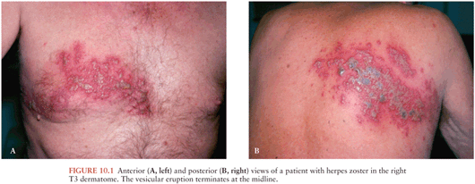
The inflammatory changes that occur in the sensory ganglion and nerve as VZV reactivates are manifested by pain in the corresponding dermatome. The patient may report sensations ranging from mild itching or tingling to severe pain that precedes the appearance of the skin lesions by 1 to 5 days (or occasionally longer). Constitutional symptoms are reported by fewer than 5% of patients during the prodromal phase. The cutaneous eruption of herpes zoster is unilateral and does not cross the midline. The rash appears in the skin segment innervated by a single sensory ganglion (Fig. 10.1). Overlap of lesions into adjacent dermatomes occurs in 20% of patients. Scattered cutaneous vesicles beyond the primary or adjacent dermatomes appear in about one third of patients with herpes zoster infection and have no prognostic significance. Disseminated VZV infection with visceral involvement is extremely rare in immunocompetent patients with herpes zoster infection, although occasional cases of encephalitis and myelitis have been reported. Herpes zoster appears with increased frequency in certain dermatomes, and this is thought to reflect the distribution of skin lesions during varicella (18). The most common sites for herpes zoster are the thoracic dermatomes (50% of cases), followed by cervical (15%), lumbar (15%), and sacral (5%) dermatomes (18). About 15% of zoster cases occur in cranial nerve dermatomes, with most cases involving the first division of the trigeminal nerve (HZO). Ocular involvement can be a serious complication of HZO. Conjunctivitis is common and benign, but more serious ocular manifestations, such as keratitis, scleritis, and iridocyclitis, may develop in 50% to 70% of patients with HZO (40).
Skin changes begin with an erythematous maculopapular rash, followed by the appearance of clear vesicles. New vesicle formation typically continues for 3 to 5 days, followed by lesion pustulation and scabbing. The extent of involvement can range from a few vesicles to a confluent eruption filling the entire dermatome. Most lesions are crusted by day 10, but complete skin healing may require 2 to 4 weeks. Unlike varicella, herpes zoster is associated with a significant risk of skin scarring and permanent pigmentation changes. Skin necrosis and gangrene in the involved dermatome can occur but are more commonly encountered in immunocompromised patients.
During the acute phase of herpes zoster, most patients experience dermatomal pruritus and pain, which can be severe. The acute neuritis is variably described as an aching, burning, or stabbing pain. Many patients complain of headache, photophobia, and malaise, but significant fever is rare. CSF examination (which is not routinely necessary) reveals a lymphocytic pleocytosis and increased protein concentration; VZV can occasionally be cultured from the CSF. Although the skin usually heals in 2 to 4 weeks, pain (postherpetic neuralgia [PHN]) persists for longer than 1 month in 20% to 70% of patients with herpes zoster, with elderly patients experiencing the highest frequency of chronic pain. Although herpes zoster is most common among older adults, the disease can occur in patients of any age. Immunocompetent children and young adults with herpes zoster tend to have less extensive cutaneous eruptions, less severe pain, and a much lower risk for chronic pain.
Patients with deficiencies in cell-mediated immunity have an increased incidence of herpes zoster and a higher risk for complications. In immunocompromised patients, zoster causes more severe skin involvement within the dermatome and may be accompanied by viremia with cutaneous or visceral dissemination. Without antiviral chemotherapy, cessation of new vesicle formation does not occur until about day 8, pustulation on day 9, and scabbing on day 18 (41). Patients with herpes zoster infection at highest risk for dissemination are those with lymphoproliferative malignancies or those who recently received induction chemotherapy (8,42). Without antiviral treatment, the reported incidence of cutaneous dissemination in immunocompromised populations is 6% to 26% (43). In most patients, dissemination is limited to the skin and does not substantially alter the prognosis. However, 10% to 50% of patients who develop cutaneous dissemination also have evidence of visceral dissemination (such as pneumonitis, meningoencephalitis, or hepatitis), which carries a much more serious prognosis. Even with the availability of antiviral chemotherapy, the mortality rate for zoster with visceral dissemination is 5% to 15%, with most deaths attributable to pneumonitis (44). Visceral VZV infection occurring without any evidence of skin lesions is uncommon. However, several patients with acquired immunodeficiency syndrome (AIDS) and VZV myelitis or encephalitis but without clinically apparent zoster have been reported. Another syndrome unique to the immunocompromised host is chronic cutaneous VZV infection, sometimes associated with acyclovir-resistant virus.
Neurologic Complications of Herpes Zoster
Neurologic complications of herpes zoster can occur during the acute eruption (e.g., segmental motor paresis) or may appear weeks to months after the herpes zoster rash has resolved (e.g., delayed contralateral hemiparesis) (45). Neurologic complications appear more often in immunocompromised patients, including patients with HIV infection. Several pathologically distinct syndromes have been defined, including large and small vessel vasculopathies and ventriculitis (46). Investigators have also described myelitis and polyradiculitis, as well as a variety of cranial and peripheral nerve palsies in association with herpes zoster (47). The most common neurologic complication of herpes zoster is chronic pain, or PHN, which occurs with sufficient frequency that it can be considered a part of the natural history of the disease (48).
Encephalitis
Herpes zoster can be complicated by acute encephalitis that usually occurs a few days after the onset of rash but has been reported from days to weeks before or after the skin eruption (49). Encephalitis has occasionally been documented in the absence of apparent cutaneous zoster and in patients who received appropriate antiviral therapy during the acute episode of herpes zoster (50,51). Immunocompromised patients are clearly at increased risk for the development of encephalitis (49
Related posts:

Stay updated, free articles. Join our Telegram channel

Full access? Get Clinical Tree


