Neck, Axillary, Ilioinguinal, and Other Lymph Node Dissections


Neck, Axillary, Ilioinguinal, and Other Lymph Node Dissections


NECK LYMPH NODE DISSECTION


CRITICAL ELEMENTS


  • Boundaries of an Oncologically Appropriate Neck Dissection


  • Superficial or Total Parotidectomy


  • Removal of a Sufficient Number of Lymph Nodes for Adequate Staging


1. BOUNDARIES OF AN ONCOLOGICALLY APPROPRIATE NECK DISSECTION

Recommendation: A selective neck dissection based on anatomic drainage patterns should be performed for microscopic or low-volume clinical nodal metastatic disease. Bulky clinical nodal metastatic disease should typically be treated with a modified radical neck dissection. In the setting of extracapsular extension, resection of contiguous structures directly invaded by tumor is clinically appropriate.

Type of Data: Observational studies.

Grade of Recommendation: Strong recommendation, low-quality evidence.


Rationale

The head and neck anatomy is the most complex and variable of all the melanoma regional lymph node basins. The lymphatic drainage is also fairly predictable based on the location of the primary tumor. Classical drainage patterns have been described by O’Brien et al114 and reproduced by others.37 The studies have demonstrated discordant drainage pathways in 8% to 34% of patients. The most common discrepancies are drainage to the postauricular, suboccipital, contralateral neck, and noncervical lymph
node basins. As noted in chapter 9, the addition of single photon-emission computed tomography and computed tomography (SPECT/CT) to traditional two-dimensional planar lymphoscintigraphy can be very helpful in identifying both traditional and discordant lymphatic drainage patterns in the head and neck.115 The standard nodal basin levels of the neck with corresponding anatomic boundaries are defined in Table 10-1 and illustrated in Figure 10-1.

There are two goals of neck dissection for nodal metastatic melanoma. The first is to obtain regional nodal basin control. Approximately 13% to 20% of completion lymph node dissection (CLND) specimens for a positive head and neck sentinel lymph node biopsy (SLNB) will contain additional positive lymph nodes.33,116 When an intraparotid lymph node is positive, the risk for additional positive cervical lymph nodes is as high as 27%.117 In contrast to breast cancer, melanoma can have lymph node metastases that become very large and develop extracapsular extension. Secondary to the limited anatomic space within the neck, this can lead to direct invasion of adjacent structures, such as musculature, the internal jugular (IJ) vein, the carotid artery, and critical nerves (facial, vagus, phrenic, and spinal accessory [SAN]). The second goal of neck dissection is prognosis, because the total number of positive lymph nodes is a predictor of both the risk of regional nodal basin relapse as well as distant metastatic disease. The extent of lymphadenectomy necessary to achieve these goals is based on the nodal metastatic tumor burden, the anatomic location of the lymph nodes within the neck, and the lymphatic drainage pattern of the primary tumor site.

Similar to neck dissections for other head and neck malignancies, melanoma dissections are categorized according to the lymph node levels and the anatomic structures removed. Radical or comprehensive neck dissection (CND) is classically defined as resection of level I-V lymph nodes, the sternoclavicular muscle (SCM), SAN, and IJ vein. CND can also include other structures of the neck based on direct extension (e.g., the phrenic nerve or part of the trapezius). Modified radical neck dissection (MRND) is defined as resection of level I-V nodes with preservation of one or more classic nonlymphatic anatomic structures typically resected with a CND (SCM, SAN, or IJ vein). Selective node dissection (SND) is defined as removal of only certain lymph node levels based on lymphatic drainage patterns and the location anticipated to be most proximal to metastatic disease. All nonlymphatic anatomic structures are preserved.

SND is routinely performed for microscopic positive SLNB or low-volume clinical nodal metastatic disease. Bulky clinical nodal metastatic disease is typically treated with an MRND. Resection of contiguous nonlymphatic anatomic structures directly invaded by extracapsular tumor extension is clinically appropriate to remove all gross disease.

The overall type (CND, MRND, or SND) and extent (levels) of neck dissection are based on several features: microscopic versus macroscopic, subclinical versus bulky clinical nodal metastatic disease; the anticipated lymphatic drainage pattern based on the primary tumor location; and additional information obtained from any prior sentinel lymph node lymphoscintigraphy or SPECT/CT. In general, the appropriate levels for SND based on the primary tumor site are listed in Table 10-2.


TABLE 10-1 Anatomic Boundaries of Nodal Basins by Level


Nodal Basin Level


Anatomic Boundaries


Level IA


Lymph nodes within the boundary of the anterior belly of digastric muscles and the hyoid bone.


Level IB


Lymph nodes within the boundaries of the posterior belly of the digastric muscles, the stylohyoid muscle, and body of the mandible.


Level IIA/B


Lymph nodes encompassing the upper third of the internal jugular vein region and adjacent to the spinal accessory nerve extending from the level of the skull base to the level of the inferior border of the hyoid bone. The medial boundary is represented by the lateral border of the sternohyoid muscle and the stylohyoid muscle. The lateral boundary is defined by the posterior belly of the sternocleidomastoid muscle.


Level III


Lymph nodes encompassing the middle third of the internal jugular vein extending from the inferior border of the hyoid bone to the inferior border of the cricoid cartilage. The anterior boundary is the lateral border of the sternohyoid muscle. The posterior boundary is the posterior border of the sternocleidomastoid muscle.


Level IV


Lymph nodes encompassing the lower third of the internal jugular vein and extending from the inferior border of the cricoid cartilage to the clavicle. The anterior boundary is the lateral border of the sternohyoid muscle. The posterior boundary is the posterior border of the sternocleidomastoid muscle. Of note, Virchow’s node is within level IV.


Level VA/B


Lymph nodes along the lower half of the spinal accessory nerve and the transverse cervical artery. The supraclavicular nodes are also included in this posterior triangle group. The superior boundary is the formed by the intersection of the sternocleidomastoid and the trapezius muscles. The inferior boundary is the clavicle. The anterior boundary is the posterior border of the sternocleidomastoid muscle. The posterior boundary is the anterior border of the trapezius muscle.


Superficial parotid


The gland anterior to and with preservation of the fascial nerve to include the invested lymph nodes within the gland.


Posterior auricular


Lymph nodes along the posterior auricular artery as it courses above the digastric muscle and stylohyoid muscle, opposite the apex of the styloid process, posterior to the ear and medial to the lateral edge of the superior extent of the insertion of the sternocleidomastoid muscle.


Occipital


Lymph nodes located in the posterior neck as defined by the lateral margin of the trapezius medially, the insertion of the semispinalis capitis superiorly, and the posterior body of the sternocleidomastoid muscle anteriorly.


FIGURE 10-1 Standard nodal basin levels of the neck.


TABLE 10-2 Appropriate Levels for Selective Neck Dissection Based on Primary Tumor Site


Primary Tumor Site


Levels for Selective Neck Dissection


Anterior and mid neck


Levels I-V


Anterior scalp, preauricular area, face


Levels I-V ± parotid


Vertex scalp


Levels I-V ± parotid ± occipital/posterior auricular area


Posterior scalp


Levels I-V, occipital/posterior auricular area


Ear


Levels I-V ± parotid ± posterior auricular area


Posterior neck


Levels II-V


2. SUPERFICIAL OR TOTAL PAROTIDECTOMY

Recommendation: A superficial parotidectomy should be combined with lymphadenectomy when there is macroscopic nodal metastatic disease involvement, a positive intraparotid sentinel lymph node, or evidence on the mapping technique used for sentinel lymph node biopsy showing that the superficial parotid gland was directly in the lymphatic drainage path from a more proximal positive sentinel node. Total parotidectomy, which is rarely clinically indicated, should be reserved only for extensive gross tumor involvement.

Type of Data: Observational studies.

Grade of Recommendation: Strong recommendation, low-quality evidence.


Rationale

In general, 25% to 30% of primary head and neck melanomas will have lymphatic drainage into the parotid gland.118 But as noted earlier in this section, parotid drainage is more frequently associated with certain primary tumor anatomic locations, such as portions of the scalp, the preauricular/ear area, and the face. Given that superficial parotidectomy can be associated with potential morbidity (facial nerve injury, salivary fistula), the addition of superficial parotidectomy to a melanoma neck dissection should be reserved for situations in which there is a high risk of parotid gland involvement. There are three clinical scenarios in which superficial parotidectomy should be considered. The first is macroscopic nodal metastatic disease in the parotid region. Older CND data have shown that 44% of lymphadenectomy specimens have parotid involvement.119 The second scenario is a positive intraparotid SLNB. The third situation is one in which sentinel node planar lymphoscintigraphy or SPECT/CT shows that the superficial parotid gland is in a direct lymphatic drainage path from a more proximal positive lymph node.

The clinical indication for a total parotidectomy is essentially gross extensive tumor involvement. In one study, even in the setting of clinical superficial parotid melanoma nodal metastases, deep parotid lobe involvement occurred in only 13% of patients.120


3. REMOVAL OF A SUFFICIENT NUMBER OF LYMPH NODES FOR ADEQUATE STAGING

Recommendation: As a quality metric, a melanoma neck dissection specimen should contain at least 15 lymph nodes.

Type of Data: Observational studies.

Grade of Recommendation: Strong recommendation, low-quality evidence.


Rationale

The purpose of a melanoma neck dissection is to remove all of the lymph nodes at risk of harboring nodal metastatic disease. Consequently, the goal of surgery should
not be to obtain a minimal number of lymph nodes. Instead, the total number of lymph nodes removed can serve as a surrogate quality metric for the adequacy of the lymphadenectomy. There are two components to this quality metric. The first is the anatomic extent of the lymphadenectomy performed by the surgeon. The second is the completeness of the pathologic assessment of the specimen, including identification and examination of all available lymph nodes.

Spillane et al121 examined the total number of lymph nodes removed at the time of neck dissection for level III or below and level IV and higher in several hundred melanoma patients as well as the number that would represent the 90th percentile. For level III and below, the 90th percentile was six lymph nodes. For level IV and above, it was 20 nodes. In an attempt to standardize the CLND, the protocols for the Multicenter Selective Lymphadenectomy Trials (MSLTs) defined minimal numbers of lymph nodes removed during lymphadenectomy for various regions of the neck: anterior cervical (including suprahyoid, jugulodigastric), 15; posterior cervical (including supraclavicular), 15; supraclavicular, 6; suprahyoid, 4; and parotid, 3. The Commission on Cancer metric is a minimum of 15 lymph nodes within a melanoma lymph node dissection specimen.

Based on these data, a melanoma neck dissection specimen should contain at least 15 lymph nodes on final pathology.


AXILLARY LYMPH NODE DISSECTION


CRITICAL ELEMENTS


  • Removal of Level I, II, and III Lymph Nodes


  • Removal of a Sufficient Number of Lymph Nodes for Axillary Staging


1. REMOVAL OF LEVEL I, II, AND III LYMPH NODES

Recommendation: Axillary lymph node dissection for nodal metastatic melanoma should routinely include all level I, II, and III lymph nodes. Except in the case of bulky nodal metastatic disease with direct invasion, the long thoracic and thoracodorsal nerves should be preserved.

Type of Data: Observational studies.

Grade of Recommendation: Strong recommendation, low-quality evidence.


Rationale

Two goals govern axillary lymph node dissection for melanoma. The first is to obtain regional control of nodal metastatic disease. The majority of melanoma patients undergoing axillary dissection will not receive adjuvant radiation. Therefore, complete removal of all potentially involved nodal tissue is important. Unlike breast cancer, melanoma lymph node metastases can become very large and matted. Extension can also lead to direct invasion into adjacent structures, including the chest wall, axillary vein, the thoracodorsal and long thoracic nerves, and the brachial plexus. The second
goal is prognostication, because the total number of positive lymph nodes is a predictor of the risk of developing distant metastatic disease.


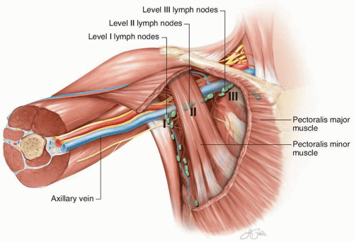
FIGURE 10-2 Standard axillary lymph node levels in relationship to the pectoralis minor muscle and other axillary structures.

As it relates to axillary dissection, the axilla is a pyramidal space defined by the axillary vein and brachial plexus superiorly, the latissimus dorsi muscle laterally, the serratus anterior muscle and musculoskeletal chest wall medially, the subscapularis muscle posteriorly, and the pectoralis major and minor muscles anteriorly. The axillary lymph node levels are determined by their position relative to the pectoralis minor muscle: level I nodes are lateral, level II nodes are deep, and level III nodes are medial (Fig. 10-2).

The traditional axillary lymph node dissection for melanoma is levels I-II-III. This remains the standard for clinically apparent lymph node metastases.122,123 Although a level I-II-III axillary dissection is frequently performed for a positive SLNB, only 3% of level III lymph nodes will be involved with metastatic disease in this situation.124,125 Therefore, consideration can be given to only a level I-II axillary dissection in select patients with a very low tumor burden, such as only sentinel node micrometastatic disease. A decision to not include level III lymph nodes in the axillary dissection should be carefully weighed against the potential morbidity of undergoing axillary surgery again if clinical level III nodal metastases developed in the future.


Technical Aspects

The removal of level I and II lymph nodes for a melanoma axillary dissection is fairly similar to what is performed for breast cancer. The interpectoral (Rotter) nodes and
the supra-axillary fat pad are routinely removed as part of this lymphadenectomy. The interpectoral lymphoareolar tissue sits between the pectoralis major and pectoralis minor muscles. Laterally, the median pectoral neurovascular bundle can be divided to facilitate anterior retraction of the pectoralis major muscle. During removal of the interpectoral nodes, care is taken to avoid injury to the lateral pectoral neurovascular bundle located medially, which can lie anterior to a portion of the medial aspect of the pectoralis minor muscle. Injury could result in complete denervation of the pectoralis major muscle.

All of the lymphoareolar tissue located inferior to the inferior edge of levels I-II of the axillary vein is removed as part of the lymphadenectomy. There is often a tonguelike supra-axillary fat pad in the region of level II that extends over the anterior aspect of the neurovascular structures (axillary vessels and brachial plexus). Because this supra-axillary fat pad can contain two to six lymph nodes, it is typically dissected free from the underlying neurovascular structures and brachial plexus and maintained en bloc with the specimen.

The long thoracic nerve originates from the cervical nerve roots, travels within a layer of areolar tissue lateral to the chest wall, and innervates the serratus anterior muscle. The nerve is not located immediately adjacent to the chest wall, but is typically ≤1 cm lateral. Denervation of the serratus anterior muscle results in decreased shoulder elevation and scapular winging. The thoracodorsal nerve arises primarily from the C7 and C8 cervical nerve roots, passes deep to the axillary vein, and then joins the thoracodorsal artery and vein to form the thoracodorsal neurovascular bundle to the latissimus dorsi muscle. Denervation of the latissimus dorsi muscle can result in decreased arm adduction strength.

For a routine axillary lymph node dissection, the long thoracic and thoracodorsal nerves should be identified and preserved. However, if there is direct invasion or encasement by bulky nodal metastatic disease, the nerves can be resected en bloc with acceptable postoperative morbidity.

Level III lymph nodes are located inferior to the medial aspect of the axillary vein. All nodal tissue is removed to where the axillary vein enters the chest wall. Given that the pectoralis minor muscle is deep and below the pectoralis major muscle and that the level III lymph nodes are medial to the pectoralis minor, the ability to adduct the arm during the axillary dissection is important for relieving tension on the pectoralis major muscle and allowing adequate retraction. Consequently, the entire arm is usually prepped within the surgical field for an axillary lymph node dissection. Although the pectoralis major muscle can be retracted with a hand-held instrument, a selfretaining retractor system on the pectoralis major can provide excellent exposure to the pectoralis minor muscle and levels II-III of the axilla.

For bulky level II and III nodal metastatic disease, it is sometimes necessary to divide the pectoralis minor muscle to provide adequate exposure to the lymph nodes and the axillary vein (Fig. 10-3). In this situation, the medial pectoral nerve, which innervates the lateral aspects of the pectoralis minor and pectoralis major muscles, also often needs to be divided. For clinically uninvolved level II and III lymph nodes, it may be possible to preserve both the medial pectoral nerve and the pectoralis minor
muscle. In this case, the pectoralis minor muscle is often dissected free circumferentially and retracted with an instrument or a Penrose drain.


FIGURE 10-3 Division of the pectoralis minor muscle at the time of a left axillary lymph node dissection to provide access to level II-III lymph nodes. The entire axillary vein to the level of the chest wall medially is visible in the upper portion of the photograph (arrows).


2. REMOVAL OF A SUFFICIENT NUMBER OF LYMPH NODES FOR AXILLARY STAGING

Recommendation: As a quality metric, a melanoma axillary dissection specimen should contain at least 10 lymph nodes.

Type of Data: Observational studies.

Grade of Recommendation: Strong recommendation, low-quality evidence.


Rationale

Removal of a specified number of lymph nodes is not the goal of the axillary dissection; it is the clearance of all lymphatic tissue within the anatomic boundaries of the axilla. Consequently, a minimum number of obtained lymph nodes should be considered a quality metric to ensure an adequate lymphadenectomy. In the MSLT-I trial, the minimum number of lymph nodes for an adequate axillary dissection was 15.81 To meet the American College of Surgeons Commission on Cancer quality metric, surgeons should include at least 10 lymph nodes in the final pathology specimen for a melanoma axillary lymph node dissection. Based on these data, a melanoma axillary dissection specimen should contain at least 10 lymph nodes on final pathology.


ILIOINGUINAL LYMPH NODE DISSECTION


CRITICAL ELEMENTS

Ilioinguinal lymph node dissection includes superficial and deep groin dissection.


  • Resection of Superficial Groin Lymph Nodes


  • Removal of a Sufficient Number of Lymph Nodes for Inguinal Staging


  • Resection of Deep Groin Lymph Nodes


  • Sartorius Muscle Rotation Flap


  • Minimally Invasive Groin Dissection


1. RESECTION OF SUPERFICIAL GROIN LYMPH NODES

Recommendation: Complete removal of superficial (inguinal) lymph nodes includes all lymphoareolar tissue overlying the lower abdominal wall at least 5 cm proximal to the inguinal ligament to the apex of the femoral triangle.

Type of Data: Observational studies.

Grade of Recommendation: Strong recommendation, low-quality evidence.


Rationale

The definition of the superficial (inguinal) groin for a melanoma lymphadenectomy is more extensive than that required for gynecologic or urologic malignancies. The lymph node-bearing tissue begins just below the superficial fasciae of the abdomen (Camper’s fasciae) and the anterior thigh. Proximally, lymph nodes can be located up to 5 cm proximal to the inguinal ligament. The distal extent is the apex of the femoral triangle, formed by the intersection of the sartorius and gracilis muscles. The lateral boundary of node-bearing tissue is the lateral anterior abdominal wall and anterior superior iliac spine proximal to the inguinal ligament and the sartorius muscle distal to the inguinal ligament. The medial extent is the pubic tubercle proximal to the inguinal ligament and the medial edge of the gracilis muscle distal to the inguinal ligament. The deep aspect of the superficial groin is defined by the external oblique fascia proximally; the inguinal ligament; the femoral artery; the femoral vein; and the sartorius, pectineus, and adductor longus muscles. The anatomic boundaries of the superficial (inguinal) groin are outlined in Figure 10-4.


Technical Aspects

As noted in the SLNB incision placement section of the Sentinel Lymph Node Biopsy chapter, the location of the SLNB incision should have taken into consideration the ability to facilitate en bloc removal of the prior biopsy scar with the lymphadenectomy specimen. This is because of a theoretical concern for seeding of the biopsy scar with microscopic tumor, although rigorous evidence to support this notion is lacking. Within the constraints of acceptable morbidity, the SLNB scar should also be excised en bloc with axillary and ilioinguinal lymphadenectomy specimens.


FIGURE 10-4 The inguinal lymph nodes and their relationship to anatomic landmarks, including the inguinal ligament and the femoral vessels.

A longitudinal incision with or without a “lazy S” portion across the groin crease provides significant exposure to the node-bearing tissue. Although transversely oriented incisions can avoid crossing the groin crease, these can potentially compromise exposure for complete resection of all in-field lymph nodes. There is also no reported significant difference in wound healing complications between longitudinal and transverse incisions.126 The incision placement is typically approximately 3 to 4 cm medial to the anterior superior iliac spine, extending from 5 cm superior to the inguinal ligament proximally to near the apex of the femoral triangle distally.

Skin flaps are raised medially and laterally to the boundaries of the superficial groin. In general, there is no lymph node-bearing tissue superficial to Scarpa’s fascia and the superficial subcutaneous fascia. Therefore, the plane for raising the skin flaps should be just superficial to these fascial layers. In the case of bulky nodal metastatic disease with extracapsular extension into the overlying skin, where flap creation would compromise the oncologic aspects of the surgery, the overlying involved skin should be removed en bloc with the lymph node dissection specimen.

Once the flaps have been created, the lymphoareolar node-containing tissue is dissected from the external oblique fascia, the inguinal ligament, and the deep structures of the femoral triangle. Traditionally, the saphenous vein from the saphenofemoral junction to the apex of the femoral triangle is resected en bloc with the node dissection specimen. Although some surgeons preserve the saphenous vein in the setting of micrometastatic disease because of the theoretical possibility that preserving it could reduce the risk of
lymphedema, scientific data to support this assumption with melanoma lymphadenectomy are lacking. If preservation of the saphenous vein is being considered, then it is oncologically important to remove all of the surrounding lymph node-bearing tissue.

The femoral artery and vein are skeletonized and the adjacent lymphoareolar tissue is maintained en bloc with the specimen. The femoral nerve is not routinely visualized, because its trunk lies deep to the iliacus fascia lateral to the femoral artery. However, awareness of its anatomic location helps avoid inadvertent injury. At the apex of the femoral triangle, femoral nerve branches, such as the greater saphenous nerve, should be preserved if uninvolved by tumor. The lateral femoral cutaneous nerve, which supplies sensation to the lateral thigh, courses in a lateral direction anterior to the proximal sartorius muscle. This is often divided as part of the lymphadenectomy, but it should be preserved if identified and uninvolved by tumor. A completed superficial groin dissection is illustrated in Figure 10-5.


FIGURE 10-5 A completed left superficial groin dissection with removal of the lymphoareolar tissue overlying the lower abdominal wall, inguinal ligament, and the femoral vessels.


Within the femoral canal (medial to the femoral vein) is Cloquet’s node, the most cephalad inguinal lymph node. The node lies slightly posterior and medial to the external iliac vein just inside the pelvis. Frequently, there is a small vein that also sits just inside the pelvis that needs to be retracted or ligated. To gain access to Cloquet’s node, the surgeon creates a femoral hernia. Generally, this is repaired at the end of the lymphadenectomy by approximating the medial inguinal ligament to Cooper’s ligament or the fascia of the pectineus muscle. The potential value of intraoperative assessment of Cloquet’s node is discussed in the next section.


2. REMOVAL OF A SUFFICIENT NUMBER OF LYMPH NODES FOR INGUINAL STAGING

Recommendation: As a quality metric, a melanoma inguinal (superficial groin) dissection specimen should contain at least five lymph nodes.

Type of Data: Observational studies.

Grade of Recommendation: Strong recommendation, low-quality evidence.


Rationale

The completeness of a groin dissection is defined by anatomic considerations. However, lymph node number has increasingly been considered an important surrogate for the adequacy of lymph node staging or the “completeness of lymphadenectomy” in many cancers, including melanoma. Additionally, some data support the notion that a minimum lymph node ratio (number of metastatic lymph nodes divided by the number of lymph nodes excised) may serve as a valuable method for improving staging in patients undergoing lymphadenectomy.127,128,129,130

A large Surveillance, Epidemiology, and End Results study identified a lymph node ratio of 0.18 for inguinal node dissection as a reasonable metric, corresponding to approximately six nodes examined for each positive node.130 Although there is some interinstitutional variability, six to seven lymph nodes examined for an inguinal lymph node dissection represents the 10th percentile of the distribution in some larger institutional experiences.121,131 The American College of Surgeons Commission on Cancer quality metric requires at least five lymph nodes in the final pathology specimen for a melanoma superficial groin lymph node dissection. According to these data, a melanoma inguinal (superficial groin) dissection specimen should contain at least five lymph nodes on final pathology.


3. RESECTION OF DEEP GROIN LYMPH NODES

Recommendation: Complete removal of deep groin (iliac and obturator) lymph nodes requires removal of all lymphoareolar tissue along the external iliac vessels as well as the obturator lymph nodes. A more extended proximal dissection to include common iliac lymph nodes may be indicated for clinical nodal metastatic disease.

Type of Data: Observational studies.

Grade of Recommendation: Strong recommendation, low-quality evidence.


Rationale

In the SLNB era, the majority of patients will have a limited number of positive lymph nodes, typically containing only micrometastatic disease. Consequently, if the positive sentinel lymph node was located in the superficial (inguinal) groin and there was no suggestion of immediate secondary drainage to pelvic lymph nodes at the time of lymphoscintigraphy, a superficial (inguinal) lymphadenectomy alone is frequently oncologically appropriate. In contrast, a positive iliac sentinel lymph node would be an indication for a deep lymph node dissection.

Although several studies have demonstrated that the presence of pelvic nodal metastatic melanoma is associated with a poorer prognosis than that of superficial groin lymph node metastases alone, 5-year survival after combined superficial and deep lymph node dissection can still be as high as 28%.132,133,134 Therefore, there is a role for a deep (iliac and obturator) lymphadenectomy in certain clinical situations.

There is an association between the presence of clinically palpable inguinal nodal metastatic disease and the presence of concomitant pelvic nodal disease.133,135,136 In a series of patients studied before SLNB was common, the presence of pelvic nodal metastases in patients with palpable inguinal nodal metastases was as high as 43%.133 In more contemporary series, the incidence of pelvic nodal metastatic disease in patients with microscopic inguinal nodal disease is only 12% to 17%.136,137 Secondary to the high rate of iliac and obturator lymph node involvement in the setting of bulky clinical inguinal nodal metastatic disease, a deep lymph node dissection is routinely performed in this situation. Similarly, patients with radiographically suspicious pelvic lymph nodes on staging imaging with known superficial (inguinal) lymph node metastases would also be candidates for a combined superficial and deep lymphadenectomy.

The number of metastatic inguinal lymph nodes has also shown a strong correlation with the presence of pelvic nodal metastases. The presence of three or more metastatic nodes appears to confer a higher risk of pelvic nodal disease, serving as an additional indication for also pursuing a deep lymph node dissection.137

The value of Cloquet’s node and lymphoscintigraphy patterns in predicting pelvic nodal metastatic disease has been variable.138,139 The sensitivity of Cloquet’s node for predicting positive iliac and obturator lymph nodes is in the 55% to 71% range, which increases to 65% to 82% with the use of immunohistochemistry techniques.138,139 In addition, Cloquet’s node may be absent in some patients.140 Although intraoperative frozen-section or touch-preparation analysis of Cloquet’s node has low sensitivity for detecting micrometastatic disease, the addition of a deep lymphadenectomy for a positive Cloquet’s node would be clinically appropriate.138,139

Only gold members can continue reading. Log In or Register to continue

Stay updated, free articles. Join our Telegram channel

May 7, 2019 | Posted by in ONCOLOGY | Comments Off on Neck, Axillary, Ilioinguinal, and Other Lymph Node Dissections

Full access? Get Clinical Tree

Get Clinical Tree app for offline access