Gastrectomy


Gastrectomy


CRITICAL ELEMENTS


  • Intraoperative Staging


  • Resection of Primary Tumor


    • Endoscopic Resection


    • Partial and Total Gastrectomy


    • Minimally Invasive Gastrectomy


  • Assessment of Surgical Margins


  • Regional Lymphadenectomy


  • Reconstruction of the Gastrointestinal Tract


    • After Total Gastrectomy


    • After Distal and Subtotal Gastrectomy


    • After Proximal Gastrectomy


  • Placement of Drains and Tubes


    • Placement of Intraperitoneal Drain


    • Placement of Small-Bowel Feeding Tube


1. INTRAOPERATIVE STAGING

Recommendation: Staging laparoscopy that includes peritoneal lavage and acquisition of fluid for cytologic examination should be routinely performed for patients with operable (especially locally advanced T3/4, N+) gastric cancer prior to gastrectomy. There is low yield for patients with early stage disease (T1, T2, N0).

Type of Data: Retrospective studies.

Grade of Recommendation: Strong recommendation, moderate-quality evidence.


Rationale

Gastric cancer is an aggressive solid-organ cancer, and as many as 23% of patients with this disease are found to harbor peritoneal disease or subdiaphragmatic metastases at the time of gastric cancer surgery.1 Laparoscopic examination of the peritoneal surfaces and solid organs before gastrectomy is undertaken, including the performance of peritoneal lavage to obtain fluid for cytologic examination, is a staging study that is critical to the management of gastric cancer. Because laparoscopy may overcome the limitations of cross-sectional radiologic imaging studies to detect low-volume peritoneal or hepatic disease, the appropriate use of laparoscopy can potentially spare patients who have radiographically occult metastases from undergoing unnecessary laparotomy and nontherapeutic surgery.


Staging Laparoscopy

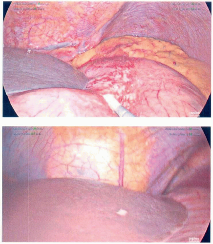
Staging laparoscopy is a minimally invasive procedure that can be used to influence treatment decisions made for patients with gastric cancer both upon diagnosis and during treatment. Staging laparoscopy has sensitivity (86%) and specificity (100%) rates2,3,4 for detecting metastatic deposits that are superior to those for computed tomography (CT) scans (81% to 83%) and functional imaging studies, such as positron emission tomography (PET) scans (65% to 78%).5,6 Indeed, staging laparoscopy performed at presentation may detect radiographically occult metastases in 16% to 37% of patients with apparently localized cancers, even after adequate cross-sectional imaging studies have been performed (Table 3-1, Fig. 3-1).7 Therefore, by using this procedure, approximately one-third of patients who present with radiographically localized gastric cancers can be spared a nontherapeutic laparotomy. Staging laparoscopy, with or without peritoneal lavage,
can also be performed after the administration of preoperative systemic therapy to measure the therapeutic response and to confirm the absence of occult metastatic disease prior to gastric cancer surgery. And at the same time, other adjunct procedures can be performed, such as placement of a feeding jejunostomy tube in advance of neoadjuvant therapy or placement of a central line catheter for systemic therapy.8,9,10


TABLE 3-1 Incidence of Occult Metastases at the Time of Diagnostic Laparoscopy for Patients Eligible for Curative Gastric Resection on the Basis of Preoperative Imaging Studies


Author, Year


Number of Patients


Preoperative Imaging Modality Used


Incidence of Occult Metastases (%)


Lowy et al, 199616


71


CT


23


Burke et al, 19972


111


CT


37


D’Ugo et al, 200322


100


US, CT


21


Sarela et al, 200617


657


CT


31


Mezhir et al, 20101


1,241


EUS, CT


16


Convie et al, 20155


317


CT/PET, CT/EUS


22


CT, computed tomography; US, ultrasound; EUS, endoscopic ultrasound; PET, positron emission tomography.


FIGURE 3-1 Intraoperative staging during diagnostic laparoscopy. Peritoneal carcinomatosis identified during laparoscopy with a tumor on the serosal surface of the stomach (top), and peritoneal nodules on the surface of the liver (bottom) that had not been detected preoperatively on cross-sectional imaging.

Staging laparoscopy is associated with a low risk of perioperative adverse events, which may include port-site infection, bleeding, and bowel injury during entry into the peritoneum. Despite its potential benefits, it remains underused in
the management of gastric cancer. A recent analysis of national data showed that diagnostic laparoscopy was performed in only 8% of a population of older patients with gastric cancer.11


Timing of Staging Laparoscopy

Staging laparoscopy should be performed prior to gastrectomy and lymphadenectomy for cancer in patients whose treatment plan calls for either the administration of preoperative therapy or gastrectomy as a first step. Because analysis of the peritoneal lavage specimen may require several days, staging laparoscopy that includes cytology evaluation is typically performed as a separate procedure.


Current Indications for Staging Laparoscopy

National Comprehensive Cancer Network (NCCN) guidelines currently support the use of staging laparoscopy to exclude metastatic disease prior to gastrectomy or perioperative chemoradiation (CRT) for clinical stage T1b or higher disease because there can be nearly 14% occult peritoneal dissemination in patients with T2 lesions.12 Staging laparoscopy should also be performed before initiating any preoperative chemotherapy. The role of repeat staging laparoscopy is unclear, but there may be some benefit to repeating the examination after preoperative therapy to exclude occult progression, which may occur in 7% to 24% of patients.13,14,15 On the other hand, staging laparoscopy is clearly not indicated for patients who require an immediate, palliative resection for symptoms of obstruction, hemorrhage, or perforation.

Although staging laparoscopy is strongly recommended for all patients with ostensibly operable, gastric cancer, its selective usage has been specifically advocated in certain subsets of gastric cancer patients perceived to be particularly likely to harbor subclinical metastases.3,16 In one study, the presence of gastroesophageal junction (GEJ) tumors, entire stomach involvement, or lymphadenopathy >1 cm on highquality cross-sectional imaging was an adequate trigger for diagnostic laparoscopy to identify occult M1 or peritoneal disease.17


Peritoneal Lavage as Part of Staging Laparoscopy

Peritoneal lavage and acquisition of fluid for cytologic analysis adds to the clinical utility of staging laparoscopy. A key benefit to the evaluation of peritoneal fluid is that it serves as a risk-stratification tool in patients perceived to be at high metastatic risk, such as those with linitis plastica, bulky radiographic adenopathy, or borderline performance status. The detection of cancer cells in the absence of grossly macroscopic metastases (i.e., positive cytology, C1) has been shown to be an indicator of poor prognosis and has been associated with a median overall survival (OS) duration of 14 to 20 months, far below the median OS duration of 98.5 months in patients whose studies were negative (C0).18,19 Furthermore, peritoneal cytology can be used to assess for response to systemic chemotherapy; for example, patients who present initially with C1 cytology but then are found to “convert” to C0 on repeat laparoscopy would be considered to have responded favorably.20 Conversely, occult metastatic disease detected after neoadjuvant therapy in 7% to 24% of patients with cytology initially classified C013,14,15 would
be spared an unnecessary laparotomy. Thus, sequential laparoscopic procedures and peritoneal sampling may have a role in specific scenarios, such as selecting patients for a potentially curative resection after neoadjuvant therapy and sparing those for whom the gastric resection would be futile. As noted, cytologic analysis of the samples acquired at peritoneal lavage may require several days; therefore, preoperative therapy or gastrectomy may have to be suspended until results are received.


Technical Aspects

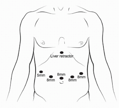
Once pneumoperitoneum has been obtained, the staging laparoscopy should be performed by using a “reverse” tumor-node-metastasis (TNM) approach. First, a 360-degree inspection of the peritoneal surfaces for peritoneal carcinomatosis, hepatic metastases, or ascites should be performed. Next, additional 5-mm trocars can be introduced to facilitate the systematic inspection of the peritoneal cavity, including its parietal and visceral surfaces, the diaphragm, liver, spleen, the small bowel, and the omentum. Any suspicious lesions should be incised/excised and sent for immediate histopathologic analysis. One should be sure to retract the left lateral lobe of the liver off the stomach to inspect the perigastric lymph nodes along the greater and lesser curvatures as well as to inspect the porta hepatis, gastrohepatic ligament, and the lesser sac.21 Laparoscopic sonography (5 to 12 MHz) may also be used to enhance visualization of deeper hepatic lesions. However, the successful use of ultrasound is limited by the expertise of the operator, and little data exist to support its routine use.

If gross ascites is identified, it should be aspirated and sent for cytologic analysis. In the absence of ascites, peritoneal lavage should be conducted: 200 to 500 mL normal saline may be irrigated into the peritoneum and allowed to dwell for 5 to 7 minutes while the patient is gently rotated to the left and right via bed positioning and external physical manipulation. The fluid should then be aspirated and collected via a suction trap assembly from dependent portions of the peritoneum, including the bilateral pelvis and subdiaphragmatic spaces, to detect the cytologic presence of metastatic gastric cancer.22 Peritoneal lavage and acquisition of fluid for cytologic analysis should be conducted before any other biopsy specimens are taken to avoid contamination from any bleeding that may result.

If metastatic disease is confirmed, this opportunity may be used to perform any necessary palliative procedures or to facilitate the subsequent delivery of nonoperative therapies by placing either feeding jejunostomy or gastrojejunostomy tubes or intraperitoneal reservoir ports.12

In conclusion, the use of laparoscopy and the selective use of peritoneal cytologic evaluation are safe and effective tools that play a vital role in the diagnosis, staging, and management of gastric cancer. The high sensitivity and specificity of laparoscopy are better than those of cross-sectional imaging for assessment of metastatic involvement of the peritoneum, and this spares the patient from undergoing a nontherapeutic laparotomy. We recommend routine use of staging laparoscopy, with or without peritoneal cytologic evaluation, in the treatment of patients with operable gastric cancer.


2. RESECTION OF PRIMARY TUMOR


2A. Endoscopic Resection

Recommendation: Removal of small early stage gastric cancers is readily accomplished by using endoscopic techniques in some patients with such cancers, but strict criteria should be used to select patients for these approaches. In routine clinical practice, endoscopic resection should be limited to patients with flat or superficially elevated early gastric cancer lesions <2 cm.

Type of Data: Retrospective studies and case series.

Grade of Recommendation: Strong recommendation, moderate-quality evidence.


Rationale

Some small gastric cancers without deep penetration into the visceral wall are associated with a low incidence of lymph node metastases. Modern endoscopic techniques that may spare some patients the risks of formal gastrectomy have been developed for removal of these tumors.


Indications for Endoscopic Resection

The Japanese Society of Gastrointestinal Endoscopy initially developed the definition of Early gastric cancer (EGC) in 1962 as adenocarcinoma involving the gastric mucosa or submucosa, irrespective of lymph node status. Until the early 2000s the standard treatment approach to EGC was oncologic resection with lymphadenectomy. This paradigm shifted after the initiation of national gastric cancer screening programs in Japan and Korea, when EGC was found to account for approximately 50% of all gastric cancer diagnoses and both screening and endoscopic treatment options began to be further explored.

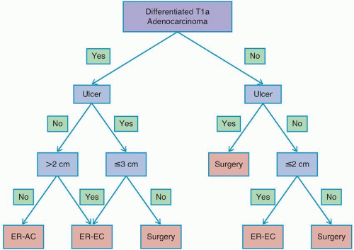
After initiation of gastric cancer screening in Asian nations, patients with EGC limited to the mucosa or superficial submucosa with no lymph node metastasis were found to have 5-year survival rates in excess of 95%.23 Moreover, these studies identified tumor characteristics that were associated with negligible (<1%) risk of lymph node metastasis and thus well suited for endoscopic resection.23,24 Using these data, the Japanese Gastric Cancer Association treatment guidelines defined absolute indications for endoscopic resection of EGC as differentiated-type adenocarcinoma limited to the mucosa, without ulceration, and with a diameter of ≤2 cm.25 With increased experience in the endoscopic treatment of EGC, expanded indications for endoscopic resection of gastric cancer have also been proposed, focusing on patients with EGC and <3% risk of lymph node metastasis (Fig. 3-2).23,24,25 In Japan and Korea, patients with EGC fulfilling these criteria have been treated with endoscopic resection for more than a decade. Retrospective Asian studies have demonstrated excellent long-term outcomes after endoscopic resection for patients with EGC, with overall and diseasefree survival rates in excess of 95% reported at 3-year follow-up for patients meeting both the standard and expanded criteria.26,27,28

Endoscopic resection for EGC is less well established in the West, with the first Western case series having been published in 2009. This is not surprising, given that EGC accounts for only 10% to 20% of all gastric cancers in the Western world.
Several promising reports have described short-term outcomes after endoscopic resection of EGC at Western centers, with curative resection rates in excess of 80%.29,30 However, recent epidemiologic data have indicated significant racial and ethnic disparities in the risk of lymph node metastasis in patients with EGC and have raised concern about the use of endoscopic resection versus surgery in Western patients.27,31 In a review of 923 patients with T1a gastric cancer from the US Surveillance, Epidemiology, and End Results Program (SEER) database, it was observed that the overall rate of lymph node metastasis was 7.8%, more than double that observed in the Asian literature. One important note is that the incidence of lymph node metastasis differed significantly when stratified by race, with patients of Asian origin having a lower rate of lymph node metastasis than non-Asian patients (4.4% vs. 9.1%).27


FIGURE 3-2 Absolute and expanded indications for endoscopic treatment of early gastric cancer. ER, endoscopic resection; AC, adjuvant chemotherapy; EC, endoscopic coagulation. Adapted from Japanese Gastric Cancer Association. Japanese gastric cancer treatment guidelines (ver.3). Gastric Cancer 2011;14:113-123.


Identifying EGC Lesions Suitable for Endoscopic Resection

The crucial step in identifying patients who may benefit from endoscopic resection of EGC is evaluation of deep submucosal tumor invasion, because this dramatically increases the risk of lymphatic spread and lymphatic metastasis. Such invasive lesions cannot be cured with endoscopic resection, and those patients should proceed directly to formal, anatomic gastrectomy. Prior to consideration of endoscopic resection, gross lymph node metastasis should be ruled out with CT and endoscopic sonography.32 In most cases, the
determination of suitability for endoscopic resection is based on assessment of the morphologic characteristics of the lesion on endoscopy, because endoscopic sonography and cross-sectional imaging cannot reliably exclude submucosal invasion.33


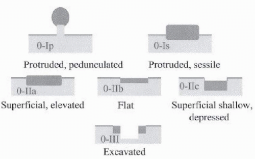
FIGURE 3-3 Paris classification of superficial neoplastic lesions of the stomach. Reprinted from The Paris endoscopic classification of superficial neoplastic lesions: Esophagus, stomach, and colon: November 30 to December 1, 2002. Gastrointest Endosc 2003;58:S3-S43, with permission.

The Paris classification of superficial gastrointestinal neoplasia is the best-studied and most-used system for identifying lesions suitable for mucosal resection. This classification system can be used to predict the depth of invasion and lymphatic metastases. Lesions are broadly classified as follows: Paris class I if protruding, Paris class II if flat or superficially elevated, and Paris class III if depressed or ulcerated (Fig. 3-3).34 EGC Paris class I and IIc lesions that are protruding or depressed and/or ulcerated have a >30% risk of submucosal invasion and are thus not suitable for endoscopic resection (Table 3-2).35 The size of an EGC lesion also has a significant effect on the risk of lymphatic metastasis: Lymph node metastases are present in as many as 16% of American patients with T1a cancers >2 cm, compared with 4.4% with tumors <2 cm.27


TABLE 3-2 Risk of Submucosal Invasion in Japanese Patients with Stage 0 (In Situ) Gastric Adenocarcinoma


Tumor Subtype


Risk of Submucosal Invasion (%)


0-I


57


0-IIa


29


0-IIb


20


0-IIc


40


Source: Endoscopic Classification Review Group. Update on the Paris classification of superficial neoplastic lesions in the digestive tract. Endoscopy 2005;37:570-578.


Given these data as well as the limited experience with endoscopic resection of EGC in Western countries, careful patient selection is mandatory because endoscopic resection risks the undertreatment of patients whose cancer could be cured with gastrectomy and lymphadenectomy. Thus, in routine clinical practice, endoscopic resection should be limited to patients with flat or superficially elevated EGC lesions <2 cm. Patients with tumors >2 cm with ulceration or submucosal invasion should not be offered endoscopic resection outside of a clinical trial setting. In cases of borderline tumors, endoscopic resection can be considered for staging EGC lesions. Then, if high-risk features are identified on histology, patients should be referred for surgical resection, and low-risk patients can undergo close endoscopic surveillance.


Technical Aspects

Endoscopic resection of EGC evolved as an extension of snare polypectomy with a high-frequency electrosurgical generator.36 Resection of EGC was initially preformed via endoscopic mucosal resection (EMR), but endoscopic submucosal dissection (ESD) has been used more recently. This newer technique allows for en bloc resection of lesions >3 cm and is associated with low local recurrence rates.36,37


Endoscopic Mucosal Resection

The core principle of EMR involves three steps: lifting the lesion by the submucosal injection of fluid, entrapment of the lesion with a snare, and removal of the lesion with electrocautery. The purpose of the submucosal fluid injection prior to resection is twofold: (1) to expand the submucosal layer and protect the muscularis propria from injury during cautery and cutting, and (2) to elevate flat or depressed lesions for entrapment with a snare.36 Multiple solutions have been used for submucosal injections during EMR; most commonly, normal saline, hyaluronic acid, and glycerol. Hyaluronic acid is used most frequently for resection of EGC because it provides a longer-lasting cushion than the other fluids do. The volume of solution to be injected varies according to the size and location of the lesion. Repeated injections may be required for larger lesions. Dilute epinephrine (1:100,000) is often added to the injectate to decrease intraprocedural bleeding and to help maintain the submucosal lift. Additionally, staining dyes, such as methylene blue or indigo carmine, help facilitate identification of the lesion margins during resection and improve recognition of intraprocedural injury to the muscularis propria.

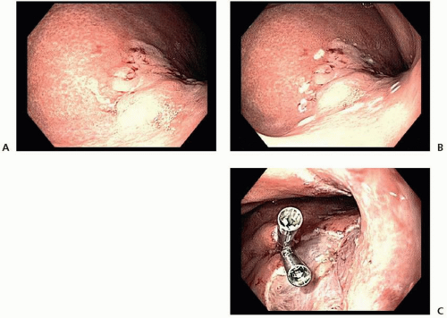
Because of the flat or depressed nature of most EGCs, submucosal injection alone is typically insufficient to permit entrapment of the lesion with a snare. As such, adjunctive techniques have been developed to help facilitate the resection. In cap-assisted EMR, a transparent plastic cap is attached to the distal end of the endoscope; after the submucosal injection of fluid, the lesion is suctioned into the cap, creating a pseudopolyp that is then captured with a snare and resected. An alternative method is band ligation-assisted EMR. In this technique, a cap with rubber-band ligatures (similar to a bander used for treatment of esophageal varices) is fitted to the distal end of the endoscope. The banding cap is then positioned over the lesion, and a band is deployed, creating a pseudopolyp that is then removed with a snare (Fig. 3-4).36


FIGURE 3-4 Piecemeal endoscopic mucosal resection of a Paris 0-IIa lesion in the gastric cardia with the band ligation-assisted method. A: Endoscopic appearance of the lesion. B: The resection margin marked by cautery prior to resection. C: The lesion site after endoscopic resection and clip placement on a visible vessel in the resection bed.

EMR is highly successful in en bloc resection of lesions <10 mm. Larger lesions require piecemeal EMR, which is complicated by the inability to assess the lateral histologic margins for completeness of the resection. This potentially can lead to local recurrence of EGC. Local recurrence after piecemeal EMR of EGC is seen in 6.4% of patients who undergo that procedure, making intensive postresection surveillance mandatory.38


Endoscopic Submucosal Dissection

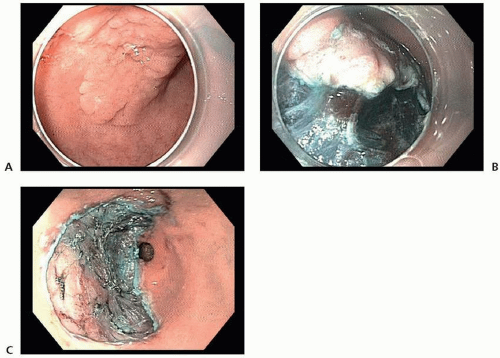
ESD is an endoscopic-resection technique that evolved from EMR; it allows for en bloc resection of lesions regardless of size. ESD is performed in a stepwise process: (1) marking the lateral edges of the lesion with electrocautery, (2) submucosal injection and lifting of the lesion and surrounding normal mucosa, (3) circumferential incision of the mucosa surrounding the lesion with an endoscopic knife, and (4) freehand dissection of the submucosa beneath the lesion with an endoscopic knife (Fig. 3-5). In expert hands, ESD is highly effective in removing EGC, with en bloc and R0 resection rates greater than 90%.36,37

ESD was originally developed and is most widely used for the treatment of EGC in Korea and Japan, where EGC accounts for more than 50% of newly diagnosed gastric cancers. The adoption of ESD by physicians in the United States and Europe has been slow not only because of the low incidence of gastric cancer in the West but also because of the steep learning curve required to master the procedure: Unlike EMR, which uses standard endoscopic techniques and accessories, ESD requires a new skill set and extensive training.32,39


FIGURE 3-5 Endoscopic submucosal dissection of a Paris 0-IIa+c lesion in the gastric antrum. A: Endoscopic appearance of the lesion. B: Appearance of the lesion after the circumferential incision while submucosal dissection is being undertaken. C: Appearance of the lesion site after the submucosal dissection.


Comparison of EMR and ESD in the Treatment of EGC

The literature on the efficacy of endoscopic resection of EGC is limited to large singlecenter series from expert Asian centers. However, two recent meta-analyses have compared the immediate and long-term outcomes of EMR and ESD in treating EGC.38,40 The rates of en bloc and R0 resections are significantly higher for ESD than they are for EMR (Table 3-3); moreover, local recurrence rates are significantly lower with ESD than they are with EMR. Despite the higher local recurrence rate after EMR, the disease-specific survival rate after 36 months does not appear to differ between EMR
and ESD (98.5% vs. 99.7%).41 Thus, on the basis of its higher en bloc resection rates and decreased local recurrence rates, ESD is the preferred technique for endoscopic resection of EGC when adequate expertise is available.32,36


TABLE 3-3 Oncologic Outcomes of Endoscopic Resection of Early Gastric Cancer


Outcome


EMR (%)


ESD (%)


En bloc resection32,34


52


92


R0 resection32,34


42


82-92


Local recurrence32,34


5-6


0.8


36-mo disease-specific survival35


98.5


99.7


EMR, endoscopic mucosal resection; ESD, endoscopic submucosal dissection.


Comparison of Endoscopic Resection and Gastrectomy for EGC

To date there have been no direct comparisons between ESD and gastrectomy for EGC. The available literature consists of multiple retrospective reports and meta-analyses. In one recent propensity score-matched retrospective study from a Korean center, Choi et al27 found that patients with EGC undergoing endoscopic resection and surgery had similar 5-year overall (95.7% vs. 93.6%, respectively, for the two techniques) and disease-free (90.7% vs. 92.8%) survival rates. No gastric cancer-related deaths occurred in either group. Metachronous gastric cancer developed more frequently in the patients who had undergone endoscopic resection—6.1% versus 0.9%—but 93.8% of those lesions were curatively treated with repeat endoscopic resection. Adverse events were observed more frequently in the patients who had undergone surgery (7.9% vs. 2.7%).27 Similarly, three recent meta-analyses found no difference in overall or disease-specific survival between patients with EGC who had undergone gastrectomy and those who had undergone endoscopic resection. Postoperative morbidity and length of hospital stay were significantly lower in the endoscopic-resection groups. However, local recurrences and metachronous cancers were more common after endoscopic resection than they were after surgery.28,42,43

It needs to be emphasized that virtually all data on outcomes of endoscopic resection of EGC come from expert centers in Asia, and to date, there have been no randomized controlled trials (RCTs) comparing endoscopic resection and surgery. It also bears repeating that ESD is technically demanding, even for endoscopists experienced with EMR. Mastery of ESD requires extensive training. Most studies have demonstrated that a minimum of 50 supervised ESD procedures is required to achieve acceptable oncologic (R0 resection) and safety outcomes.37,39,44 Although no prospective data are available on surveillance after endoscopic resection, the Japanese Society of Gastroenterology recommends performing surveillance endoscopy at 6- to 12-month intervals. This strategy enables early detection of both local recurrences and metachronous gastric cancers that can then be successfully treated with repeated endoscopic resection in more than 80% of cases.27,45


2B. Partial and Total Gastrectomy

Recommendation: Partial gastrectomy is recommended for cancers of the middle third and distal third of the stomach, and total gastrectomy is generally recommended for cancers of the proximal third. Removal of adjacent organs when needed because of local invasion is recommended when the operative intent is potentially curative. Resection of the omental bursa, although advocated by some for middle- or distal-third bulky gastric cancers, is not considered standard of care.

Type of Data: Primarily retrospective studies; some prospective and randomized controlled clinical trials.

Grade of Recommendation: Strong recommendation, moderate-quality evidence.


Rationale

Gastric cancer is generally an aggressive malignancy with a predilection for serosal penetration, local invasion, and lymphatic metastasis. Surgical removal of the primary gastric tumor is required for potential cure in patients who do not have metastatic disease. The choice of performing formal, anatomic gastrectomy on the basis of tumor location is inconsistent, and standards for best practice are needed. For gastric adenocarcinomas, wedge or segmental resections are generally considered inadequate since lymphatic drainage patterns do not allow for adequate lymphadenectomy.


Formal Gastrectomy

The type of formal gastrectomy that should be performed depends primarily on the tumor’s location and the presence or absence of local invasion into adjacent organs. It is well established that less than a total gastrectomy (partial, subtotal gastrectomy) is reasonable for most tumors. One French multicenter randomized trial that compared partial and total gastrectomy for cancers in the gastric antrum found no difference in the 5-year survival rates of about 50% for both groups.46,47 Robertson et al48 compared partial gastrectomy with radical total gastrectomy including splenectomy and distal pancreatectomy for antral gastric cancer and found that the OS was better and the complication rate was lower in the subtotal gastrectomy group.

Perhaps the strongest evidence for this question about which type of gastrectomy to use comes from the Italian Gastrointestinal Tumor Study Group.49 Those investigators initially compared the incidence of complications and mortality in a trial of 624 patients with distal gastric cancer who had been randomly assigned to undergo either partial (distal) or total gastrectomy. All patients underwent D2 lymphadenectomy. The complication and mortality rates were 9% and 1%, respectively, in the partial gastrectomy group and 13% and 2%, respectively, in the total gastrectomy group. The resection of other organs when required was associated with more complications. A follow-up study by the same investigators published later reported similar 5-year OS rates of 65% after partial gastrectomy and 62% after total gastrectomy.50 The authors concluded that a 3- to 6-cm margin of resection is adequate on the basis of their inclusion criteria and that partial gastrectomy is their procedure of choice for distal gastric cancers. They also emphasized that better quality of life and nutritional status were associated with partial gastrectomy, relative to those associated with total gastrectomy described in other studies.51 The outcomes of these major studies comparing partial and total gastrectomy are summarized in Table 3-4.46,48,49,50

Although this large amount of evidence exists to support the performance of distal partial gastrectomy, studies comparing proximal with total gastrectomy for proximal gastric cancers are limited to retrospective series. Generally, these studies have shown comparable outcomes.52,53 However, many surgeons have abandoned the use of proximal gastrectomy because of concerns about regurgitation esophagitis.54 The incidence of severe esophagitis can be reduced by using alternative techniques such as jejunal interposition55 or fundoplication.56


Bursectomy

Complete removal of the omental bursa (i.e., bursectomy) is defined as the removal of the peritoneum overlying the pancreas and anterior transverse mesocolon with omentectomy during radical gastrectomy for cancer. Imamura et al57 evaluated the

safety of bursectomy in an RCT in Japan among patients with T2/3 gastric cancer. In that study, 210 patients were randomly assigned to include bursectomy or not with radical gastrectomy and D2 lymphadenectomy. The morbidity (14%) and mortality (1%) rates were similar for the two groups, and complications included pancreatic fistula, abscess, and anastomotic leakage. The survival outcomes were recently published in a follow-up report.58 After a median follow-up time of 80 months, the 5-year OS (77.5% with bursectomy vs. 71.3% without bursectomy) and recurrence-free survival (RFS; 73.7% vs. 66.6%) rates were similar for the two groups. Bursectomy was associated with a 10% improved OS rate for the subgroup of patients with middleand distal-third cancers and with 20% survival in patients with serosal-penetrating tumors (T3/T4). Based on this subset analysis, the authors of this study suggest that there might be some utility of bursectomy in T3/T4 middle to distal-third cancers. However, the trial was not powered for this outcome measure. Thus, although some surgeons, predominantly in Asia, routinely perform and advocate for bursectomy, it cannot be recommended based on the available data and should not be considered a necessary component of gastrectomy for adenocarcinoma. A larger trial that includes a quality-control aspect for the bursectomy procedure is currently underway.


TABLE 3-4 Comparison of Outcomes after Total Versus Partial Gastrectomy for Cancers in the Distal Stomach


Author, Year, and Study Location


Number of Patients


Tumor Location


Type of Gastrectomy (N)


Mortality Rate (%)


Morbidity Rate (%)


Lymph Node Positivity (%)


OS


Gouzi et al, 1989,46 France


169


Antrum


A: Total (76) B: Partial (93)


A: 1 B: 3


A: 33 B: 34


A: 54 B: 56


5-y A: 48% B: 48%


Robertson et al, 1994,48 Hong Kong


54


Antrum


A: Total radical (29) B: Partial (25)


A: 3 B: 0


A: 10 leak, 24 ROR B: 0 leak or ROR


A: 52 B: 44


A: Median, 30 mo B: Median, 50 mo


Bozzetti et al, 199749 and 1999,50 Italy


624


Distal half


A: Total +D2 (304) B: Partial +D2 (320)


A: 2 B: 1


A: 13 B: 9


A: 58 B: 51


5-y A: 62% B: 65%


ROR, unplanned reoperation; +D2, D2 lymphadenectomy OS, overall survival


Resection of Adjacent Organs and Structures

For patients with T4 gastric cancer, multivisceral resection may be required to achieve complete surgical removal of all disease. Although the literature is heterogeneous concerning whether multivisceral resection increases the risk of complications, it is now generally accepted that this procedure is indicated when needed to completely remove all gross tumor in the setting of an otherwise potentially curative operation for gastric cancer.59,60 Thus, surgical exploration for patients with gastric adenocarcinoma should be done by those proficient in all foregut procedures, including pancreatectomy. The literature also lacks concordance on whether removal of certain organs, such as the pancreas or liver, is associated with a higher risk of morbidity than splenectomy or partial colectomy.61 The initial report in 1960 by Appleby62 on resection of the celiac artery in a patient with locally advanced gastric cancer is interesting historically.


Technical Aspects

During radical gastrectomy for the potential cure of gastric cancer, the distal stomach is typically divided at the level of the duodenal bulb beyond the pylorus. Local invasion beyond this point is rare, but if this occurs, the addition of pancreatoduodenectomy is appropriate in selected cases. Total gastrectomy also requires division of the lower esophagus in the posterior mediastinum. In a partial gastrectomy, the proximal or middle stomach is classically divided at a level at least 5 cm proximal to the grossly palpable mass. Division of the stomach for proximal gastrectomy is classically done at both the level of the distal esophagus and across the stomach along a line between the distal three-fourths of the lesser curvature and one-half of the greater curvature. However, significant decision-making with respect to the proper margin of resection should guide the formal resection technique, as is detailed in Gastrectomy Critical Element number 3 (Assessment of Surgical Margins).


Other important technical aspects of radical gastrectomy include removal of the greater omentum and ligation of the right gastric and right gastroepiploic arteries. Ligation of the left gastric artery and removal of the lesser omentum are required for a total gastrectomy, but this is done selectively for a partial gastrectomy. Some surgeons advise removal of lymph node tissue from the left gastric vessels even if they are spared. Similarly, ligation of all the short gastric arteries is required for a total gastrectomy, but ligation of some or all of the short gastric arteries is practiced selectively in the setting of partial gastrectomy, depending on whether the left gastric artery is preserved and the level of stomach division. Routine splenectomy or distal pancreatectomy is not required, although the addition of these procedures is indicated in cases of local invasion without metastatic disease. The extent of lymphadenectomy and restoration of the intestinal continuity are described in other chapters.

Sometimes termed wide omentobursectomy, the bursectomy procedure involves en bloc removal of peritoneal tissues anterior to the pancreas and along the portion of transverse mesocolon facing the stomach (also anterior). The rationale for the procedure in part arises from the concern that microscopic metastatic disease may be limited to the peritoneal tissues of the omental bursa at the time of potentially curative gastrectomy.57 During removal of the peritoneal lining from the anterior transverse mesocolon and pancreas, the dissection along the left side of the transverse mesocolon extends to the left gastroepiploic artery. For distal gastrectomy, the dissection of the pancreatic capsule extends to the proximal half of the splenic artery.


3. ASSESSMENT OF SURGICAL MARGINS

Recommendation: The following recommendations govern intraoperative assessment of the surgical margins at gastrectomy:


  • For resections of gastric adenocarcinoma, a microscopically negative (R0) resection margin should be sought.


  • For patients with early stage cancers of the distal stomach, the primary tumor should be resected with intent to achieve a final proximal margin of 3cm or more. For patients with later-stage distal cancers, and for patients with proximal tumors, data do not support extension of the margin to obtain any specific margin length.


  • Achieving greater proximal margin length must be balanced with the potential of increased morbidity with an extended resection, especially when other poor prognostic factors, such as nodal metastases, are present.


  • Frozen-section analysis is an appropriate method of margin evaluation intraoperatively, but gastric re-resection in the setting of a positive frozen-section margin is controversial.

Type of Data: Retrospective studies.

Grade of Recommendation: Strong recommendation, low-quality evidence.


Rationale

Although the goal of gastrectomy for cancer involves achieving a complete (R0) resection, this goal ultimately must be balanced against its cost. Specifically, surgeons must determine when the morbidity of further gastric or esophageal resection outweighs the potential oncologic benefit when other negative prognostic variables simultaneously indicate a high likelihood of systemic disease.


Margin Distance

The submucosal spread of gastric cancer can allow microscopic disease to extend into tumor margins beyond the macroscopic tumor. Data from studies conducted more than three decades ago in patients undergoing resection of gastric adenocarcinoma suggest that a gross margin <3 to 6 cm from the tumor is often associated with a microscopically positive (R1) margin.122,123,124 This evidence from the 1980s is the foundation for the pervasive surgical doctrine that says that achieving an R0 margin requires gross resection margins of at least 5 cm. Beyond this dogma, expert consensus guidelines on margin length differ. Although US NCCN practice guidelines endorse a margin >4 cm, Japanese gastric cancer treatment guidelines recommend a 2- to 5-cm margin contingent on the T stage and tumor growth pattern.25,125 Although these guidelines aim to decrease the probability of an R1 margin, they do not address the
question of whether there are improved outcomes associated with any resection margin result other than a microscopically negative (R0) margin.

To evaluate the literature on the topic of margin length, one must appreciate a few of the key methodologic differences across studies: tumor location, proximal versus distal margin, and margin definitions.126,127,128,129,130,131,132 As the surgical approach to tumors of the distal stomach (body and antrum) is distinct from proximal tumors (GEJ, cardia, and fundus), the issue of margin length must be considered separately between the two locations. Similarly, the proximal and distal margins on each specimen are anatomically distinct and cannot be considered equivalent. The focus of the majority of the existing literature is on the proximal margin. Finally, margin lengths are differentially defined across studies. Tissue elasticity leads to a decrease in margin length between in vivo and resected specimen measurements, and formalin fixation yields even shorter margin measurements.133 Thus, it becomes difficult to compare outcomes directly when the methods of specimen measurement were not uniform.


Distal Tumors of the Body and Antrum: Proximal Margin Length

For patients with distal gastric cancer, the extent of surgical resection (i.e., total or subtotal gastrectomy) is typically determined by tumor location and length of the resection margin. Prospective data suggest that, if oncologically appropriate, subtotal gastrectomy yields oncologic outcomes similar to those of total gastrectomy but with decreased morbidity.46,50,134,135

Retrospective studies of patients undergoing distal gastric cancer resection in Asia reported no differences in long-term survival when comparing differential lengths of the proximal margin.136,137 However, historically in the gastric cancer literature, outcomes in the East have not always translated to Western outcomes, and thus these data may not be applicable to a Western population.138 Until recently, Western studies have been limited by small numbers, practice-pattern changes in studies that span over an extended time period, and single-institution bias. The US Gastric Cancer Collaborative, however, which includes seven academic surgical centers across the United States, has assembled the largest (n = 965) contemporary (2000-2012) multi-institutional surgical database of Western patients who have undergone an abdominal-approach resection of gastric adenocarcinoma. In the US Gastric Cancer Collaborative study evaluating the proximal margin length for distal tumors, a final microscopic margin <3 cm measured after formalin fixation was associated with decreased RFS and OS compared with specimens >3 cm. Yet stage-specific analyses revealed that this relationship of poorer outcomes with a decreased margin length <3 cm was only observed in early stage disease (stage I). In patients with more advanced stages of gastric adenocarcinoma (stage II and III), the margin length was no longer associated with RFS or OS. This stage-specific relationship implies that progression of more advanced disease may be attributed to aggressive tumor biology instead of surgical technique.126 Thus, for patients with early stage cancers, the primary tumor should be resected with intent to achieve a final proximal margin of 3 cm or more. For stage II/III disease, a negative microscopic margin is the goal, because a specific length does not seem to be associated with any recurrence or survival advantage. It is important to note that the final microscopic margin distance must account for the specimen ‘shrinkage’ factor
when determining the gross in vivo margin and intraoperative decision of where to transect the stomach.


Proximal Tumors of the Gastroesophageal Junction, Cardia, and Fundus: Proximal Margin Length

In the treatment of proximal gastric tumors, one must consider the relationship between the tumor and the GEJ: the Siewert classification (see Table 3-15 and Figure 3-17 on pages 152-153).139 Typically, the most proximal Siewert I tumors are treated as esophageal tumors. The most recent NCCN guidelines counsel that Siewert II tumors also be approached as esophageal tumors; however, historically, esophageal or gastric cancer approaches have been utilized.125 Lastly, Siewert III tumors are approached as gastric cancers. Recent studies of proximal gastric tumors suggested that a gross margin of 6 to 8 cm was associated with decreased risk of R1 margins, specifically in more advanced tumors.127,140 These findings, similar to those from studies three decades ago, do not address the question of whether obtaining a particular margin distance beyond the negative margin is associated with improved patient outcomes.

Consequently, a Japanese study evaluated patients who had Siewert II and III tumors with R0 margins and found that gross margin lengths >2.8 cm were independently associated with improved survival.128 Comparably, in 100 patients from a single US center (1985-2003), a gross margin distance >5 cm was associated with increased OS; however, upon subset analysis, these findings were specific only to patients with T2 tumors with fewer than six lymph node metastases.141 Furthermore, extending resection margins 3 to 5 cm beyond the negative margin could potentially increase the operative morbidity and could lead to conversion of a total gastrectomy to an esophagogastrectomy. Hence, these findings should be interpreted and applied with caution. As previously discussed, outcomes from Asia have historically been discordant from those of the West, and outcomes from a single US center from more than a decade ago may not translate to the current US population.

Again, the US Gastric Cancer Collaborative addressed this topic by examining margin status in proximal gastric adenocarcinoma utilizing a multi-institutional cohort of 151 US patients who underwent R0 resection of gastric adenocarcinoma. The margin distance, measured after formalin fixation, was associated neither with locoregional recurrence (LRR) nor with OS.132 In a similar study including both proximal and distal tumors, margin length was again not related to recurrence or survival.131 Thus, the potential for increased morbidity with an extended resection to achieve a specific margin distance for proximally located gastric tumors must be strongly considered when making intraoperative decisions.


Frozen-Section Margin Analysis

Frozen-section analysis represents one tool to evaluate the resection margin at the time of operation and could potentially influence intraoperative decision-making and extent of resection.142,143,144 Japanese treatment guidelines suggest frozen-section processing if recommended gross resection margins cannot be obtained.25 Intraoperative frozen-section evaluation is also practiced in the West with the goal of identifying R1 margins, and, if identified, converting R1 to R0 margins by additional resection; yet,
it is the effect of this conversion on recurrence and survival that must be understood to determine appropriate patient selection for this practice.

R1 margins on frozen section are associated with larger tumors (>5 cm), increased T stage, presence of signet ring cells, proximal tumor location, and more infiltrative disease.143,145,146 A Korean study compared patients who had an R1 frozen-section margin that was resected a second time to achieve an R0 margin with patients who had R0 margins both on frozen section and final pathology. Decreased survival was observed in patients who initially had R1 frozen section margins (41 vs. 93 months; P = 0.049), despite the fact that these two groups eventually both had R0 margins, suggesting that perhaps an R1 margin could be a surrogate for poor tumor biology.145 A separate study of Eastern patients with final R0 margins reported that an R1 frozen section was not independently associated with recurrence when accounting for other pathologic factors, again implying that the biologically aggressive context of an R1-positive frozen-section margin, rather than the margin itself, was most influential on patient outcomes. From these studies, the question arises about which patients could have improved outcomes by converting an R1 to an R0 margin.

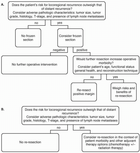
Both US and Asian studies have suggested that converting an R1 margin to an R0 margin through re-excision is associated with improved survival compared with no re-excision (Chen et al147: 44 vs. 25 months; P = 0.021); however, this appears to be true only in early stages of disease.147,148 The US Gastric Cancer Collaborative studied patients grouped by margin status on frozen section and subsequent final pathology: patients whose margins were always R0; those whose margins were R1 on frozen section, underwent re-excision, and all were R0 on final pathology; and those who were R1 on initial and final pathology.149 In early stage gastric adenocarcinoma, R1 frozen-section margins were rare (stage I, 6.2%). For all patients, margins converted from R1 to R0 were associated with decreased LRR compared with that in patients with persistently positive R1 margins (10% vs. 32%; P = 0.01). However, when accounting for other adverse pathologic factors, margin status was not independently associated with LRR. Patients with R1 margins converted to R0 had decreased RFS and OS compared with those who had R0 margins on frozen and final pathology (RFS, 20 vs. 37 months [P = 0.05]; OS, 26 vs. 50 months [P = 0.02]). There were no differences in RFS or OS when R1 margins were converted to R0 margins compared with when margins were R1 on final pathology (RFS, 25 vs. 20 months [P = 0.49]; OS, 36 vs. 26 months [P = 0.14]).149 Therefore, positive margins on frozen-section analysis are rare in early stages of disease, because a positive frozen section appears to be a surrogate for more aggressive tumor biology. Re-excision to convert an R1 margin to an R0 margin does not address the inherent biology of the tumor. A simplified decision tree for pursuit of a frozen section is depicted in Figure 3-9.


The Distal Resection Margin

The majority of the gastric cancer literature focuses on either the proximal margin or both margins. As such, data specific to the distal, or duodenal, margin are limited. Studies from more than three decades ago recommended a distal gross resection margin of >5 cm because microscopic tumor extension was observed up to 5 cm distal to the gross tumor in some cases.123 The clinical context of this positive distal margin
was found to be similar to that of the proximal margin, because it was associated with the presence of lymphovascular invasion, larger tumor size, and one or more positive lymph nodes.150 Although some have reported that an R1 duodenal margin was related to shortened survival, others have found it not to be associated with LRR, other recurrence, or OS.131,150,151 Re-excision of the distal margin in gastric surgery typically requires a pancreaticoduodenectomy. This aggressive management has been reported in the literature; however, these studies’ small sample sizes preclude determination of the benefit of this practice in the context of increased procedural morbidity.152,153,154 Thus, again, the clinical context must be considered, and aggressive additional surgical intervention should not be pursued in the context of aggressive, advanced disease.152


FIGURE 3-9 Algorithms for proximal margin assessment at the time of gastrectomy. A: Pursuit of frozen-section analysis. B: Management of microscopically positive margins.


Clinical Context, Outcomes, and Management of Positive Margins

To interpret the literature exploring positive margins in gastric adenocarcinoma, one must be cautious of the definitions of positive margins in specific studies. Although positive margins are defined by some as microscopically positive R1 margins, others include both R1 and grossly positive (R2) margins in a single category. Certainly, leaving gross residual disease and having a palliative or incomplete resection (R2) is distinct from a curative-intent R1 resection in which any evidence of disease that remained was detected only microscopically. As such, caution is warranted in interpreting and applying data when R1 and R2 margins are included in a single category.139,140,155,156,157,158 Discussion hereafter will include only studies in which patients with R2 margins were excluded or considered a separate category, and the definition of positive margin will be restricted to margins with only microscopic disease.

Before considering prognosis of patients or making management decisions on positive margins, one must understand the clinical context in which an R1 margin typically occurs. Similar to positive frozen-section margins, as discussed earlier, a positive margin on final pathology has been associated typically with other known poor prognostic factors: tumors that are larger than average, advanced T stage, poor differentiation, diffuse tumor spread, proximal location, and increased lymph node metastases.132,140,151,159,160,161,162,163 Given this clinical context, one must attempt to distinguish whether the R1 margin drives poor outcomes or whether such outcomes should be attributed to aggressive tumor factors.

Multiple studies have attempted to answer the question of the complex relationship between R1 margins, recurrence, and survival. Some reports have suggested that there is a relationship between R1 margins and poor OS; however, on critical review, some include only univariate analysis or include multivariable models omitting clinical factors known to be associated with OS. Additionally, others do not establish that this difference in survival is attributed to oncologic etiology, as they do not include data on recurrence or disease-specific survival.161,164,165 Studies offering a more complete analysis reported that margin status was not independently associated with survival when accounting for other poor prognostic variables.163,166

Bickenbach et al151 in 2013 explored the stage-specific prognostic value of margin status in 2,384 patients who underwent gastric adenocarcinoma resection at Memorial Sloan Kettering Cancer Center. In this cohort, 4.5% of patients (n = 108) had R1 margins. Having a positive margin was independently associated with decreased disease-specific survival only in patients with early stage disease: T1 or T2 disease with metastases in fewer than three lymph nodes. Margin status was not, however, associated with patient outcomes in those who had more advanced disease. Many others have observed this same stage-specific phenomenon. The R1 margin plays a role in patient prognosis for early stage but not late-stage disease.148,159,160,162,167

To understand this stage-specific observation, one must consider the patterns of recurrence. The greatest risk of residual microscopic disease at the resection line (R1), in theory, is LRR162; yet, in patients with R1 margins, the LRR rate has been reported at only 16%, and an R1 margin was not an independent risk factor for LRR.132,151 Patients with more advanced disease are more likely to present with distant recurrence, making
an LRR less clinically relevant in this population.151,168 Conversely, in early stage disease, an isolated LRR likely dictates patient prognosis. What is important, however, is that, as previously discussed, the event of a positive margin is associated with other poor prognostic factors, and R1 margins are less common in early stage disease.162 Additionally, in early stage disease, the event of a LRR is rare.132 Therefore, the role of re-resection or other additional therapy in the context of a positive margin must be carefully considered.

Therapeutic options for a positive margin include adjuvant radiation therapy, chemotherapy, and surgical re-resection. In the aforementioned study of Bickenbach et al,151 none of these interventions were related to decreased LRR. On the other hand, comparing adjuvant CRT to surgery alone, Dikken et al169 reported decreased LRR (6% vs. 26%, P = 0.02) and improved 2-year survival (66% vs. 29%, P = 0.002) in patients who received CRT. Neither of these studies, however, addressed stage-specific outcomes.

As such, in making treatment decisions for patients with positive margins, the potential risks and benefits must be weighed, and the comprehensive clinical picture of each patient must be taken into account. Candidacy for radiation therapy, chemotherapy, or surgical re-resection must be considered in the context of performance status and whether patients could tolerate a specific therapy. Next, one must consider biologically whether a particular intervention could improve outcomes based on oncologic factors. In later stages of disease, for example, when aggressive tumor biology, rather than margin status, is defining outcomes, perhaps aggressive margin management is not appropriate. Yet, in early stages of disease where margin status has been associated with increased recurrence and decreased survival, treatment of the positive margin should perhaps be pursued (see the treatment algorithm in Fig. 3-9).


4. REGIONAL LYMPHADENECTOMY

Recommendation: At least 16 regional lymph nodes should be removed and examined at gastrectomy. A D2 dissection is the minimum lymph node dissection that would enable routine resection and assessment of at least 16 regional nodes.

Type of Data: Prospective trials and meta-analyses.

Grade of Recommendation: Strong recommendation, high-quality evidence.


INTRODUCTION

Gastric cancer is an invasive cancer that spreads through lymphatics to the regional lymph nodes. Lymphadenectomy is recommended as a main component of radical gastrectomy. Historically, a majority of recurrences (54% to 63%) after curative resection of gastric cancer were noted to occur locoregionally.170,171 Even though more recent studies have shown a higher proportion of distant sites of first recurrence, lymph node metastasis remains a strong independent predictor of distant tumor spread.33,172,173,174,175,176 Eastern surgeons have traditionally focused surgical techniques on radical control of the lymph node basins with the purpose of adequately staging patients, minimizing locoregional recurrence, and improving OS. Western surgeons have been hesitant to adopt this rationale on the basis of several studies that have shown
increased morbidity and perioperative mortality with this approach, but little oncologic benefit. As highlighted in Key Question 1, the debate over the optimal extent of lymphadenectomy therefore continues.


Evolution of the Definitions and Classifications of Lymph Node Stations and Lymphadenectomy

Prior to the establishment of the American Joint Committee on Cancer (AJCC) classification system, the Japanese Research Society for Gastric Cancer published general rules for classification of gastric carcinoma in 1962 to standardize pathologic classification of gastric cancer.177 It issued three English editions of the classification system, beginning almost two decades later in 1981 and followed by publications in 1995 and 1998, that corresponded, respectively, to the 10th, 12th, and 13th Japanese editions.178 The terms D1, D2, and D3 as originally defined in the second English edition of the Japanese Classification of Gastric Cancer (JCGC) were used in most large RCTs of gastric cancer surgery, including trials of the Dutch, the British Medical Research Council (MRC), and the Taipei (Taiwan) veterans.179,180,181,182,183,184

Based on the seminal work by Sasako et al,185 a complex definition of the nodal groups was established. The regional lymph nodes were classified into stations based on their anatomic position in relation to the geographic location of the primary tumor. The primary tumor was classified as one of five types (various combinations of the three equal portions of the stomach—upper, middle, and lower third). Each primary tumor location type was in turn associated with certain lymph node stations grouped into one of three N “compartments” (N 1, 2, 3, or M).178 These lymph node compartments also correlated with the extent of lymphadenectomy (D1, 2, and 3 dissections corresponding with N1, 2, and 3 lymph node stations). However, the terms D1, D2, and D3 have since been used more loosely, with N1 referring to perigastric nodes and the N2 nodes denoting those along the celiac artery and its branches, and the corresponding dissections of these designated as D1 and D2, respectively.

Therefore, in 2010, to simplify classification, the Japanese Gastric Cancer Association published an integrated version of the classification system and the Japanese Gastric Cancer Treatment Guidelines, which adopted definitions and classifications that were similar to those of the seventh edition of the AJCC Cancer Staging Manual.186 In this version of the classification system, nodal grouping was replaced by the number of metastatic lymph nodes as determined in the TNM classification.186,187 Additionally, the definition of lymphadenectomy was markedly simplified with the lymph node dissection in D1, D1+, and D2 defined for total and distal gastrectomy, regardless of the tumor location.22 D3 (also called “para-aortic lymph node dissection”) was not included after the negative results of the Japan Clinical Oncology Group (JCOG) 9501 trial comparing D2 with D3 dissection.188

Nodal dissections are therefore now defined based on the type of operation performed rather than the location of the primary tumor. The eighth edition of the AJCC manual, released in 2017 by Springer, made several changes to the staging classification and groupings, as highlighted in the introduction to this chapter.189 See Table 3-8187 and Figure 3-10 for definitions of lymph node stations. Figure 3-11 illustrates the extent of lymphadenectomy based on the type of gastrectomy performed.


TABLE 3-8 Anatomic Definitions of Lymph Node Stations


Number


Definition


1


Right paracardial LNs, including those along the first branch of the ascending limb of the left gastric artery


2


Left paracardial LNs including those along the esophagocardiac branch of the left subphrenic artery


3a


Lesser curvature LNs along the branches of the left gastric artery


3b


Lesser curvature LNs along the second branch and distal part of the right gastric artery


4sa


Left greater curvature LNs along the short gastric arteries (perigastric area)


4sb


Left greater curvature LNs along the left gastroepiploic artery (perigastric area)


4d


Right greater curvature LNs along the second branch and distal part of the right gastroepiploic artery


5


Suprapyloric LNs along the first branch and proximal part of the right gastric artery


6


Infrapyloric LNs along the first branch and proximal part of the right gastroepiploic artery down to the confluence of the right gastroepiploic vein and the anterior superior pancreatoduodenal vein


7


LNs along the trunk of left gastric artery between its root and the origin of its ascending branch


8a


Anterosuperior LNs along the CHA


8p


Posterior LNs along the CHA


9


Celiac artery LNs


10


Splenic hilar LNs including those adjacent to the splenic artery distal to the pancreatic tail, and those on the roots of the short gastric arteries and those along the left gastroepiploic artery proximal to its first gastric branch


11p


Proximal splenic artery LNs from its origin to halfway between its origin and the pancreatic tail end


11d


Distal splenic artery LNs from halfway between its origin and the pancreatic tail end to the end of the pancreatic tail


12a


Hepatoduodenal ligament LNs along the proper hepatic artery, in the caudal half between the confluence of the right and left hepatic ducts and the upper border of the pancreas


12b


Hepatoduodenal ligament LNs along the bile duct, in the caudal half between the confluence of the right and left hepatic ducts and the upper border of the pancreas


12p


Hepatoduodenal ligament LNs along the portal vein in the caudal half between the confluence of the right and left hepatic ducts and the upper border of the pancreas


13


LNs on the posterior surface of the pancreatic head cranial to the duodenal papilla


14v


LNs along the SMV


15


LNs along the middle colic vessels


16a1


Para-aortic LNs in the diaphragmatic aortic hiatus


16a2


Para-aortic LNs between the upper margin of the origin of the celiac artery and the lower border of the left renal vein


16b1


Para-aortic LNs between the lower border of the left renal vein and the upper border of the origin of the inferior mesenteric artery


16b2


Para-aortic LNs between the upper border of the origin of the inferior mesenteric artery and the aortic bifurcation


17


LNs on the anterior surface of the pancreatic head beneath the pancreatic sheath


18


LNs along the inferior border of the pancreatic body


19


Infradiaphragmatic LNs predominantly along the subphrenic artery


20


Paraesophageal LNs in the diaphragmatic esophageal hiatus


110


Paraesophageal LNs in the lower thorax


111


Supradiaphragmatic LNs separate from the esophagus


112


Posterior mediastinal LNs separate from the esophagus and the esophageal hiatus


LN, lymph node; CHA, common hepatic artery; SMV, superior mesenteric vein.


Source: Reprinted from Japanese Gastric Cancer Association. Japanese classification of gastric carcinoma: 3rd English edition. Gastric Cancer 2011;14:101-112 (Table 6-5),21 with permission.


In short, because the number of N1 lymph nodes (those lymph nodes that would be assessed with a D1 lymph node dissection) would often be <15, the most oncologically appropriate extent of lymph node dissection for gastric adenocarcinoma is a D2 lymph node dissection that includes N2 nodes, as shown in Figures 3-10 and 3-11. The AJCC 8th edition thus defines an adequate D2 lymph node dissection as including the appropriate lymph nodes to be congruent with this principle.

Only gold members can continue reading. Log In or Register to continue

Stay updated, free articles. Join our Telegram channel

May 7, 2019 | Posted by in ONCOLOGY | Comments Off on Gastrectomy

Full access? Get Clinical Tree

Get Clinical Tree app for offline access