INTRODUCTION
Melanoma is the most aggressive form of skin cancer. Although its incidence pales in comparison to basal cell carcinoma and squamous cell carcinoma (SCC), melanoma is the cause of approximately 75% of all skin cancer–related deaths. The majority of patients who are diagnosed with early-stage melanoma have very good outcomes with appropriate surgical management. In contrast, patients with regional and distant metastases have historically had poorer outcomes, because agents that have proven efficacious in other malignancies (eg, chemotherapy) generally have had limited activity in this disease. However, the management of melanoma is evolving rapidly due to parallel breakthroughs in the understanding and targeting of the molecular drivers of this disease and the regulators of the antitumor immune response. These advances are rapidly translating into improved outcomes in patients with advanced melanoma and the consideration of new diagnostic and therapeutic approaches across the full continuum of this disease.
EPIDEMIOLOGY AND RISK FACTORS
Melanoma is the fifth most common cancer in men and the sixth most common cancer in women in the United States (1). The age-adjusted incidence for cutaneous melanoma from 2007 to 2011 was 21.3 per 100,000 per year in the United States (2). In contrast to the favorable trends that have been observed with almost all other major cancers, the annual incidence of melanoma continues to rise by approximately 2% to 3% per year and has increased overall more than 500-fold since the 1950s (3).
A number of factors have been identified that correlate with an increased risk of being diagnosed with melanoma (Table 41-1). Many of these factors reflect the strong association between melanoma and ultraviolet radiation (UVR) exposure, which is supported by epidemiologic studies (4). More recently, whole-exome sequencing studies have demonstrated that melanomas are characterized by a higher rate of somatic mutations than almost all other solid tumors and that the majority of mutations that are identified bear the molecular signature of UVR-related DNA damage (5). Several risk assessment aids have been developed to identify high-risk individuals, including the Melanoma Risk Assessment Tool (MRAT), which is available online (http://www.cancer.gov/melanomarisktool/). Individuals at increased risk of developing melanoma should have awareness of the signs of melanoma and regular screening examinations.
| Risk Factor | Features |
|---|---|
| Personal history of melanoma | 9× increased risk of developing a second melanoma (vs general population) |
| Family history of melanoma | First-degree relatives have a higher risk, and 10% of all melanomas are familial (FAMMM syndrome and dysplastic nevus syndrome) |
| Total number of nevi | Relative risk of 5 to 17 with presence of >50 nevi |
| Congenital nevi | 6% lifetime risk with large (>20 cm) congenital nevi |
| Dysplastic nevi | 3- to 20-fold higher risk of developing melanoma than general population |
| Immunosuppression | Chronic immune suppressant use, HIV infection, and organ transplantation |
| MC1R variants | Associated with fair skin, red hair, and freckles |
| Exposure to ultraviolet light | Tanning bed use, sunburn |
Patients with dysplastic (atypical) nevi with irregular borders, multiple colors, and >5 mm diameter have a 3- to 20-fold higher risk of developing melanomas than the general population (6). Although the majority of melanomas are sporadic, these nevi can be inherited in a familial pattern. The familial atypical multiple mole and melanoma (FAMMM) syndrome is an autosomal dominant disorder characterized by the occurrence of melanoma in one or more first- or second-degree relatives and the presence of a high number of acquired nevi or atypical nevi. This syndrome is associated with germline mutations of the CDKN2A gene and is also associated with an increased risk of other cancers, especially pancreatic cancer (7). Congenital nevi can also be a precursor of melanoma, and individuals with large congenital nevi (>20 cm) have been shown to be at increased risk of developing melanoma (8).
CLASSIFICATION
Cutaneous melanomas, which are the most common manifestation of this disease, arise from melanocytes in the skin. The four major types of cutaneous melanoma are superficial spreading, nodular, lentigo maligna, and acral lentiginous melanoma. Melanomas can also arise from melanocytes in other areas, including the uveal tract of the eye (uveal melanomas) and mucosal surfaces throughout the body (mucosal melanoma). Desmoplastic melanomas represent a distinct subtype that arise from melanocytes in the skin, generally in areas with chronic sun exposure, that are characterized by highly invasive local growth that often tracks along nerves. Very rare subtypes include primary central nervous system (CNS) melanomas, which arise from melanocytes in the leptomeninges, and melanomas of soft parts (also known as clear cell sarcoma), which arise in soft tissues and dermis. Although the melanoma subtypes are not independent prognostic factors, they can be associated with distinct clinical (Table 41-2) and molecular (Table 41-3) features (9).
| Type | Frequency | Sites | Features |
|---|---|---|---|
| Cutaneous | |||
| Superficial Spreading | 70% | Any site (more common on the upper back in both sexes and the lower extremities in women) | Most common subtype of cutaneous melanomas |
| Nodular | 15%-30% | Any site (common on the trunk or legs) | Presents with vertical growth phase without radial growth phase |
| Lentigo maligna | 4%-15% | Sun-exposed area (the head and neck and arms) | |
| Acral lentiginous | 2%-8% | The palms, soles, and beneath the nail plate | More common in African Americans and Asians |
| Uveal | Rare | Uveal tract of the eye (iris, ciliary body, and the choroid) | Frequent, and often exclusive, metastatic involvement of the liver |
| Mucosal | Rare | Mucosal surfaces (head and neck, respiratory, gastrointestinal, and genitourinary tracts) | Poor prognosis, potentially due to delayed diagnosis and the rich lymphovascular supply of the mucosa |
| Desmoplastic | Rare | Areas with chronic sun exposure, especially head and neck | High risk for local recurrence and growth along nerves |
| Primary CNS | Rare | Leptomeninges | |
| Melanoma of soft parts (clear cell sarcoma) | Rare | Soft tissues, dermis | Associated with fusions involving the EWSR1 gene |
MOLECULAR BIOLOGY
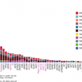
Cutaneous melanomas are characterized by an extremely high rate of somatic mutations. The first cutaneous melanoma analyzed by whole-genome sequencing identified more than 33,000 somatic changes in the tumor (10). The majority of the somatic changes detected in cutaneous melanomas are typical of DNA damage induced by UVR. Despite the challenges presented by this overall high background mutation rate, confirmed driver mutations are detectable in the majority of cutaneous melanomas. A number of important molecular events have also been identified in other melanoma subtypes.
The RAS-RAF-MEK-ERK pathway promotes cellular proliferation and survival, and activation of this pathway has been implicated in multiple tumor types. Genetic events that activate this pathway are detected in almost all cutaneous melanomas (11). The most common alterations detected are point mutations in the BRAF gene. BRAF encodes a serine-threonine kinase in the RAS-RAF-MEK-ERK cascade. Point mutations in BRAF are detected in approximately 45% of cutaneous melanomas, and approximately 95% of these mutations result in substitutions for valine at position 600 in the BRAF protein (12). The most common mutations result in the substitution of a glutamic acid (BRAFV600E, 70%) or lysine (BRAFV600K, 20%) residue. These and other substitutions at codon 600 increase the kinase activity of the BRAF protein ≥200-fold and result in constitutive activation of downstream components of the RAS-RAF-MEK-ERK pathway. The BRAFV600E mutation is also frequently detected in benign nevi, supporting that this molecular event occurs very early in melanoma development (13). Consistent with this theory, BRAF mutation status is highly concordant between primary melanomas and their metastases. Mutations at other sites in BRAF (BRAFNon-V600) are detected in approximately 5% of cutaneous melanomas. These mutations have variable effects on BRAF’s catalytic activity, but preclinical data supports that they still activate the RAS-RAF-MEK-ERK pathway (14). Recently, translocations involving the BRAF locus have also been identified as rare events in melanoma (15). These translocations generate fusion proteins that again appear to activate the RAS-RAF-MEK-ERK pathway.
Mutations in NRAS are detected in 20% of cutaneous melanomas (11). These mutations overwhelmingly occur in hotspot regions that result in substitutions at amino acid residues Q61 (approximately 80%) or G12/G13 (approximately 20%). Similar to BRAFV600E, the mutant NRAS proteins potently activate the RAS-RAF-MEK-ERK pathway, and they are also commonly detected in benign nevi. Notably, hotspot mutations in NRAS are essentially mutually exclusive (<1% co-occurrence) with BRAFV600E mutations, but they co-occur relatively frequently with BRAFNon-V600 mutations (16). Mutations in KRAS and HRAS, which are members of the RAS family of GTPases, are also detected as rare events (<2%) in melanoma. Loss-of-function mutations of NF1, a negative regulator of RAS, are detected in approximately 15% of cutaneous melanomas, predominantly in melanomas without NRAS or BRAFV600 mutations.
The PI3K-AKT pathway is a key regulator of many cellular processes, including proliferation, survival, motility, and metabolism. As studies in multiple cancer types have shown that oncogenic RAS mutations use this pathway in addition to RAS-RAF-MEK-ERK to transform cells, the identification of NRAS mutations was the first evidence supporting a role for PI3K-AKT activation in melanoma. This pathway may also be activated by loss of function of PTEN, which is a lipid phosphatase that normally inhibits the activation of the PI3K-AKT pathway. Loss-of-function mutations and deletions involving PTEN are detected in up to 30% of cutaneous melanomas (11). Loss of PTEN appears to be largely mutually exclusive in melanomas with NRAS mutations, but it occurs frequently in tumors with concurrent BRAFV600 mutations. Experiments in preclinical models have shown that loss of PTEN cooperates with BRAFV600 mutations to promote transformation, invasiveness, and metastasis of melanocytes, providing functional support for this clinical association (17). Rare (<2% each) activating mutations in AKT1, AKT3, and PIK3CA have also been detected in cutaneous melanomas, again generally in tumors with BRAFV600 mutations.
Alterations in key cell cycle regulators are also pervasive in cutaneous melanomas. Germline loss-of-function mutations in the CDKN2A gene are the most common events detected in familial melanoma. The CDKN2A gene encodes two different proteins, P14ARF and P16INK4A. P16INK4A regulates cell cycle progression by binding to cyclin-dependent kinase 4 (CDK4). Point mutations in the gene that encodes CDK4 are the most common germline event detected in familial melanoma cases without CDKN2A mutations. The mutations alter the site in CDK4 that P16INK4A normally binds to, thereby promoting cell cycle progression (18). Somatic mutations and copy number changes in both CDKN2A and CDK4 are detected commonly in cutaneous melanomas. Cyclin D1, which forms a protein complex with CDK4 or CDK6 to promote cell cycle progression, can also be amplified in this disease (19). Loss of function of P14ARF inhibits the function of TP53 via increased MDM2 activity. In addition, mutations in TP53 are present in approximately 20% of cutaneous melanomas (11).
Activating mutations in BRAF are relatively rare events in acral lentiginous (approximately 15%) and mucosal (approximately 5%) melanomas, and they are not detected at all in uveal melanomas (9). However, several other prevalent oncogenic events have been identified in these melanoma subtypes.
KIT is a type III transmembrane receptor tyrosine kinase that activates several pro-survival signaling pathways following binding of its ligand, stem cell factor (SCF). KIT amplifications and mutations are frequently (20%-30%) detected in acral, mucosal, and cutaneous melanoma with chronic sun-induced damage, whereas few are detected in cutaneous melanoma without chronic sun-induced damage (<5%) (20). The two most common KIT mutations in melanoma are L576P (34% of KIT mutations) and K642E (15%) in exons 11 and 13, respectively, and overall, 70% of KIT mutations occur in exon 11, which encodes the juxtamembrane domain (21). KIT mutations in exon 11 prevent the juxtamembrane domain’s inhibitory function and induce the constitutive activation of KIT and its associated pathways.
GNAQ and GNA11 encode regulatory subunits of G-protein-coupled receptors that are frequently mutated in uveal melanomas. Hotspot mutations affecting residues Q209 (exon 5) and R183 (exon 4) in these genes are mutually exclusive events. Combined, mutations in either GNAQ or GNA11 are present in 85% of uveal melanomas (22). The mutant GNAQ/11 proteins hyperactivate a number of cellular signaling pathways, including RAS-RAF-MEK-ERK and PI3K-AKT. GNAQ and GNA11 mutations appear to be extremely rare (≤1%) in cutaneous melanomas, but they have been detected in both blue nevi and primary CNS melanoma.
STAGING
Once patients are diagnosed with melanoma, staging of melanoma is important for prognosis and treatment. The seventh edition of the American Joint Committee on Cancer (AJCC) staging system for cutaneous melanoma based on the primary tumor (T), regional lymph node (N), and distant metastasis (M) was published in 2009 and took effect in 2010 (Tables 41-4 and 41-5) (23). Prognostic factors for primary tumor staging are Breslow thickness, ulceration, and (for melanomas with Breslow thickness <1 mm) mitoses (24). For regional lymph nodal staging, the number of involved lymph nodes is the strongest predictor of outcome, but tumor burden (microscopic vs macroscopic) and pattern of involvement (lymph nodes, in-transit disease) are also prognostic (24). In patients with distant metastasis, staging is organized into three subgroups (M1a, M1b, and M1c) reflecting the site(s) of metastasis and serum lactate dehydrogenase (LDH) levels.
| T Classification | Primary Tumor (Breslow) Thickness (mm) | Ulceration Status/Mitoses |
| Tis | NA | melanoma in situ |
| T1 | ≤1.00 | a: without ulceration and mitosis <1/mm2 b: with ulceration and/or mitoses ≥1/mm2 |
| T2 | 1.01-2.00 | a: without ulceration b: with ulceration |
| T3 | 2.01-4.00 | a: without ulceration b: with ulceration |
| T4 | >4.00 | a: without ulceration b: with ulceration |
| N Classification | No. of Metastatic Nodes | Nodal Metastatic Burden |
| N1 | 1 | a: micrometastasisa b: macrometastasisb |
| N2 | 2-3 | a: micrometastasisa b: macrometastasisb c: in-transit met(s)/satellite(s) without metastatic lymph nodes |
| N3 | 4+ metastatic nodes, or matted lymph nodes, or in-transit met(s)/satellite(s) with metastatic node(s) | |
| M Classification | Distant Metastatic Site(s) | Serum Lactate Dehydrogenase (LDH) Level |
| M1a | Distant skin, subcutaneous, or nodal met(s) | Normal |
| M1b | Lung met(s) | Normal |
| M1c | All other visceral met(s) Any distant met(s) | Normal Elevated |
| Clinical Staginga | Pathologic Stagingb | ||||||
|---|---|---|---|---|---|---|---|
| T | N | M | T | N | M | ||
| 0 | Tis | N0 | M0 | 0 | Tis | N0 | M0 |
| IA | T1a | N0 | M0 | IA | T1a | N0 | M0 |
| IB | T1b | N0 | M0 | IB | T1b | N0 | M0 |
| T2a | N0 | M0 | T2a | N0 | M0 | ||
| IIA | T2b | N0 | M0 | IIA | T2b | N0 | M0 |
| T3a | N0 | M0 | T3a | N0 | M0 | ||
| IIB | T3b | N0 | M0 | IIB | T3b | N0 | M0 |
| T4a | N0 | M0 | T4a | N0 | M0 | ||
| IIC | T4b | N0 | M0 | IIC | T4b | N0 | M0 |
| III | Any T | N > N0 | M0 | IIIA | T1-4a | N1a | M0 |
| T1-4a | N2a | M0 | |||||
| IIIB | T1-4b | N1a | M0 | ||||
| T1-4b | N2a | M0 | |||||
| T1-4a | N1b | M0 | |||||
| T1-4a | N2b | M0 | |||||
| T1-4a | N2c | M0 | |||||
| IIIC | T1-4b | N1b | M0 | ||||
| T1-4b | N2b | M0 | |||||
| T1-4b | N2c | M0 | |||||
| Any T | N3 | M0 | |||||
| IV | Any T | Any N | M1 | IV | Any T | Any N | M1 |
SURGICAL MANAGEMENT OF EARLY-STAGE MELANOMA
For patients with primary cutaneous melanoma and clinically negative regional lymph nodes, surgery represents the mainstay of initial clinical management. It is useful to consider this approach in the context of two themes: (1) wide excision and (2) approach to the regional nodal basin. Prior to definitive surgery, it is important to identify other lesions suspicious for a second primary melanoma; evidence, if any, of regional metastasis; and signs/symptoms that may raise suspicion for distant metastatic disease. Such findings may alter treatment plans.
Recommended wide excision margins are based on primary tumor thickness (Breslow thickness), and they are measured from the melanoma biopsy site edges or residual intact disease (25). Wide excision includes subcutaneous tissue down to the level of, but generally not including, underlying muscular fascia. Recommended margins of excision are summarized in Table 41-6.
The surgical approach to the regional nodal basin is guided by Breslow thickness and other tumor and host factors. The technique of lymphatic mapping and sentinel node biopsy (SNB) is based on the observation that finite regions of skin drain via afferent lymphatics to regional lymph nodes, termed sentinel nodes, and that these represent the most likely nodes to contain occult metastatic disease, if any are involved (26). Overall, for patients with clinically negative regional nodes, the risk of regional lymph node metastasis ranges from <1% for patients with very thin primary tumors to approximately 50% for patients with thick, ulcerated primary tumors. Sentinel node biopsy has been widely recommended for patients with primary cutaneous melanomas ≥1 mm in tumor thickness (27). In contrast, a selective approach to SNB is entertained for patients with thin (T1) melanomas due to the overall low risk of microscopic regional metastasis. Although indications for SNB among patients with thin melanoma continue to evolve, SNB should be considered for a patient whose primary tumor is ≥0.75 mm and/or has other high-risk features (28). Preoperative lymphoscintigraphy is generally recommended to identify regional nodal basins at risk and to localize the sentinel nodes; a dual-modality approach (ie, in the United States, isosulfan blue dye and technetium-99 sulfur colloid) along with a hand-held gamma probe is used intraoperatively (26,29). Following surgery, enhanced histologic analysis is performed, generally as a combination of step sectioning and immunohistochemical analysis. In contrast to many other solid tumors, intraoperative frozen section assessment is rarely employed.
Sentinel node pathologic status is an important independent predictor of survival (30,31). Although complete lymph node dissection (CLND; also known as completion lymphadenectomy or early therapeutic lymph node dissection) has been a standard of care for patients with a positive sentinel node for over two decades, its role continues to evolve. The recently completed Multicenter Selective Lymphadenectomy Trial-I (MSLT-I) prospective, randomized, clinical trial compared wide excision and SNB (with CLND for patients with a positive SNB) to wide excision and nodal observation (followed by lymphadenectomy for patients who developed nodal recurrence). This trial confirmed the strong prognostic significance of SNB in patients with early-stage melanoma, but it did not demonstrate an overall survival (OS) benefit for the procedure. However, subset analysis among all node-positive patients did show a survival advantage to SNB-positive patients who had CLND compared to patients who had nodal observation and subsequently recurred in regional nodes (31). The role of CLND for patients with at least one positive sentinel node is currently being investigated in the randomized international MSLT-II trial and the European Organization for Research and Treatment of Cancer (EORTC) registry-based MINITUB clinical trial.
MANAGEMENT OF REGIONAL DISEASE
Recurrences at the primary tumor site after surgery are rare and occur in <5% of cutaneous melanomas (32). However, in some cases, the risk of regional recurrence may be increased and the use of adjuvant radiation may improve local disease control. Inadequate margins due to anatomic restrictions, satellitosis in the surgical specimen, or recurrence at the primary site are relative indications for adjuvant radiation at the primary tumor site. Thick (>4 mm) tumors, especially those that originate in the head and neck region, are also sometimes considered for adjuvant radiation. In particular, desmoplastic melanomas, which overall are rare but frequently (>60%) occur in the head and neck region (33) and tend toward neurotropic spread rather than classical lymphatic spread, have a high local failure rate (20%-50%). Retrospective reports of postoperative adjuvant radiation in desmoplastic melanoma patients have demonstrated a significant decrease in recurrence rate with postoperative adjuvant radiation versus observation alone (local recurrence, 24% vs 7%; P = .009) (33).
Stay updated, free articles. Join our Telegram channel

Full access? Get Clinical Tree


