Lysosomal Abnormalities of the Monocyte-Macrophage System: Gaucher and Niemann-Pick Diseases


Lysosomal Abnormalities of the Monocyte-Macrophage System: Gaucher and Niemann-Pick Diseases


Margaret M. McGovern

Robert J. Desnick


Abnormalities of the monocyte-macrophage system include certain of the inherited lysosomal storage diseases that result from specific defects in lysosomal function. Most of these disorders are caused by the deficiency of a particular hydrolytic enzyme, but others are due to impaired receptors, transporters, or deficiencies of crucial cofactors or protective proteins. Prevalent among these disorders are the sphingolipidoses, which are a unique family of diverse diseases related by their molecular pathology. Here, the clinical, biochemical, and genetic features of two of these disorders are presented: Niemann-Pick types A and B disease, caused by defects in the acid sphingomyelinase (SMPD1) gene, and Gaucher disease, caused by defects in the acid α-glucosidase (GBA) gene. In each of these autosomal recessive disorders, mutations in the gene that encodes the lysosomal enzyme result in a defective gene product (i.e., a noncatalytic enzyme) that is unable to perform its normal function as a hydrolase. The deficiency of these lysosomal hydrolases results in abnormal metabolism of the enzyme’s specific sphingolipid substrate, and the substrate accumulates in the cells of the monocyte-macrophage system, causing the clinical manifestations. For example, in Gaucher disease and Niemann-Pick types A and B diseases, anemia, thrombocytopenia, leukopenia, and/or hepatosplenomegaly can be the presenting symptoms. Thus, these disorders are frequently diagnosed by the hematologist and must be included in the differential diagnosis for patients with monocyte-macrophage involvement.


PATHOPHYSIOLOGY OF THE LYSOSOMAL STORAGE DISEASES

The underlying defect in the inherited lipidoses is the accumulation of metabolites, including glycolipids and sphingomyelin. The glycosphingolipids, which are present in the membranes of many cell types, are formed by the addition of various carbohydrates to a ceramide backbone (Fig. 59.1). The fatty acid portion of ceramide consists primarily of stearic acid (C18) in the brain, whereas in non-neural tissues it is somewhat longer (C20 to C24). Each sphingolipid is characterized by the nature of the compound that is esterified to the first carbon of the ceramide molecule. For example, the addition of hexoses and N-acetylneuraminic acid to ceramide forms the gangliosides, which are found in brain, whereas the neutral glycolipids are found more ubiquitously in cell membranes. The blood group antigens are also glycosphingolipids. In the lysosomal storage diseases, these lipid compounds vary in amount in different cell types, so their rate and amount of accumulation because of a specific lysosomal enzyme defect will vary, leading to their cell-related manifestations (Fig. 59.2). Tables 59.1 and 59.2 outline the biochemical and phenotypic characteristics and the molecular basis of the lysosomal storage diseases, respectively.


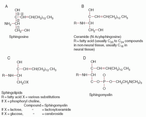
FIGURE 59.1. Formulas of some of the sphingolipids.


GAUCHER DISEASE


Definition and History

Gaucher disease is a lipid storage disease characterized by the deposition of glucocerebroside in cells of the macrophage-monocyte system. It was first described by Gaucher in 1882, and the storage of glucocerebroside was first recognized by Epstein in 1924. The metabolic defect, which is the deficiency of the lysosomal hydrolase acid β-glucosidase, or β-glucocerebrosidase, was identified by Brady et al.1 There are three clinical subtypes, which are delineated by the absence or presence and progression of neurologic involvement: type 1 or the non-neuronopathic form; type 2, the infantileonset, acute neuronopathic form; and type 3, the juvenile-onset neuronopathic form.2 All three subtypes are inherited as autosomal recessive traits. Type 1 disease is the most common lysosomal
storage disease and one of the most prevalent genetic disorders among Ashkenazi Jewish individuals, with an incidence of about 1 in 1,000 and a carrier frequency of about 1 in 15.3


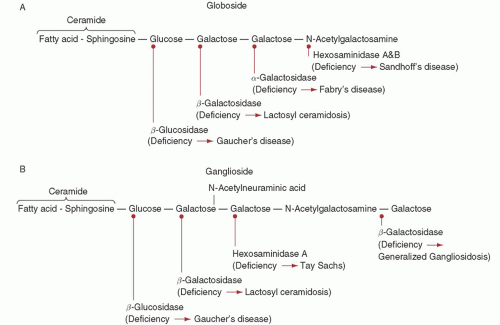
FIGURE 59.2. Schematic structure of globoside (A) and ganglioside (B) to show site of action of the several catabolic enzymes, which result in one of the storage diseases when defective.


Etiology and Pathogenesis

All three subtypes of Gaucher disease result from the deficient activity of the lysosomal hydrolase, acid β-glucosidase (Table 59.1). The major acid β-glucosidase gene mutations that cause Gaucher disease among Ashkenazi Jewish patients have been identified (Table 59.2). Genotype/phenotype correlations have been made for the different subtypes and provide insight into the molecular basis for the remarkable clinical variation in Gaucher disease. Presumably, the amount of residual enzymatic activity determines disease subtype and severity. For example, the mutations that cause the severe type 2 (infantile) disease express little, if any, enzymatic activity in vitro, whereas type 1 patients who are homozygous for the milder N370S mutation tend to have a later
onset and a milder course than patients with one N370S allele and another mutant allele. However, the wide variability in clinical presentation among Gaucher disease patients cannot be fully explained by the underlying acid β-glucosidase mutations, and presumably other “modifier” genes can influence disease severity.


TABLE 59.1 BIOCHEMICAL AND PHENOTYPIC CHARACTERISTICS OF GAUCHER AND NIEMANN-PICK DISEASES


Disease


Enzyme Deficiency


Substance Accumulated


Site


Complications


Gaucher Disease


Type 1


Acid β-glucosidase


Primarily glucosylceramide


Macrophage-monocyte system


Infiltration of bone marrow, progressive hepatosplenomegaly, skeletal complications


Type 2


Acid β-glucosidase


Primarily glucosylceramide


Macrophage-monocyte system, CNS


Infiltration of bone marrow, progressive hepatosplenomegaly, skeletal complications, neurodegeneration


Type 3


Acid β-glucosidase


Primarily glucosylceramide


Macrophage-monocyte system, CNS


Progressive neurodegeneration


Niemann-Pick Disease


Type A


Acid sphingomyelinase


Sphingomyelin


Monocyte-macrophage system, CNS


Hepatosplenomegaly, progressive neurodegeneration


Type B


Acid sphingomyelinase


Sphingomyelin


Monocyte-macrophage system


Progressive hepatosplenomegaly, infiltrative lung disease


Type C


Abnormal cholesterol transport


Primarily cholesterol


Most cells, especially liver, CNS


Hepatosplenomegaly, progressive neurodegeneration


CNS, central nervous system.


TABLE 59.2 MOLECULAR BASIS OF GAUCHER AND NIEMANN-PICK DISEASES


Disease


Chromosome Assignment


Molecular Characteristics


Comments


Gaucher disease


1q21


cDNA, functional and pseudogenomic sequences, >200 mutant alleles known


Four mutations (N370S, L444P, 84insG, IVS2+1) account for 90 to >95% of mutant alleles in Ashkenazi Jewish patients


Niemann-Pick disease


Types A and B


11p15.1 to p15.4


cDNA, entire genomic sequence, >70 mutant alleles known


Four mutations account for >95% of mutant alleles in Ashkenazi Jewish patients with type A disease


Type C


18q11-q12 region


cDNA, entire genomic sequence, >100 mutant alleles known


More than 100 mutations in NPC1 gene

Only gold members can continue reading. Log In or Register to continue

Stay updated, free articles. Join our Telegram channel

Oct 21, 2016 | Posted by in HEMATOLOGY | Comments Off on Lysosomal Abnormalities of the Monocyte-Macrophage System: Gaucher and Niemann-Pick Diseases

Full access? Get Clinical Tree

Get Clinical Tree app for offline access