, Joong Mo Ahn2 and Yusuhn Kang2
(1)
Department of Radiology, Seoul National University College of Medicine, Seoul National University Bundang Hospital, Seongnam, South Korea
(2)
Department of Radiology, Seoul National University, Bundang Hospital, Seongnam, South Korea
6.1 Plasma Cell Myeloma/Solitary Plasmacytoma of Bone
Overview
Plasma cell myeloma (multiple myeloma) and solitary plasmacytoma are clonal neoplastic proliferation of plasma cells of bone marrow derivation. Plasma cell myeloma is a multicentric disease with systemic manifestations, whereas solitary plasmacytoma is a unicentric disease, localized to a single bone. Multiple solitary plasmacytomas without evidence of multiple myeloma may occur.
Epidemiology
Plasma cell myeloma is the most common malignant primary bone tumor. It commonly occurs in patients in their sixth and seventh decades of life, and is rare under the age of 40 years. Solitary plasmacytomas tend to occur at a younger age, with a median age of 55 years at diagnosis. Plasma cell myeloma affect both genders equally, whereas solitary plasmacytomas are more common in males than in females (M:F = 2:1).
Common Locations
Plasma cell myelomas and solitary plasmacytomas are found in bones containing hematopoietic marrow, usually the axial skeleton. Solitary plasmacytomas have been reported most commonly in the vertebra, and other possible sites include ribs, skull, pelvis, and femur.
Imaging Features
Radiograph
Plasma cell myelomas may have various appearances according to the site of involvement and type of lesion. Typical plasma cell myelomas present with multiple well-demarcated osteolytic lesions with medullary location, often referred to as “punched-out” lesions (Fig. 6.1). In bones with small calibers, such as the rib, plasma cell myelomas may cause expansion of the bone (Figs. 6.2, 6.3, and 6.4). However, periosteal reactions are rare. Atypical cases may present with more aggressive features with either moth-eaten or permeative pattern of bone destruction (Fig. 6.5). Diffuse osteopenia in the spine with or without compression fractures may be the sole finding, making the diagnosis of myeloma difficult. Occasionally, myeloma may present with multiple sclerotic lesions, usually in the setting of POEMS syndrome (polyneuropathy, organomegaly, endocrinopathy, monoclonal gammopathy, skin changes) (Fig. 6.6).
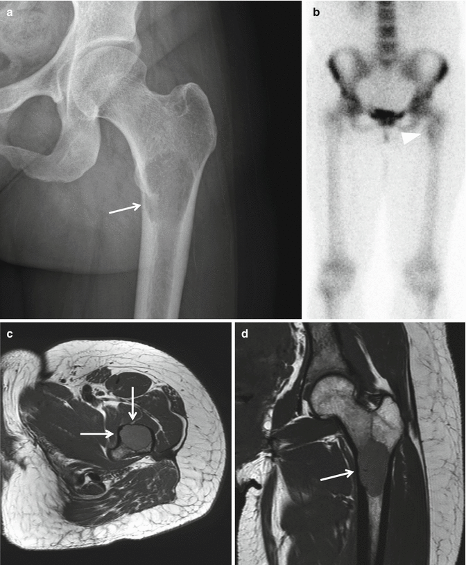
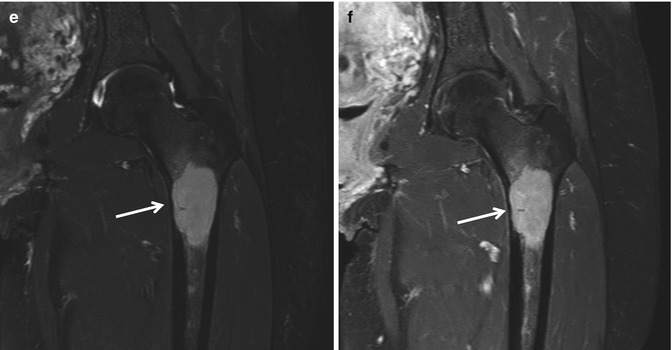
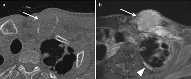
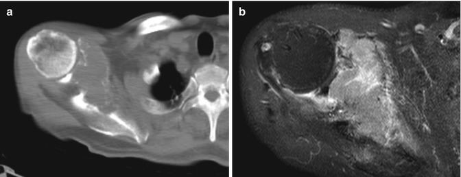
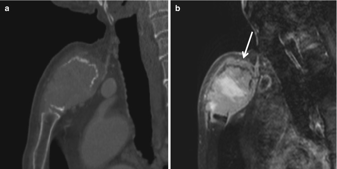
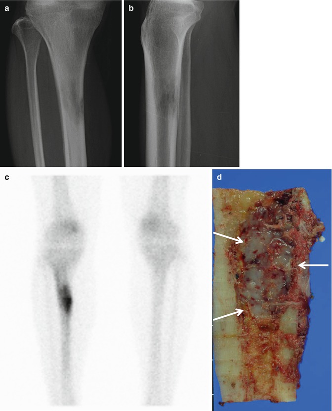


Fig. 6.1
Solitary plasmacytoma in the left femur. (a) On radiograph, a geographical osteolytic lesion with a well-defined margin (punched-out lesion) is noted at the proximal metadiaphysis of the left femur in a 39-year-old female. The femoral cortex is thinned due to endosteal scalloping (arrow). (b) Increased uptake is noted at the left proximal femur on bone scan (arrowhead). (c) Axial and (d) coronal T1-weighted images, (e) coronal T2-weighted fat-suppressed image, and (f) coronal contrast-enhanced T1-weighted fat-suppressed images are shown. The lesion shows homogeneous signal intensity on all sequences. Endosteal scalloping is noted around the lesion, resulting in cortical thinning. Based on negative bone marrow biopsy results and other laboratory tests, the lesion was diagnosed as solitary plasmacytoma

Fig. 6.2
Plasmacytoma in the left clavicle in a 58-year-old male with plasma cell myeloma. (a) Axial CT image shows an expansile osteolytic lesion at the proximal end of the left clavicle (arrow). (b) A homogeneously enhancing lesion is noted at the corresponding site on axial contrast-enhanced T1-weighted fat-suppressed image (arrow). Another enhancing lesion is noted at the posterior arc of the left third rib (arrowhead). Based on bone marrow biopsy results and other laboratory tests, the patient was diagnosed with plasma cell myeloma

Fig. 6.3
Plasmacytoma in the right scapula in a 79-year-old male with plasma cell myeloma. (a) Axial CT image shows an expansile osteolytic lesion involving the right scapula, extending from the scapular body, glenoid to the coracoid process. (b) A homogeneous enhancing mass with extraosseous extension is noted on axial contrast-enhanced T1-weighted fat-suppressed image.

Fig. 6.4
Plasmacytoma in the sternal manubrium in an 87-year-old male with plasma cell myeloma. (a) Sagittal reformatted CT image shows an expansile osteolytic lesion with cortical breaching in the manubrium. (b) Sagittal contrast-enhanced T1-weighted fat-suppressed image shows an enhancing mass lesion in the manubrium. Extraosseous extension is noted without overt cortical breaching along the superior aspect of the manubrium (arrow), favoring the diagnosis of a round cell tumor.


Fig. 6.5
Solitary plasmacytoma. (a) Anteroposterior and (b) lateral radiographs of the right lower leg show an ill-defined osteolytic lesion with a moth-eaten pattern of destruction in the proximal diaphysis of the tibia. (c) Bone scan exhibits an increased uptake at the corresponding site. (d) Sagittal section of the resected tibia shows a whitish glistening mass lesion in the medulla. (e) Axial T1-weighted image, (f) axial T2-weighted image. (g) Axial and (h) coronal contrast-enhanced T1-weighted images show an intramedullary mass lesion with endosteal scalloping (arrowheads) in the right tibia

Fig. 6.6
POEMS syndrome. A 47-year-old female patient presented with tingling sensation and weakness in the lower extremities and numbness in the upper extremities. (a) Coronal reformatted CT image shows multiple sclerotic bone lesions scattered in the vertebral column, pelvic bones, and left proximal femur (arrows). (b) Sagittal T1-weighted and (c) T2-weighted images of the thoracic spine show lesions with dark signal intensity on both sequences. (d) Axial CT image of the upper abdomen shows splenomegaly
Magnetic resonance imaging
Plasma cell myeloma and solitary plasmacytoma show nonspecific findings on MR imaging. MR may be useful in (i) detecting small lesions not readily seen on radiographs and (ii) evaluating the presence and extent of soft tissue extension.
Differential Diagnoses
- 1.
Plasma cell myeloma: metastasis, lymphoma, Langerhans’ cell histiocytosis, brown tumors of hyperparathyroidism
- 2.
Solitary plasmacytoma: metastasis
6.2 Lymphoma of Bone
Overview
Lymphoma is a disease histologically characterized by the proliferation of lymphocytes, histiocytes, and their precursors. Lymphoma of bone can be classified as primary lymphoma of bone or secondary bone lymphoma. Secondary bone lymphoma refers to the involvement of bone secondary to a systemic disease with nodal or other extranodal involvement, and is not uncommon. On the other hand, primary lymphoma of bone is defined as lymphoma confined to the bone or bone marrow without evidence of concurrent systemic disease, and is rare. The majority of primary bone lymphomas are non-Hodgkin lymphomas.
Epidemiology
Primary lymphoma of bone accounts for <5 % of primary bone tumors. It may occur in any age group, but has a peak prevalence in the sixth and seventh decades of life. It is slightly more common in males(male: female = 1.5:1).
Common Locations
The femur is the most commonly affected site, accounting for 25 % of the cases. Within the femur, the lesion is usually located in the metadiaphysis. The spine and pelvic bone, humerus, tibia, and head and neck are the other possible sites.
Stay updated, free articles. Join our Telegram channel

Full access? Get Clinical Tree


