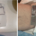
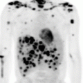
Fig. 4.1
Frequency rates of extranodal marginal zone lymphomas (MZL) according to site (excluding the skin and spleen), SEER data (Khalil et al. [40])
In this chapter, illustrative cases of a gastric and ocular MALT lymphoma, the most common EN-MZL, are presented followed by a discussion of the appropriate evaluation and management, with a focus on the role of radiotherapy.
Gastric Marginal Zone B-Cell Lymphomas (MALT Lymphomas)
The gastrointestinal tract is the most frequent site of extranodal lymphoma, and the stomach is involved in up to two-thirds of the cases [99]. Indeed, 30–45 % of all extranodal lymphomas are detected in the stomach [96]. Primary gastric lymphoma is a rare disease, representing nearly 2–8 % of all stomach tumors [56, 95]. Virtually all gastric lymphomas arise from B cells [95], as T-cell gastric lymphoma is extremely rare [64]. Marginal zone MALT lymphoma accounts for nearly 50 % of gastric lymphomas [59, 64]. Gastric MALT lymphoma is characterized by a dense lymphoid infiltrate mainly composed of small-size lymphocytes that invade and destroy gastric glands, configuring the so-called lymph epithelial lesion which is pathognomonic of lymphoma [37].
A recent systematic review including data of 2000 patients found that gastric MALT lymphoma occurs over a wide age range, with a mean of 57 years at diagnosis, and is slightly more prevalent in males (male–female ratio = 1.27:1) [97].
Clinical Case
A 61-year-old man was in his usual state of health until June 2002 when he began to suffer dyspepsia. At that time, he didn’t report any other symptoms, like drenching night sweats, fevers, weight loss, or pruritus. An upper endoscopy demonstrated the presence of gastric macroscopic lesions, and biopsies showed a diagnosis of gastric MALT lymphoma with associated Helicobacter pylori (H. pylori) infection. Further assessment including a complete physical examination; routine laboratory tests; computed tomography (CT) of the chest; abdomen, and pelvis; endoscopic ultrasonography; and bone marrow biopsy were performed and demonstrated a disease confined to one extranodal site. Therefore, the patient fell classically into a stage I disease. He underwent antibiotic eradication with resolution of the macroscopic lesion and negativity of H. pylori infection but persistence of MALT areas in the stomach. He repeated antibiotic therapy with endoscopic persistence of MALT areas in the stomach. For this reason, chemotherapy with six cycles of chlorambucil was administered. An upper endoscopy showed minimal residual disease, and patient started clinical-endoscopic follow-up with stable disease for many years. At August 2013 an endoscopy showed a progression of disease with evidence of new macroscopic MALT lesions in almost all the stomach (Fig. 4.2) and H. pylori negativity. A positron emission tomography (PET)/CT confirmed increased uptake to the whole stomach.
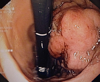

Fig. 4.2
Endoscopic finding with a well-visible massive gastric MZL lesion
Clinical-Endoscopic Presentation
The clinical presentation of gastric lymphoma is poorly specific because symptoms range from vague dyspepsia, including epigastric pain or discomfort, to, less frequently, symptoms such as gastrointestinal bleeding or persistent vomiting [64, 99]. Classic B symptoms (fever, night sweats, weight loss) are extremely rare. A gastric MALT lymphoma is often diagnosed following an upper endoscopy performed for dyspepsia.
Similarly, several different nonspecific endoscopic patterns have been described. Although it may appear at endoscopy as a clear malignant lesion (giant ulcer, vegetant mass, etc.) as shown in Fig. 4.2, gastric MALT lymphoma is frequently characterized by erosions, small nodules, thickening of gastric folds – generally suggesting a benign condition – or even by apparently normal gastric mucosa [99].
Pathology and Helicobacter pylori Infection
The histologic diagnosis can be challenging due to the heterogeneity of cells within the tumor specimen, but the presence of lymph epithelial lesions (invasion of neoplastic cells into individual glands) is a strong indicator of gastric MALT lymphoma. Once the diagnosis is confirmed, the next task is to establish the H. pylori status [99]. Gastric MALT lymphoma is uniquely associated with H. pylori infection (present in 90 % of cases). A variety of detection methods are available, including identification in the histologic specimen, a biopsy urease test, a urea breath test, a stool antigen test, and serology. Upon establishment of the diagnosis, it is then worthwhile to perform a FISH evaluation for a t(11;18)(q21;q21) [4]. This translocation is recurrent in MALT lymphomas and produces a fusion between the AIP2 gene and the MLT/MALT1 gene, generating a chimeric protein that promotes cell survival and proliferation via activation of the transcription factor NF-k. This translocation can be detected in 30–40 % of gastric MALT lymphoma cases and has clinical significance. Patients harboring t(11;18) are more likely to have widely disseminated disease, more likely to be H. pylori negative, and less likely to respond to H. pylori eradication [39]. In one series, only 2 of 44 patients with t(11;18) responded to eradication therapy and both later relapsed. The t(1;14)(p22;q32) translocation is also found in gastric MALT lymphoma but less frequently (~4 % of cases). This translocation results in deregulation of the BCL10 gene with subsequent activation of NF-k B. Gastric MALT lymphoma with t(1;14) is also unlikely to respond to eradication therapy [93].
Diagnosis and Staging
Although gastric MALT lymphoma has a typical indolent course, with a low disease-related morbidity and mortality, an accurate staging procedure is considered mandatory [14, 69]. Gastric MALT lymphoma presents as localized disease in 90 % of cases; advanced stages (III–IV) at diagnosis usually present with localization in both lymph nodes and other organs, particularly the bone marrow, lungs, and liver [2]. A comprehensive staging procedure includes a complete physical examination; routine laboratory tests; CT of the chest, abdomen, and pelvis; endoscopic ultrasonography; as well as bone marrow biopsy. Endoscopic ultrasound allows to accurately assessing both gastric wall infiltration and regional lymph node involvement [12, 38, 57]. The depth of invasion into the different layers of the gastric wall is predictive of lymphoma remission following therapy [57]. The role of PET scan in such a setting is still unclear [8].
Treatment and Outcome
The mainstay of treatment for H. pylori-positive patients is a combination of proton pump inhibitors (PPI) and antibiotics [7, 90]. Treatment with amoxicillin, clarithromycin, and PPIs results in eradication rates of 90 %. An alternative regimen of metronidazole, clarithromycin, and PPIs achieves the same result. Initial treatment with chemotherapy, surgery, or radiotherapy (alone or in combination) has not been shown to be superior to antibiotic treatment [66]. Locally advanced disease that has infiltrated the muscularis mucosa, serosa, or perigastric lymph nodes has a significantly lower response rate (RR). Such cases and H. pylori eradication failures require alternative approaches; however, there is no consensus on how to manage these patients. In some centers, H. pylori-negative patients at diagnosis also receive eradication therapy and high-dose PPIs as frontline therapy. Alternative treatment options include oral chlorambucil, radiation therapy (RT), or rituximab, without convincing evidence on the superiority of one of these strategies over the others due to the lack of prospective randomized trials. The role of surgery has been questioned, as a more conservative local approach (low-dose radiotherapy) may obtain similar results while improving quality of life. A prospective observational study of all histological subtypes of NHL of the gastrointestinal tract (GIT NHL 01/1992) included 185 patients with stage I or II gastric lymphoma; 106 of these patients underwent nonsurgical treatment, i.e., radiotherapy and/or chemotherapy. The subsequent, second prospective observational study (GITNHL02/1996) included 393 patients with localized primary gastric lymphoma who were treated with radiotherapy and/or chemotherapy only or additional surgery in the time interval 1996–2003. The survival rate at 42 months for patients treated with surgery was 86 % compared with 91 % for patients without surgery. In both these nonrandomized studies, there was no disadvantage for an organ-preserving treatment, while quality of life was certainly better in nonoperated patients. Patients with advanced-stage disease (10 %) should be treated according to the same principles as for other advanced-stage indolent B-cell lymphomas (single-agent rituximab, R-CVP, R-CHOP, R-fludarabine, etc.), without evidence in favor of one regimen over the others [39].
Role of Radiation Therapy
The efficacy of radiation therapy (RT) has been shown in several studies, with excellent results in terms of complete response rate (nearly 100 %) and long-term durability [5, 71, 91]. Historically, total abdominal radiation (20 Gy in 3–4 weeks) with a boost to the stomach, para-aortic lymph nodes, and spleen (20 Gy in 2–3 weeks) was given on the basis that the pattern of spread, i.e., to the peritoneum, was similar to its solid tumor counterpart, with 5-year survival rates quoted of 70–95% for stage I disease [1, 87]. In recent years radiation fields were progressively reduced: from the data of 72 patients who underwent radical surgery between 1991 and 2001, retrospectively reviewed by Park et al. [60], gastric MALT lymphoma emerged as localized gastric disease in the majority of cases. Of the 72 patients analyzed, 45 (62.5 %) had low-grade MALT lymphoma, and 67 % of low-grade tumors were confined to the mucosa and submucosa. Lymph node involvement was identified in 24.4 % of the low-grade MALT patients (and 63.0 % of the high-grade MLS patients). In the low-grade group, lymph node involvement was limited to the perigastric lymph nodes in all cases except for one. Thus, a rational radiation clinical treatment volume is the stomach itself and local perigastric nodes [1] (Fig. 4.3). In the experience of Princess Margaret Hospital in Toronto, RT was used in 167 patients with extranodal MALT lymphomas (involving the stomach in 25 patients). The median dose to non-orbital sites was 30 Gy (range 17.5–35 Gy). The median follow-up was 7.4 years (range 0.67–16.2 years) with a 10-year relapse-free survival of 92 % [31].
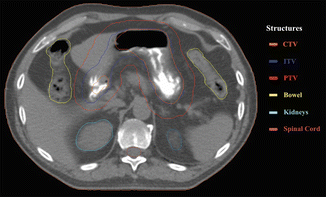

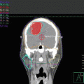
Fig. 4.3
Target delineation in our clinical case. A 4D-CT simulation was acquired to take into account gastric movements and displacement of organ at risk during respiratory phases
Radiotherapy Techniques
The optimal dose and technique of RT are currently not well defined. As already mentioned, the target volume should be limited to the stomach and perigastric nodes. In combination with reduced radiation doses, a smaller volume leads to equivalent disease control with less acute and late side effects. Excellent results were achieved after a median dose of 30 Gy in 15–20 fractions [42, 71, 87, 91]. Table 4.1 summarizes the most important experiences regarding the role of radiotherapy in gastric MALT lymphoma. A recent UK dose–response study that included extranodal lymphoma (although potential differences in efficacy at different anatomical sites have not been reported) suggested a dose of 24 Gy in 12 fractions [46]. This dose level is now accepted as a standard in most centers [22].
Table 4.1
Results of radiotherapy in patients with gastric MALT lymphoma
Reference, year | No. of patients | Treatment | RR (%) | EFS (%) | Survival (%) |
|---|---|---|---|---|---|
Schechter et al. 1998 [71] | 17 | RT to the stomach and adjacent lymph nodes: 30 Gy/20 fr (range 28.5–43.5 Gy) | ND | 27 months 100 | 27-month OS 100 |
Yahalom et al. 2002 [91] | 51 | RT to the stomach and adjacent nodes: median 30 Gy | CR 96 | 4-year FFTF 89 | 4-year OS 83 4-year CSS 100 |
Tsang et al. 2003 [84] | 10 | RT to the stomach and local nodes: 20–35 Gy | 100 | 5 years 100 | 5-year OS 100 |
Koch et al. 2005 [42] | 144 | Whole abdominal RT: 30 Gy ± 10 Gy boost | ND | 42-month FFP 88 | 42-month OS 93 |
Avilés et al. 2005 [5] | 78 | Whole abdominal RT : 30 Gy ± 10 Gy boost | CR 100 | 10-year FFP 52 | 10-year OS 75 |
Vrieling et al. 2008 [87] | 56 | Stomach + whole abdominal RT : 20 Gy + 20 Gy boost | CR 95 | ND | 5-year OS 74 10-year OS 63 10-year CSS 94 |
Wirth et al. 2013 [89] | 102 | RT to the stomach and involved nodes (61 pts) or whole abdominal RT (41 pts): median 40 Gy | CR 96 | 10-year FFTF 88 | 10-year OS 70 |
Improvements in target visualization (4D-CT scans) and radiation techniques and delivery (multi-fields 3D conformal-RT and intensity-modulated radiation therapy, IMRT) have further optimized gastric radiotherapy. Early reports suggested that 4D-CT and IMRT improve the definition of treatment volumes. Della Biancia et al. [18] analyzed 15 patients, categorized into three types depending on the geometric relationship between the planning target volume (PTV) and the kidneys. AP/PA and four-field three-dimensional conformal radiation therapy (3DCRT) plans were generated for each patient. IMRT was planned for four patients with a challenging geometric relationship. For patients without an overlap between PTV and kidneys, no benefit was shown for 3DCRT over AP/PA. However, for patients with PTVs in close proximity to the kidneys or with a high degree of overlap, the four-field 3DCRT plans were superior, reducing the volume of the left kidney receiving 15 Gy. In the selected cases, IMRT led to a further decrease in the left kidney dose as well as in the mean liver dose. This study showed that geometric relationships between the target and the kidneys should be taken into account when choosing the best beam arrangement. Van der Geld et al. [86] also showed that a reduction in kidney dose was evident with IMRT; the authors also evaluated the potential benefit of respiration-gated radiotherapy techniques, showing no differences. More recently, Watanabe et al. [88] demonstrated that both intrafractional gastric motion and interfractional variability of the stomach shape were considerable during RT and thus recommended regular verification of gastric movement and shape before and during RT in order to individualize treatment volume. Matoba et al. [50] compared a treatment planning approach using 4D-CT (plan A) and a conventional planning with a uniform margin (plan B), using dose-volume histograms of the planning target volume and the organ at risk, as well as the dose coverage of the clinical target volume (CTV) assessed by weekly online cone-beam CT during the treatment course. The mean PTV of plan A was significantly smaller than that of plan B (p = 0.008). The mean doses to the liver and heart in plan A were significantly lower than those in plan B (p = 0.02 and 0.03, respectively). The reductions of V(20Gy) of each kidney in plan A compared with those in plan B were 4.8 ± 2.4 % in the right kidney and 16.3 ± 10.4 % in the left. There was no significant difference in the dose coverage of the CTV between the plans during the treatment course. They concluded that treatment planning using 4D-CT for gastric MALT lymphoma was useful for minimizing the dose to organs at risk. In summary, in combination with treating the patient in a fasting state (to minimize stomach distension and movement), with oral contrast medium (Gastrografin), modern radiation techniques allow a homogeneous dose delivery to the stomach and perigastric lymph nodes while sparing the surrounding normal tissues such as the kidneys and liver.
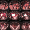
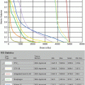
In our patient exclusive RT was utilized after progression of disease in August 2013. A CT simulation after 6 h of fasting and using oral contrast with barium sulfate was performed with patient in supine position and arms above the head. We utilized a 4D-CT to document stomach movement with free-breathing respiratory motion. The CTV encompassed the entire stomach, while the internal target volume (ITV) corresponded to CTV enlarged to account for the movement of the stomach during the respiratory phases. An isotropic margin of 10 mm was added to ITV to obtain the PTV (Fig. 4.3). A total dose of 30 Gy in 20 fractions was delivered using image-guided volumetric radiation therapy (IGRT-VMAT), as shown in Fig. 4.4. Treatment was well tolerated without any significant toxicity. PET-CT scan (Fig. 4.5) and upper endoscopy, both performed 3 months after the end of treatment, showed a complete response of the MALT lymphoma, still persisting 20 months later.
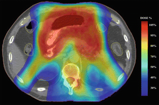
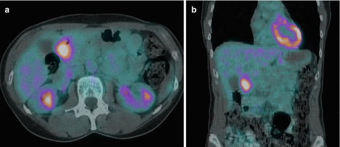

Fig. 4.4
IMRT/VMAT dose distribution generated for the RT plan. The intensive modulation plays an important role in obtaining good-quality plan for this type of case and allows a good sparing of organs at risk (e.g., kidneys, outlined with blue lines)

Fig. 4.5
Complete response at PET-CT scan 3 months after the end of radiation therapy (a) axial (b) coronal
Toxicity of Radiation Therapy
Acute toxicities are usually manageable during treatment and typically include anorexia, nausea, and vomiting. In the past, radiotherapy was often omitted because of concerns regarding the risk of perforation, hemorrhage, or late effects such as persistent renal damage. Mittal et al. [53], in a critical review of published studies in which an increased rate of gastric perforation had been described, identified that only one of the 75 gastric perforations reported (1 %) had truly occurred during or after radiotherapy. On the other hand, the risk of gastric perforation at the time of diagnosis, but before treatment, was 4 % (25 of 626 patients). It has to be noted that of the 25 patients with a gastric perforation before the starting of treatment, 16 had aggressive lymphomas and the other 9 NHL of the stomach without further subclassification. Thus an aggressive histology is probably a risk factor for perforation; the GIT NHL 01/1992 study reported no radiation-induced perforation or gastrointestinal hemorrhage among 185 patients with MALT lymphoma.
The risk of clinically significant renal damage or hypertension as a complication of primary radiotherapy of gastric lymphoma is very low. Maor et al. [48] studied 27 patients with gastric lymphoma in stage I or II, who had received at least 24 Gy of radiation to at least one third of the left kidney (unusual with current radiation volumes and dose). Only two patients developed mild hypertension (150/90 mmHg) after high-dose renal irradiation (irradiation of half, or all, of the left kidney with 40 Gy).
Summary
Radiation therapy is highly effective in the treatment of gastric MZL with excellent results in terms of complete response rate and long-term durability. A planning procedure with 4D-CT is highly recommended to account for gastric movements during the respiratory phases. GTV volume should include gross disease (if present and evaluable at CT and PET-CT scan); CTV should encompass the entire gastric volume and perigastric lymph nodes, if visible. ITV is determined in the case of 4D-CT planning. PTV volume is influenced by setup variation; we advise an isotropic expansion of 5–10 mm over the final ITV. We suggest to consider a total dose range between 24 and 30 Gy. Modern radiation techniques, including 3DCRT and IMRT, are recommended to reduce radiation of the kidneys and liver and keep the dose as low as reasonably achievable.
Ocular Adnexal Marginal Zone B-Cell Lymphomas (MALT Lymphomas)
Lymphomas of the ocular adnexa are a heterogeneous group accounting for approximately 1–2 % of all non-Hodgkin lymphomas and approximately 7–8 % of extranodal lymphomas [6, 28]. Ocular adnexa include the orbit, extraocular muscles, conjunctiva, eyelids, lacrimal gland, and apparatus. The incidence of ocular adnexal lymphoma (OAL) has increased by 6.3 % annually in the period from 1975 to 2001, more rapidly than non-Hodgkin lymphomas at other extranodal sites (Moslehi et al. [55]). Ninety-five percent to 100 % of reported cases of OAL are B-cell neoplasms, and the majority are low grade. Extranodal marginal zone lymphoma of MALT type is the most common histologic subtype of primary OAL, constituting about 35–80 % of cases [51].
Clinical Case
A 68-year-old woman was in her usual state of health until July 2014 when she first appreciated a swelling on the left eyelid. At that time, she didn’t report any symptoms, like drenching night sweats, fevers, weight loss, or pruritus. After an ophthalmologic examination, an orbit CT demonstrated a dimensional enlargement of the left lacrimal gland. A magnetic resonance imaging (MRI) confirmed a superior-lateral mass of the left orbit that originated from the lacrimal gland and that caused deviation of the superior and lateral rectus muscles (Fig. 4.6). She underwent an incisional biopsy with diagnosis of ocular adnexal MALT lymphoma (OAML). Further assessment including a complete history and physical examination, routine laboratory studies, serum protein electrophoresis, serum LDH, β2-microglobulin, chest x-ray, and CT of the chest, abdomen, and pelvis were performed. Furthermore, she was evaluated for a Chlamydophila psittaci (Cp) infection resulting negative.
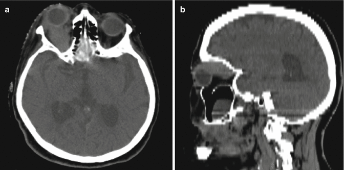

Fig. 4.6
Axial (a) and sagittal (b) view of the left orbital lesion at the MRI scan
Clinical Presentation
OAML are mostly seen in the fifth to seventh decade of life (median age, 65 years), with female predominance (male–female = 1:1.5/2). In contrast, studies in Korean populations reveal a significantly younger age (median, 46 years) at the time of diagnosis, with male rather than female predominance [15, 94].
The most frequent site of origin is the orbit (40 %), followed by conjunctiva (35–40 %), lacrimal gland (10–15 %), and eyelid (10 %) [55]. Bilateral involvement occurs in 10–15 % of cases (80 % simultaneous, 20 % sequential events).
Conjunctival lesions typically present as mobile pink infiltrates in the substantia propria (“salmon-pink patch”), causing conjunctival swelling, redness, and irritation. Orbital lymphoid proliferations are characterized by a palpable, firm, or rubbery mass causing progressive proptosis, occasionally associated with periorbital edema, decreased visual acuity, motility disturbances, and diplopia. The median interval between the onset of symptoms and time of diagnosis is variable, ranging from 1 month to 10 years (median, 7 months). This delay can be attributed to the slow evolution of symptoms, especially in conjunctival lymphomas, which may masquerade as chronic conjunctivitis and frequently show an impressive initial response to topical steroids. Steroids may mask the clinical presentation and render pathologic diagnosis more difficult, frequently resulting in a need for repeat biopsy to establish the diagnosis [76].
Pathology and Chlamydophila psittaci Infection
In the clinical case, the final pathology demonstrated OAML. OAML show similar histology and immunophenotype to MALT lymphoma in other sites. Tumor cells typically express IgM, and less often IgA or IgG, and show light chain restriction. The tumor cells of MALT lymphoma are CD20+, CD79a+, CD5−, CD10−, CD23−, CD43±, and CD11c± (weak). Infrequent cases are CD5+. The lymphoma cells express the marginal zone cell-associated antigens CD21 and CD35. Staining for CD21 and CD35 also typically reveals expanded mesh works of follicular dendritic cells corresponding to colonized follicles [79]. The most frequent translocation in OAML, observed in 15–40 % of patients, is t(11;18)(q21;q21). The t(14;18)(q32;q21), t(1;14)(p22;q34), and t(3;14)(p14;q32) translocations are found in up to 38 %, less than 5 % and 20 % of patients, respectively [76].
Cp is the etiologic agent of psittacosis, an infection caused by exposure to infected animals [11, 61]. Cp infection is detected in tumor tissue of 11 % of B-cell lymphomas [24]. The most important Cp-related tumor is OAML of the ocular adnexae, with prevalence rates between 47 and 80 % in countries like Austria, Germany, Italy, and Korea [25].
Diagnosis and Staging
The initial evaluation of patients with OAL requires careful ophthalmologic examination and adequate tissue sampling for histopathologic diagnosis. Further assessment for accurate staging and therapy planning includes a complete history and physical examination; routine laboratory studies; serum protein electrophoresis; serum LDH; β2-microglobulin; chest x-ray; CT of the chest, abdomen, and pelvis; and bone marrow biopsy (controversial). CT and MRI with contrast enhancement are the primary imaging tools in the evaluation of ocular adnexal proliferations (Fig. 4.6). They aid in the assessment of location, size, and degree of infiltration; however, they cannot reliably distinguish between benign and malignant processes. Typical imaging appearance of a lymphoid lesion is a unifocal, homogeneous, well-circumscribed lesion of isodensity to slight hyper-density, with mild to moderate contrast enhancement, and smooth, distinct borders, molding into adjacent tissues and displacing rather than infiltrating orbital structures [78].
Traditional staging of OAL has used the Ann Arbor system [13]. However, the limitations of this system for staging extranodal non-Hodgkin lymphomas, such as OAL, have long been recognized, as these show different dissemination patterns from nodal lymphomas. Under the auspices of the American Joint Committee on Cancer, a new TNM-based staging system has been developed to overcome the shortcomings of the Ann Arbor System [17].
The majority (85–90 %) of patients with OAML present with localized disease (stage I). Nodal involvement is reported in approximately 5 % of patients. In various case series, 10–15 % of patients have disseminated disease (stage IV) at initial presentation, including bone marrow involvement in approximately 5–8 % [76].
Our patient presented with disease confined to one extranodal site, and therefore, she fell classically into a stage I disease.
Treatment and Outcome
In the determination of optimal therapy, several factors must be taken into account: (1) extension of disease, within and outside the periocular region, (2) prognostic factors related to the patient and the disease, and (3) additional consideration of the functional impact of the treatment on the eye. Several standard treatment modalities are available for OAL, including surgical excision, RT, chemotherapy (single agent or combination regimens), and immunotherapy. Newer options such as radioimmunotherapy, “wait-and-see” policy, and doxycycline antibiotic therapy have been recently proposed and tested in small studies.
Surgical biopsy, mandatory to determine the histologic subtype of OAL, can be incisional or excisional, especially for localized and/or encapsulated lesions of the conjunctiva or lacrimal gland. Although a number of studies have included patients treated with surgery alone, local relapse has been reported more commonly in these patients compared with those who also received RT [15, 20, 44]. Two studies from Japan evaluated a “watch-and-wait” policy or no initial adjuvant therapy after surgical removal of a MALT lymphoma of the ocular adnexa [47, 80]. In Tanimoto series, 36 patients with a median age of 63 years were followed for a median of 7.1 years. Of these, 17 patients progressed (47 %), but only 11 actually required treatment. All others were free of progression (19/36, 53 %). In another retrospective analysis, Mannami et al. observed 12 patients with stage I OAML for a median duration of 50 months. None of the patients progressed during this time period. This strategy remains controversial but may be appropriate in frail elderly patients with asymptomatic disease or in the setting of severe comorbidities that preclude an aggressive therapeutic approach. However, even in this population, local radiation therapy can usually be given [76].
There are limited data on chemotherapy for patients with OAML. A small number of single-center, retrospective case series have included patients treated with different chemotherapy regimens, such as COP/cyclophosphamide, vincristine, and prednisone (CVP ); cyclophosphamide, Adriamycin, vincristine, and prednisone (CHOP); cyclophosphamide, vincristine, procarbazine, and prednisone (C-MOPP); and chlorambucil, either alone or in combination with other modalities [30, 52, 68, 70], but only few reports contain detailed response rates and outcomes [30, 67, 74, 81]. Chlorambucil, given in a variety of doses, schedules, and combinations, is the most frequently used chemotherapy agent and has a highly favorable toxicity profile, making it a suitable agent for the treatment of frail, elderly patients. Irrespective of chemotherapy regimens, complete responses are observed in 67–100 % of patients; however, long-term outcome data suggest that local recurrence is the predominant cause of treatment failure, occurring in up to 29 % of patients [76]. Several authors report successful salvage with RT in patients who experience local failure after initial treatment with chemotherapy [30, 74].
Phase 2 data by Conconi et al. [16], Raderer et al. [65], and Lossos et al. [45] using single-agent rituximab in previously untreated patients demonstrated overall response rates between 50 and 87 %, but median time to disease progression was less than 1 year. Only a few case reports using single-agent rituximab in patients with OAML have been published [9, 16, 27, 35]. They confirm the high activity of rituximab in both newly diagnosed and relapsed disease, but early recurrence is common, particularly in pretreated patients. An exciting new therapy has been developed in the form of the radioimmunoconjugate of 90Y ibritumomab tiuxetan, which links the specific anti-CD20 antibody to the radioactive moiety. Prospective clinical trial of 90Y ibritumomab tiuxetan for frontline treatment of stage IE indolent ocular adnexal lymphoma in 12 patients showed complete response in ten patients and partial response in the other two, with no cases of cataract, dry eye syndrome, or radiation retinopathy [21, 72].
Based on the association between Cp infection and OAML, Cp-eradicating antibiotic therapy has received attention as a novel treatment approach for this disease. Ferreri et al. [26] conducted a prospective phase 2 clinical trial of 27 patients (15 newly diagnosed and 12 relapsed) with OAML, using doxycycline 100 mg orally twice daily for 3 weeks. Cp infection was confirmed in 11 patients. Partial or complete lymphoma regression after antibiotic therapy was observed in 7 of 11 Cp-positive and 6 of 16 Cp-negative patients, with an overall response rate of 48 %. The 2-year failure-free survival rate among patients treated with doxycycline was 66 %. Given the variable prevalence of Cp infection in patients with OAML, empiric antibiotic treatment without prior testing for chlamydial infection cannot be generally recommended [33]. To shed more light on the efficacy of antibiotics in the treatment of patients with OAML, Husain et al. [36] conducted a meta-analysis, identifying four studies with a total of 42 patients who had been treated with oral doxycycline. Objective documentation of response was available for only 3 of 20 patients who reportedly achieved some response, whereas other 20 patients had stable disease and 2 progressed during antibiotic therapy. After antibiotic therapy, seven patients developed disease recurrence, six of them within 12 months of follow-up. Prospective trials with standardized objective response criteria and longer follow-up will be necessary to further evaluate the role of antibiotics in the treatment of OAML in different geographic regions.
Stay updated, free articles. Join our Telegram channel

Full access? Get Clinical Tree


