Esophagectomy


Esophagectomy


CRITICAL ELEMENTS


  • Resection of the Primary Tumor to Negative Margins


  • Lymph Node Dissection


  • Gastrointestinal Reconstruction


    • Construction of Gastric Conduit


    • Performance of a Gastric-Emptying Procedure


    • Alternate Conduits


    • Cervical Versus Intrathoracic Anastomosis


    • Hand-Sewn Versus Stapled Anastomosis


    • End-to-End Versus End-to-Side Anastomosis


1. RESECTION OF THE PRIMARY TUMOR TO NEGATIVE MARGINS

Recommendation: To achieve an optimal oncologic outcome, the proximal, distal, and circumferential esophageal margins should be free of cancer both macroscopically and microscopically. Although most surgeons attempt to divide the esophagus 5 to 8 cm proximal and 5 cm distal to the gross tumor to maximize the likelihood of a microscopically negative longitudinal margin, a firm recommendation in this regard cannot be made based on existing data. To ensure a microscopically negative proximal margin, intraoperative pathologic evaluation of the margin should be considered unless resection of additional proximal esophagus would be impossible. Intraoperative assessment of the gastric margin should also be considered when the microscopic status of the margin is of concern. Skeletonization of the esophagus should be avoided to maximize the likelihood of a microscopically negative circumferential margin as the status of this margin cannot be effectively assessed intraoperatively. In all cases, the goal of negative margins should be balanced with the ability to complete the operation safely and to reestablish gastrointestinal continuity successfully.


Type of Data: Retrospective data.

Grade of Recommendation: Strong recommendation, low-quality evidence.


Rationale

Multiple factors may affect the survival of patients who undergo esophagectomy. Among them, the status of the proximal, distal, and circumferential surgical margins is recognized as particularly important.1 Because the esophagus lacks a serosal layer, cancer of the esophagus can infiltrate into important adjacent structures such as the pericardium, heart, great vessels, trachea, and lung. Cancer of the esophagus is also characterized by microscopic submucosal extension, which may not be apparent grossly at the resection margins. To achieve an optimal oncologic outcome, proximal, distal, and circumferential margins must be free of cancer both macroscopically and microscopically (R0). However, no scientific evidence or published guideline supports the acquisition of any length of tissue between cancer cells and the inked margins beyond an R0 resection. Likewise, there is little evidence to support one surgical technique over another to ensure macroscopically and microscopically negative margins.


Proximal Margin

Few articles report on the prognostic value of the status of the proximal resection margin. Mirnezami et al1 showed that the incidence of microscopically positive proximal/distal resection margins was 7% and was a robust predictor of survival. Barbour et al2 retrospectively examined reports of 352 patients with Siewert I, II, or III adenocarcinoma in an effort to determine whether length of esophageal resection or operative approach influenced outcome. Patients had undergone complete gross resection without neoadjuvant therapy. Proximal margin lengths were recorded ex vivo as the distance from the gross tumor edge to the esophageal transection line. Their results indicated that a 3.8-cm (approximately 5 cm in situ) clear proximal margin was an independent prognostic factor. Mariette et al3 showed that, of 94 patients who underwent esophagectomy, 8 had a positive proximal resection margin with a median survival of 11.1 months versus a survival of 36.3 months in the group that had a negative proximal margin (P = 0.02). There was infiltration of tumor cells in two patients with proximal margin >6 cm, but no infiltration was observed in patients with margins >7 cm. Tam et al4 reported a prospective study on 95 esophagectomies and found a 20% anastomotic recurrence when the clear margin was <5 cm, an 8% recurrence rate when the clear margin was between 5 and 10 cm, and 0% recurrence when the clear margin was >10 cm. Hennessy and O’Connell5 reported a series of 62 esophagectomies in which a clearance of 5 cm was attempted and found that the macroscopically negative proximal resection margin was microscopically positive in 15% of cases. Migliore et al6 performed a systematic review that included many of the studies described above. Their analysis determined the optimal proximal margin should be at least 8.5 cm from the apex of the tumor.6

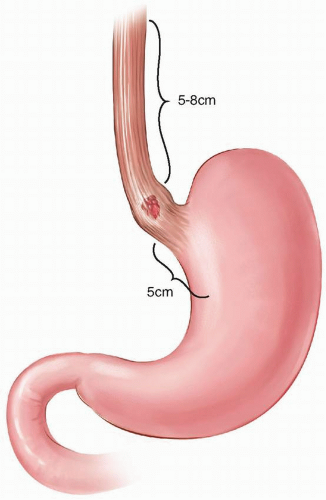
Based on these data, most surgeons generally attempt to achieve a gross proximal margin of 5 to 8 cm in an attempt to maximize the likelihood of a microscopically negative margin (Fig. 7-1). However, in all cases, the surgeon must balance the goal of
a negative margin with the ability to complete the operation safely and to reestablish GI continuity. In this regard, intraoperative pathologic evaluation of the proximal margin should be considered unless resection of additional proximal esophagus would be ill-advised.


FIGURE 7-1 A proximal distance of 5 to 8 cm and distal length of 5 cm from the respective tumor edge maximizes likelihood of a microscopically negative margin.


Distal (Gastric) Margin

Even fewer authors have considered the significance of a positive distal resection margin. In 2007, DiMusto and Orringer7 demonstrated that 1.8% of 1,044 patients undergoing transhiatal esophagectomy had a positive distal resection margin. They showed that transection of the esophagus 4 to 6 cm distal to the palpable esophageal mass led to a histopathologically negative margin in 98% of cases. Casson et al8 argued that to consistently achieve a negative distal resection margin, at least 5 cm of macroscopically normal foregut below the distal margin of the primary tumor is necessary (Fig. 7-1). They concluded that there was a trend toward reduced postoperative survival for patients with a histologically positive distal resection margin, in particular for patients with involvement of the cardia (median, 5.7 vs 15.4 months if negative; P = 0.0001).


Intraoperative frozen-section evaluation of the distal gastric margin should be considered when the status of the margin is of concern based on intraoperative findings or tumor location. In such cases, the surgeon should be prepared to perform a proximal or total gastrectomy and have a strategy for an alternate conduit (see “Alternate Conduits”).


Circumferential (Radial) Margin

The College of American Pathologists (CAP) defines a positive circumferential margin as the presence of esophageal cancer directly at the inked margin. However, the United Kingdom Royal College of Pathologists (RCP) defines a positive circumferential margin as the presence of esophageal cancer at or within 1 mm of the inked margin.9,10 A meta-analysis of 14 cohort studies, including 3,566 patients with resectable esophageal cancer, found the overall 5-year mortality rates were higher for patients with a positive circumferential margin compared with rates of those with a negative circumferential margin.11 Rates of margin positivity were 15.3% and 36.5%, according to the CAP and RCP criteria, respectively. Overall, 5-year mortality rates were significantly higher in patients with a positive circumferential margin compared with those who had a negative circumferential margin according to both (odds ratio [OR], 4.02; P <0.001) and RCP (OR, 2.52; P <0.001) criteria. Wu et al12 performed a systematic review and meta-analysis to assess the prognostic significance of a positive circumferential margin on overall survival in patients with esophageal cancer and found that a positive circumferential margin was associated with a lower survival rate in patients with stage T3 disease and in patients receiving neoadjuvant therapy.

Because a negative circumferential margin has an impact on survival of patients with esophageal cancer, one would reason that surgery should be conducted by using a wider resection that includes the healthy tissue surrounding the lesion. An en bloc esophagectomy, as classically defined by Altorki et al,13 was developed on this principle. In this procedure, the tumor is excised within a wide envelope of surrounding tissue, including both pleural surfaces laterally and the thoracic duct posteriorly. For tumors that traverse the hiatus, the envelope of tissue includes a 2.54-cm (1-inch) circumferential cuff of diaphragm as well.13 Lymph node dissection includes all lymphatic and areolar tissue between the tracheal bifurcation and the celiac axis, including the retroperitoneal, common hepatic, and splenic nodal tissue.13 The GI tract is divided with 8- to 10-cm proximal and distal margins.13

Altorki et al13 and Skinner et al14 compared results of en bloc esophagectomy versus standard esophagectomy in patients with stage III disease. These studies showed that a standard esophagectomy had a narrower circumferential margin, a less radical lymphadenectomy, and a lower median survival (standard, 12 months; en bloc, 27 months; P <0.001).13,14,15,16 Some studies have reported reduced local recurrence with en bloc (1%,17 8%,18 5%19) in comparison with standard esophagectomy (21%,20 25%,21 35%,22 and 38% with surgery alone when performed as described in the Chemoradiotherapy for Oesophageal Cancer Followed by Surgery Study [CROSS]23). However, there is significant selection bias at play and potential stage migration. This remains a controversial topic, and insufficient data exist to support a specific technical approach. At present, attention should be paid to excision of a generous circumferential margin by avoiding skeletonization of the esophagus, especially at the level of the tumor.


Technical Aspects

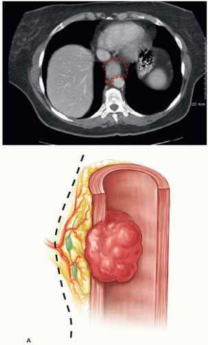
During mobilization, the gastrohepatic ligament is incised, taking a wide breadth to maximize the circumferential margin. The peritoneum overlying the hiatus is incised and the crural pillars and esophagogastric junction identified. Some surgeons advocate taking a few millimeters or more of the diaphragmatic hiatus, at least including the peritoneal lining of the crura, again for a wide circumferential margin. The esophageal mass is palpated and assessed for resectability and mobility to ensure that it is not fixed to the prevertebral fascia, aorta, or surrounding mediastinal structures. The stomach is further mobilized (Fig. 7-2A and B; also see Construction of Gastric Conduit).

A lymphadenectomy at the celiac axis is performed (see Lymph Node Dissection). Retractors can be placed into the hiatus to allow ligation of the periesophageal tissues to the level of the carina under direct vision. At least 10 cm of the distal esophagus can be mobilized under direct vision. This is also a critical step in preserving the circumferential margin. The maneuver is facilitated by retracting the esophagus from one side to the other in the lower mediastinum to create tension on the tissues.

Here exists a critical point in the operation. Local invasion into the diaphragm or nonvital mediastinal structures (pericardium, thoracic duct) should prompt surgical resection of these structures to achieve a negative circumferential margin. If the tumor is immobile and/or fixed to surrounding structures (aorta, airway, prevertebral fascia), then resection should be aborted. The esophagus is divided 5 to 8 cm from the apex of the tumor, either sharply or by firing a stapler (Fig. 7-1), and the margins are inspected grossly. Areas for concern on margins may be sent for immediate histopathologic evaluation at the time of operation to ensure an R0 resection, as detailed previously.


2. LYMPH NODE DISSECTION

Recommendation: Radical dissection of all the lymphatic tissue in the periesophagogastric, mediastinal, and celiac axis distributions should be performed regardless of current stage, use of induction therapy, or surgical approach.

Type of Data: One randomized controlled study, multiple retrospective cohort studies.

Grade of Recommendation: Strong recommendation, moderate-quality evidence.


Rationale

Lymph node involvement in the setting of resectable esophageal cancer has been shown to be an indicator of poor prognosis. To date, the majority of data on extent of lymph node dissection and number of lymph nodes removed for esophagectomy for esophageal cancer have been generated from large retrospective case series by highvolume centers. The only randomized data available are a study by Hulscher et al,24 who compared transthoracic esophagectomy to transthoracic esophagectomy with extended en bloc lymphadenectomy in a study of 220 randomly assigned patients. This trial demonstrated that even with an en bloc lymphadenectomy the increased number of nodes dissected did not result in a statistically significant improvement in disease-free or overall survival. Other large, single-center experiences have been able

to consistently demonstrate 5-year survival rates between 25% and 30%.25,26,27 These series mostly involve two-field (thoracic and abdominal) lymph node dissections or sampling through either a transhiatal or transthoracic approach. Altorki et al28 demonstrated that a three-field approach with associated three-field lymph node dissection (cervical, thoracic, and abdominal) can be safely performed with low mortality and 5-year survival rates approaching 50%. These outstanding results have not been replicated in other centers, limiting the applicability of these data.


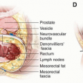
FIGURE 7-2 Circumferential dissection plane of the esophagus during esophagectomy for malignancy. A: Axial view of computed tomography scan (top) and illustration (bottom) show tumor in the esophagus and the suggested resection plane (black dotted line) to facilitate negative radial margin.


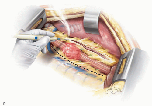
FIGURE 7-2 (continued) B: Illustration shows suggested resection plane to include mediastinal and periesophageal tissue but to avoid trauma to the adjacent structure, such as the airway, to obtain negative radial margins.

The number of nodes needed to resect for optimal prognostic value has also been intensely debated, and recently Rizk et al29 attempted to quantify the number of nodes that should be resected by reviewing the Worldwide Esophageal Cancer Collaboration database, which contains patient data on esophagectomy for cancer from a consortium of institutions. Based on their review of data from more than 4,000 patients, they recommended that the number of nodes resected be based on the T stage of the cancer: 10 nodes for pT1, 20 nodes for pT2, and >30 nodes for pT3/4.29 This study was limited in that there was no standardized protocol for extent of lymphadenectomy and it was highly variable. There was also no standardized protocol for pathology review, so the true number of lymph nodes resected may not have been known for many cases.

Given the heterogeneity in technique of lymphadenectomy, in addition to the optimal number of lymph nodes resected and the specific lymph node stations dissected,
consensus regarding the optimal approach to and extent of lymphadenectomy has not yet been achieved.

Despite the suggested “minimum” number of lymph nodes, the current recommendation by the National Comprehensive Cancer Network (NCCN) for resected esophageal adenocarcinoma with positive lymph node involvement in patients who have been treated with neoadjuvant chemotherapy and radiation is surveillance, regardless of the number of resected lymph nodes. For those patients who have not received preoperative therapy, the recommended treatment is postoperative chemotherapy and radiation. Patients with resected squamous cell cancer who have positive lymph nodes at time of resection are treated with surveillance, regardless of whether the patient has received neoadjuvant therapy.


Technical Aspects

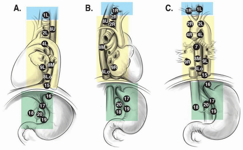
There are three primary nodal fields from which lymph nodes may be excised during esophagectomy: the abdominal field, the mediastinal/thoracic field, and the cervical field. The lymph node fields correspond to the course of the esophagus, distally from the abdomen, through the mediastinum, and into the neck. The extent of dissection and the number of fields examined are very much dependent on the operative approach. In the following section we describe the anatomic considerations for each of the lymph node fields (Fig. 7-3).


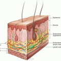
FIGURE 7-3 Regional lymph node stations for staging esophageal cancer. Cervical (blue), mediastinal/thoracic(yellow) and abdominal (green) lymph nodes. Two field lymphadenectomy includes the mediastinal/thoracic and abdominal lymph nodes while three field lymphadenectomy includes the cervical, mediastinal/thoracic and abdominal lymph nodes. The eighth edition of the AJCC uses the number of metastatic lymph nodes from any of the fields as the basis for N staging. A = left sided, B = right sided, C = anterior


Abdominal Field

The abdominal lymph node field consists of a broad range of nodes that extend along the lesser curve of the stomach, which include nodes along the celiac artery, left gastric artery, splenic artery, and common hepatic artery. During gastric mobilization, nodes along the retroperitoneum and the diaphragm should be included in the dissection.


Mediastinal/Thoracic Field

The mediastinal/thoracic field includes the nodes along the length of the thoracic esophagus and within the mediastinum. Included within this dissection are the subcarinal nodes as well as nodes associated with the thoracic duct. Paratracheal and paraesophageal lymph nodes above the carina may also be resected from the right thoracotomy approach.


Cervical Field

The addition of this “third field” was first suggested by Japanese surgeons who discovered that up to one-third of patients had recurrences in this lymph node station after a two-field lymphadenectomy. The cervical field includes the nodes along the upper esophagus and nodes that course along the recurrent laryngeal nerve as it moves into the neck. Nodes along the jugular chain and supraclavicular region are also included in this dissection.


3. GASTROINTESTINAL RECONSTRUCTION


3A. CONSTRUCTION OF GASTRIC CONDUIT

Recommendation: Existing evidence does not support gastrointestinal reconstruction using any specific width of gastric conduit. Irrespective of the specific technique chosen, the submucosal collateral blood supply to the gastric conduit should be preserved to minimize the risk for anastomotic leak.

Type of Data: Retrospective studies.

Grade of Recommendation: Weak recommendation, low-quality evidence.


Rationale

The stomach is the most common and reliable conduit for reconstruction after esophageal resection. Attention to the technical aspects of conduit formation may minimize the complications of anastomotic leak and conduit necrosis. Several critical steps in gastric conduit preparation can be used to minimize ischemia, anastomotic leak, and conduit necrosis as well as to obtain optimal function of the conduit.


Vascular Anatomy of the Gastric Conduit

To achieve an adequate lymphadenectomy when performing an esophagectomy for an esophageal or gastroesophageal junction malignancy, the surgeon must divide the left gastric artery at its origin from the celiac trunk. Division of the short gastric arteries and the left gastroepiploic vessel is also required to facilitate mobilization
of the stomach into the chest or neck for anastomosis with the proximal esophagus. Therefore, with two of the four major vessels supplying the stomach divided, the most proximal end of the mobilized stomach is always relatively ischemic. Previous anatomic studies investigating the vasculature of cadaveric adult stomachs with angiography have demonstrated that the arterial supply to the gastric conduit is derived entirely from the caudal end of the conduit.30,31,32,33,34,35 The right gastroepiploic arcade provides the vast majority, if not all, of the arterial inflow to the conduit. Both the celiac axis through the gastroduodenal artery and the superior mesenteric artery through the pancreaticoduodenal arteries supply the gastroepiploic artery. The right gastric artery also provides inflow to the conduit from the celiac axis through the hepatic artery. The left gastroepiploic arcade and short gastric vessels arise from the splenic artery. Retrograde flow from intramural and highly variable anastomoses between the right and left gastroepiploic arcades may play a role in perfusion of the cranial aspect of the gastric conduit.32,35

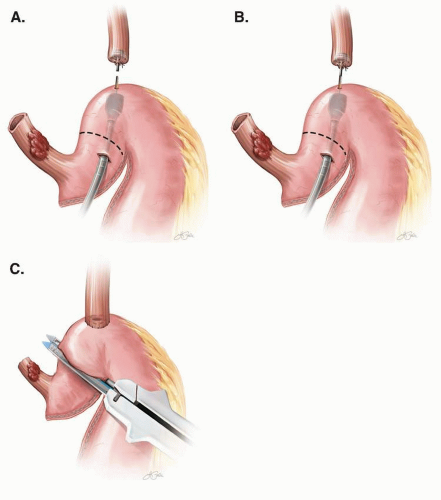
The optimal morphology of the gastric conduit remains a topic of debate. The entire stomach may be used as an esophageal substitute, or the stomach can be fashioned into a “wide” or “narrow” gastric tube. Anatomic cadaveric studies indicate that whole-stomach conduits have a more robust vascular supply because of preservation of intramural collateral flow. The definitions of wide and narrow in terms of conduit diameter or width remain ill-defined and vary in the literature. When a gastric tube is created (Fig. 7-4), the width of the conduit is typically measured from the greater curve to the line of division of the stomach on the lesser curve. A tube that measures <4 cm in width may disrupt intramural gastric collateral circulation in some patients sufficiently to compromise flow to the cranial third of the conduit. Resection of a portion of the lesser curve that maintains a conduit width of at least 4 to 5 cm preserves reliable collateral flow that conduit perfusion is maintained. Therefore, a narrow conduit may be best defined as a width <4 cm, whereas a wide conduit would measure at least 4 to 5 cm from the greater curve.

Leakage from the esophagogastric anastomosis remains one of the most common and potentially devastating major complications reported after esophagectomy.
Technical errors and poor perfusion of the gastric conduit at the anastomotic site likely account for the majority of these leaks. Based on the previously mentioned anatomic studies, inadequate perfusion of the gastric conduit and resulting anastomotic leaks may be related to the manner in which the conduit is fashioned.30,31,32,33,34,35 However, only a limited number of studies have assessed the potential association of gastric conduit width and anastomotic leak.


FIGURE 7-4 A conduit width that is <4 cm in diameter increases the preservation of submucosal blood supply and viability.

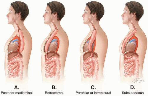
Two prospective randomized studies assessed the association of gastric conduit width and anastomotic leak, and both failed to note a significant difference between wide and narrow conduits. Tabira et al36 carried out a prospective randomized trial comparing use of a subtotal stomach (n = 22) with a narrow gastric conduit (n = 22) for reconstruction after esophagectomy for the management of esophageal cancer. A McKeown approach with retrosternal passage of the conduit and a onelayer, hand-sewn cervical anastomosis was performed in all patients. The subtotal stomach conduit was created by resecting the proximal half of the lesser curvature and cardia, which likely results in a wide gastric conduit. The narrow conduit was 3 cm. Minor leaks, all of which were treated conservatively, occurred in six patients (13.6%). No significant difference was noted between cohorts in the anastomotic leak rate (subtotal, 4.5%; narrow, 22.7%; P = 0.19). In a second prospective randomized study, Zhang et al37 compared outcomes after esophagectomy and reconstruction with a whole-stomach conduit (n = 52) and narrow gastric tube conduit (n = 52). An open McKeown approach was used for all proximal tumors with a hand-sewn cervical anastomosis (whole, 15.4%; narrow, 9.6%). An open Ivor Lewis approach was used for distal tumors (whole, 84.6%; narrow, 90.4%), and a circular stapler was used for the intrathoracic anastomosis. All conduits were brought up through the posterior mediastinum. There was no significant difference between the two groups in approach or anastomotic technique. The line of division for the whole-stomach cohort was not clearly defined. The narrow gastric conduit was 3 to 4 cm in width. No difference in the anastomotic leak rate was found (8% of anastomoses leaking in each group).

Four additional, larger retrospective series were identified, reporting conflicting results. Shu et al38 reported the results of 850 patients who underwent esophagectomy with an open McKeown approach and a two-layer, hand-sewn cervical anastomosis. In this large retrospective study, the whole stomach was used in 397 patients and a tubular stomach measuring 4 to 6 cm in diameter in 453 patients. The groups were comparable in terms of tumor stage and location. Anastomotic leak rates were significantly higher in the whole-stomach cohort (9.3% vs. 5.5%; P = 0.033). In another retrospective study from China, Shen et al39 reported the outcomes of 259 patients undergoing minimally invasive McKeown esophagectomy using a narrow (n =126) or wide gastric conduit (n = 133). Anastomotic leaks occurred more frequently when a wide conduit was used (17.3% vs. 8.7%; P = 0.041). Collard et al40 performed a retrospective review of 212 patients undergoing esophagectomy for both benign and malignant indications using a variety of approaches and conduit routes—posterior mediastinal, retrosternal, or intrapleural. The authors compared the results of a whole-stomach conduit to those with a tubular gastric conduit. However, the dimensions of the gastric tube were not reported. Contrary to the results from Shu et al,38 the incidence of anastomotic leak was significantly less when the
whole stomach was used as a conduit (1% vs. 7.9%; P = 0.021). Subgroup analysis by route of the conduit revealed consistent results favoring a whole-stomach conduit. Finley41 retrospectively reviewed single-institution outcomes of 295 patients treated with esophagectomy for management of malignant disease using a transhiatal approach with a 4-cm gastric tube, using a McKeown approach with a whole-stomach conduit and a thoracoabdominal approach with anastomosis to the “distal stomach.” Cervical anastomoses were performed with the transhiatal and McKeown approaches, whereas an intrathoracic anastomosis was used with the thoracoabdominal approach. The majority of patients had a gastric-emptying procedure with either a pyloroplasty or pyloromyotomy. Anastomotic leak rates were similar irrespective of operative approach or conduit width (tubular/transhiatal, 16%; whole stomach/McKeown, 22%; distal stomach/thoracoabdominal, 14%; P = not significant [NS]).

Strong evidence is lacking to support a recommendation of use of either a wholestomach gastric conduit or a wide 4- to 5-cm tubularized conduit. There is limited evidence supporting the use of a tubularized narrow conduit <4 cm in size. Regardless of the technique chosen, the submucosal collateral blood supply should be preserved to minimize anastomotic leak.


3B. PERFORMANCE OF A GASTRIC-EMPTYING PROCEDURE

Recommendation: A gastric-emptying procedure should be performed routinely at esophagectomy. Existing evidence does not support the exclusive use of any single procedure.

Type of Data: Retrospective.

Grade of Recommendation: Weak recommendation, low-quality evidence.


Rationale

Delayed gastric emptying (DGE) is a relatively common cause of morbidity after esophagectomy and is strongly associated with postoperative respiratory complications, primarily aspiration pneumonia.42 Typically both proximal vagus nerves are divided during standard esophagectomy, significantly altering the gastric motor activity and causing both impaired relaxation and contraction of the conduit. However some intrinsic motor activity of the stomach appears to recover at least partially over time.43,44 Emptying of the stomach in its normal anatomic position is thought to be facilitated by the positive pressure of the abdomen. However, transposition of the gastric conduit into the chest results in a shift to a negative-pressure environment, which may also alter transit through the stomach.45 Gastric emptying may be facilitated by a transient increase in intraluminal pressure resulting from passage of a food bolus through the conduit. In general, however, the neo-esophagus is thought to be an inert conduit that allows passage of oral intake primarily by the forces of gravity.

Several aspects of conduit preparation may have an effect on emptying and have been studied, particularly conduit width and employment of a drainage procedure. Some authors have proposed that a narrower conduit may result in improved gastric-conduit emptying by generating more intraluminal pressure.46,47 Additionally,
gastric-emptying procedures have been proposed to decrease the resistance at the level of the pylorus, thereby facilitating emptying of the denervated conduit. Despite the literature on these topics, definitive answers have not emerged.

Few studies have investigated the association of gastric conduit width and DGE. Variability in operative technique, definition, and manner in which DGE was assessed and the interval between esophagectomy and assessment of gastric emptying make it difficult to draw any sound conclusions. Three of the previously mentioned studies also reported the incidence of DGE they encountered. Zhang et al37 observed no difference in the incidence of DGE when a narrow 3- to 4-cm conduit (6%) or wholestomach conduit (6%) was used in their prospective randomized study with routine pyloroplasty. Shen et al39 reported only one case of DGE in their narrow conduit cohort (conduit, 3 cm; n = 126), whereas none of the patients in the wide-conduit cohort (conduit, 5 cm; n = 133) developed symptoms of delayed emptying after minimally invasive McKeown esophagectomy without a pyloric emptying procedure. Although Finley et al41 did not observe a significant difference in anastomotic leaks when comparing approach and conduit width in their single-institution experience (n = 295), they did find a higher incidence of DGE in six patients (11%) after Ivor Lewis esophagectomy using a whole-stomach conduit, nine patients (4%) after a transhiatal approach with a 4-cm conduit, and in one patient (5%) after a thoracoabdominal approach with anastomosis to the distal stomach (P <0.05). None of these studies provided a clear definition of DGE or the manner in which this outcome was assessed.

Lee et al48 retrospectively reviewed their outcomes for 56 patients who had gastric emptying assessed with a solid-food radionuclide study after esophagectomy for malignant disease using an Ivor Lewis (n = 51), McKeown (n = 2), or transhiatal (n = 3) approach. Emptying could not be measured in six patients because of rapid passage of the meal into the duodenum. Of the 50 patients analyzed, a whole-stomach conduit was used in 34 and a tubular gastric conduit in 16. The width of tubular conduit was not reported. All patients had some form of pyloric emptying procedure (finger fracture, 46; pyloroplasty, 4). DGE was defined as 50% emptying time (T50) >180 minutes. No difference in the incidence of DGE was noted when comparing the two types of conduits (16 had a whole conduit, 47.1%; 5 had tubular conduits, 31.3%; P = 0.365). Interestingly, the incidence of DGE was significantly lower in patients studied 6 months or later after esophagectomy than in those studied earlier than 6 months postsurgery (2 [14%] had DGE at >6 months, but 19 [52.9%] had DGE at <6 months; P = 0.02). However, the proportion of patients with tubular or wholestomach conduits was not clarified in terms of early or late assessment. Bemelman et al49 performed a retrospective study of 140 patients who underwent esophagectomy with reconstruction using a whole-stomach conduit (n = 40), distal two-thirds or wide gastric conduit (n = 65), or narrow gastric conduit measuring 2 to 3 cm in diameter (n = 35). Cervical anastomoses were performed in those with wholestomach and narrow conduits, whereas intrathoracic anastomoses were created for those with a wide gastric conduit. None of the patients with a narrow conduit had a pyloric emptying procedure, whereas pyloroplasty was performed in 22.5% (n = 9) of those with a whole-stomach conduit and in 30.8% (n = 20) of those with a wide conduit. In this study, DGE was defined as an inability to resume a diet of solid
foods within 1 week after normal findings on an esophagram routinely performed on postoperative day 7. DGE was significantly higher when the whole stomach was used (n = 15; 37.5%) than with wide (n = 9; 13.8%; P = 0.05) and narrow (n = 1; 2.8%; P = 0.007) gastric conduits. No difference in DGE was noted comparing wide with narrow conduits (P = 0.02), nor was there a difference with or without a pyloroplasty in the whole-stomach and wide-conduit cohorts.

Multiple retrospective studies have been performed to determine if patients developed DGE less frequently if a pyloric drainage procedure was performed at the time of esophagectomy. Six retrospective studies were identified over the time period of 1990 to 2016 that directly compared outcomes of esophagectomy patients with or without a drainage procedure. A reduction in DGE was demonstrated by Cerfolio et al (2009),50 who used botulinum toxin A (Botox) (n = 221; P = 0.002), and Deng et al (2010),51 who used finger fracture (n = 78; P =0.05); however, no statistically significant difference in DGE was noted by Lanuti et al (2011)52 (n = 242; P = 0.078); Palmes et al (2007),53 who used pyloroplasty or myotomy (n = 198; P = not reported [NR]); Nguyen et al (2010),54 who used pyloroplasty (n = 140; P = NR); or Mehran et al (2011),55 who used pyloroplasty (n = 88, P = 0.219). It is not possible to determine from these retrospective studies if pyloric drainage is necessary at the time of esophagectomy to reduce DGE.

Two randomized controlled trials (RCTs) that employed pyloroplasty were also performed during this time period and published in English. Mannell et al (1990)56 (n = 40; P = 0.1) found no difference in DGE on long-term follow-up, whereas Fok et al (1991)57 (n = 200; P = 0.001) did demonstrate a significant difference in emptying. Two meta-analyses examined the question of pyloric drainage, and both concluded there was a nonsignificant trend toward improved DGE with drainage.58,59

Several confounding factors were then considered while reviewing the above studies. First, tubularization of the conduit has been shown to be associated with a reduced incidence of DGE.50,54 The methods in the aforementioned studies were not consistently using whole stomach or tubularized stomach; therefore, it is difficult to compare the studies directly. Secondly, the definition of DGE is not standardized. Some studies used water soluble contrast swallows from postoperative day 4 (early DGE), patient questionnaires, and nuclear medicine gastric-emptying studies. Given that DGE can improve with time, the data suggest that the most clinically relevant evaluation is validated patient questionnaires combined with objective testing done >7 days from the index procedure (late DGE). Finally, a well-designed randomized study is needed to remove some of the bias inherent in retrospective studies and give greater clarity to the discussion.

Part of the rationale for performing a pyloric drainage procedure is that providing protection for DGE early in the postoperative period may protect against aspiration and pulmonary complications. Some studies in the early 1990s showed an increase in pulmonary complications without drainage: Fok et al57 found that 4% had complications without drainage, whereas Mannell et al56 reported 15% experienced complications. No patients in either study who had drainage experienced complications. However, more recent studies have not demonstrated an increase in pulmonary complications related to a lack of drainage: those with pulmonary complications along
the drainage-no drainage divide included studies from Mehran et al55 (34% vs. 32%), Cerfolio et al50 (17% vs. 22%), and Palmes et al53 (16% vs. 13%). These studies reported perioperative mortality rates of 1% to 3%, which included patients who died of pulmonary complications.

Because the incidence of DGE in this setting is estimated to be 15% to 25% and the complications can be devastating, a gastric-emptying procedure should be performed routinely at the time of esophagectomy. However, existing evidence does not support the exclusive use of any single procedure.


Technical Aspects


Botox Injection of the Pylorus

At the outset, 100 units of Botox are dissolved in 4 mL of saline, and 1 mL of this is primed into an endoscopic needle. A standard adult endoscope combined with an endoscopic 25-gauge needle is then used to inject 1 mL of Botox into each of the four quadrants of the pylorus. A 1-mL saline “chaser,” or follow-up injection, can be used to deliver the last milliliter. Botox treatments usually last 3 months, and patients may require more treatments in the future.


Pyloromyotomy

During the initial operation, the serosa at the gastroduodenal junction is scored transversely. The pylorus muscle is then divided. If there is any breach of the mucosa, then conversion to pyloroplasty is recommended. Overlaying an adjacent patch of omentum on the suture line to buttress the area of the myotomy is simple to do.


Pyloroplasty

Pyloroplasty can be accomplished laparoscopically or by open approach. After an esophagectomy and cervical esophagogastric anastomosis, the pylorus typically comes to rest 3 to 4 cm below the level of the hiatus. If done at a second operation, the pylorus is exposed by applying a liver retractor and performing lysis of adhesions until the hiatus is exposed. Great care is required to prevent injury to the right gastroepiploic vascular arcade. A 3- to 4-cm longitudinal transmural incision is made across the pylorus. A Heineke-Mikulicz pyloroplasty is then performed to close the gastrotomy in a transverse fashion.


Key Question: Esophagectomy

What constitutes optimal management of T1N0 esophageal adenocarcinoma?


INTRODUCTION

Esophageal adenocarcinoma remains the fastest increasing cancer in the United States and develops as a consequence of gastroesophageal reflux disease and Barrett’s esophagus. Increasingly, patients with early stage disease are being identified, either fortuitously related perhaps to more liberal use of upper endoscopy or as part of a Barrett’s surveillance program. In contrast to combination therapy with chemotherapy or concurrent chemoradiotherapy followed by esophagectomy, which has been demonstrated to improve survival in local and regionally advanced tumors,91,92 the optimal treatment strategy for early stage disease is still being defined. A great majority of those with early stage esophageal cancer will be cured of their disease. The introduction of endoscopic resection and ablation techniques has revolutionized the therapy for superficial esophageal cancer and allowed organ preservation rather than esophagectomy in appropriate patients.93 Therefore, a critically important concept guiding therapy is that long-term QOL and alimentary satisfaction are necessary considerations in these patients.

From an oncologic standpoint, endoscopic eradication therapy can be applied safely only when patients are at low risk of lymph node metastases.94,95,96,97,98,99,100,101,102,103,104,105 Unfortunately, typical staging studies, such as endoscopic ultrasound (EUS) and CT with positron-emission therapy (CT-PET), are of limited utility in patients with superficial tumors106,107,108,109; therefore, the risk of nodal metastases must be evaluated on the basis of tumor characteristics including size, depth of invasion, degree of differentiation, and presence of lymphovascular invasion. The decision for endotherapy or esophagectomy also should depend on patient factors, including the length of the Barrett’s esophagus segment, severity of gastroesophageal reflux, presence of comorbid conditions, and patient compliance and willingness to undergo multiple procedures.93,94,96,110 The following sections will review the evaluation, treatment, and outcomes in patients with T1 esophageal adenocarcinoma and serve to guide clinicians in the selection of the most appropriate therapy for each patient.


METHODOLOGY

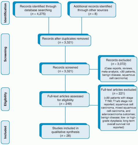
The English-language literature was queried via PubMed to include articles published between 1990 and 2016. Search terms were generated from the key statements: “Endoscopic eradication therapy results in equivalent overall survival when compared to esophagectomy for clinical T1aN0 esophageal adenocarcinoma,” and “Esophagectomy provides better overall survival than endoscopic eradication therapy for cT1b esophageal adenocarcinoma.” Specific search terms included adenocarcinoma, Barrett esophagus, esophageal neoplasms, ablation techniques, catheter ablation, cryosurgery, esophagoscopy, photochemotherapy, and esophagectomy (see Fig. 7-12). No RCTs comparing endoscopic eradication therapy with surgery for clinical T1N0 esophageal adenocarcinoma or subgroups (T1a or T1b) were found with this strategy. Filters were
then applied to include nonrandomized clinical trials including retrospective case series and comparative studies. Abstracts were reviewed and included if they met criteria of adenocarcinoma in all or most of the cohort, had >30 patients, provided a comparison between endoscopic eradication therapy and surgery, and described long-term outcomes after treatment with either endoscopic eradication therapy or surgery. Studies were excluded if they were case reports or reviews, contained cohorts of either all or mostly squamous cell carcinoma, did not contain patients with invasive adenocarcinoma, or did not report either long-term overall survival or disease-related survival. Full-text review of the selected articles applied the same inclusion and exclusion criteria. Articles selected were assessed according to the Grading of Recommendations, Assessment, Development, and Evaluation (GRADE) system, and Table 7-2 was created. 111 Recommendations were made based on the strength of available evidence.


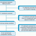
FIGURE 7-12 Preferred Reporting Items for Systematic Reviews and Meta-Analyses flow diagram: T1N0 esophageal adenocarcinoma.


TABLE 7-2 Results and Characteristics of Observational studies of T1N0 Esophageal Adenocarcinoma


Author, Year (Study Years)


Patients


Study Type


Procedure(s)


Outcomes


Results


Conclusions


Comments


Ancona et al, 200894 (1980-2006)


Total: 98; T1a: 27; T1b: 71; SM1: 36; SM2: 7; SM3:28; SCC: 67; ADC: 31


Retrospective


Ivor Lewis and McKeown


R0 resection


Lymph node metastasis


Angiolymphatic invasion


Lymphocytic infiltration


5-yr overall survival (%)


10-yr overall survival (%)


95 (97%)


T1a: 0; T1b: 20 (28%)


Absent: 53 (54%); present: 45 (45.9%)


High grade: 34 (34.7%); low grade: 64 (65.3%)


T1: 56.7; T1a: 77.7; T1b: 53.3; T1a/SM1: 67.3; SM2/3: 44.2; SCC: 43.3; ADC: 100


SCC: 37.3; ADC: 90.9


Independent predictive factors for lymph node metastasis were angiolymphatic invasion, depth of invasion, low-grade lymphocytic invasion, and neural invasion.


Survival reporting by depth of invasion was of a mixed population of SCC and ADC. Survival reported according to histologic subtype did not further segregate by depth of invasion.


Badreddine et al, 2010143 (1997-2007)


Total: 80; T1b: 80; sm1:31; sm2: 23; sm3: 26; ADC: 100%


Retrospective


Ivor Lewis: 56; THE: 24


Mean follow-up (mo)


In-hospital mortality


30-d mortality


Lymph node metastasis


LVI


Overall survival (mo)


Disease-free survival (mo)


40.5 ± 4


5 (6 %)


5 (6%)


T1b: 14 (17.5%); SM1: 4 (12.9%); SM2/3: 10 (20.4%)


T1b: 27 (34%); SM1:10 (32%); SM2/3: 17 (35%)


T1b: 46.5; SM1: 52; SM2/3: 38


T1b: 30; SM1: 35.3; SM2/3: 27.8


Superficial submucosal (sm1) invasion is a risk factor for lymph node metastasis (12.9%). Overall and cancer-free survival were not different between superficial submucosal (sm1) and deeper submucosal (sm2/3) invasion.


All cases were adenocarcinoma. Adjuvant chemoradiation was administered to five patients with metastatic lymphadenopathy.


Barbour, 2010113 (1991-2008)


Tot: 85; T1a: 35; T1b: 49; Unknown: 1; ADC: 100%


Retrospective


Ivor Lewis: 6; MIE: 73; total gastrectomy: 5; proximal gastrectomy: 1


Median follow-up (mo)


In-hospital mortality


R0 resection


Lymph node metastasis


LVI


5-yr overall survival (%)


Disease-free survival (%)


59


0


85 (100%)


T1a: 0; T1b: 8; Unknown: 1 No: 71 (83.5%); Yes: 14 (16.5%); T1a: 1; T1b: 13


T1a: 97; T1b: 65; SM1: 70 (NS); SM2: 60; SM3:71


T1a: 100; T1b: 70; SM1: 82 (NS); SM2: 60; SM3: 82


Four risk groups for lymph node metastases—I: T1a (0%); II: T1b, well/moderate differentiation/no LVI (4%); III: T1b, poor differentiation/no LVI (22%); IV: T1b with LVI (46%)


Overall and diseasefree survival reported for the T1b cohort included eight patients with nodal metastasis (N1).


Bogoevski et al, 201195 (1992-2007)


Total: 113; HGIEN: 16; T1a: 41 T1b: 56 SCC: 56 ADC: 57


Retrospective


THE (LR): 51; LR of the esophagogastric junction: 21; thoracoabdominal (ER): 41


Median follow-up (mo)


In-hospital mortality


R0 resection


Lymph node metastasis


Overall survival (all)


Overall survival T1b


39


3 (2.7%)


113 (100%)


T1a: 0; T1b: 15 (27%)


LR: 203 mo, 5-yr 67.7%; ER: 88 mo, 5-yr 57.8%


LR: 63 mo, 5-yr 51.5%; ER: 85 mo, 5-yr 53.3%


Patients with T1b submucosal invasion have a high (27%) rate of lymph node metastasis making them unsuitable for endoscopic treatment. LR esophagectomy is a valuable alternative to extended esophagectomy for T1b tumors with no difference in overall survival.


The survival outcome reported mixed ADC and SCC histologies. Survival was not reported for T1a tumors and overall survival included 16 patients with HGIEN.


Bolton et al, 200996 (1979-2007)


Total: 133; T1a: 64; T1b: 69; ADC: 133


Retrospective


Ivor Lewis: 57; THE: 63; 3-field: 7; MIE: 6


Median follow-up (mo)


R0 resection


Lymph node metastasis


LVI


3-yr overall survival (%)


5-yr overall survival (%)


42


131 (98.5%)


T1a: 5%; T1b:23%


No: 102 (77%); Yes: 23 (17%); Missing: 8 (6%); T1a: 7%; T1b: 29%


T1a: 100; T1b: 90


T1a: 90; T1b: 69


In combination with submucosal invasion, tumor length >3 cm identifies a group of patients with T1 esophageal adenocarcinoma at high risk of lymph node involvement and decreased survival.


All adenocarcinoma. Survival reported according to tumor depth included patients with nodal disease.


Estrella et al, 201198 (1997-2010)


Total: 99; T1a: 69; T1b: 30; ADC: 99


Retrospective


THE: 43; Ivor Lewis: 41; MIE: 10; 3-field: 5


Lymph node metastasis


LVI


Mean RFS (mo)


T1a: 1 (1.4%); T1b: 10 (33%)


All: 23 (23%); T1b: 18 (60%)


T1a: 108; T1b: 124


Kaneshiro et al, 2011114 (1983-2008)


Total: 185; T1a: 150; T1b: 35 (sm1 only)


Retrospective


Esophagectomy


Mean follow-up (mo)


Lymph node metastasis


Overall survival


T1a: 7.7±4.6; T1b: 7.1±4


T1a: 1 (0.7%); T1b: 3 (8.6%)


No difference between groups


No survival difference seen according to depth of invasion. T1a survival was reported according to the histologic sublayer and not in total.


Kauppi et al, 2013115 (1984-2001)


Total: 85; T1a: 35; T1b: 44; Unknown: 6; ADC: 85


Retrospective


En bloc transthoracic: 36; THE: 39; vagalsparing esophagectomy: 5; EMR: 5


Median follow-up (yr)


In-hospital mortality


R0 resection (%)


Lymph node metastasis


5-yr overall survival (%)


10-yr overall survival (%)


5-yr disease-specific survival (%)


10-yr disease-specific survival (%)


5-yr RFS (%)


10-yr RFS (%)


5


4 (5%)


Surgery: 100; EMR: 100 (number of resections: median 2, range 2-7)


T1a: 1/35 (3%); T1b: 6/44 (14%)


T1a: 73; T1b: 62


T1a: 58; T1b: 40


T1a: 92; T1b: 72


T1a: 86


T1b: 68


T1a: 89; T1b: 70


T1a: 89; T1b: 70


Recurrence of superficial ADC was the most important determinant of 5-year survival. Risk of recurrence of intramucosal tumors may be higher than reported. Beyond 5 years age-related disease is most predictive of mortality.


All adenocarcinoma. Survival reported according to tumor depth included patients with nodal disease.


Leers et al, 201199 (1985-2008)


Total: 126; T1a: 75; T1b: 51; ADC: 126


Retrospective


En bloc esophagectomy: 64; THE: 56; transthoracic esophagectomy: 6


Median follow up (mo)


LVI present


Lymph node metastasis


5-yr overall survival (%)


5-yr disease-specific survival (%)


50


T1a: 6 (8%); T1b: 22 (43%)


T1a: 1 (1.3%); T1b: 11 (22%) (P = 0.0003); SM1: 21%; SM3: 26%


T1: 78; T1a: 82; T1b:71 (P = 0.25)


T1a: 98; T1a: 78 (P = 0.001); N0: 92; N1: 70 (P = 0.001)


No significant difference in overall survival between T1a and T1b tumors. No difference in lymph node metastases between SM1 and SM3.


All ADC. Main outcome measure was prevalence of lymph node metastases. Patients undergoing esophagectomy where < six lymph nodes were harvested were excluded.


Lorenz et al, 2014100 (2000-2012)


Total: 168; T1a: 42; m1-2: 10; m3-4: 32; T1b: 126; sm1: 37; sm2: 33; sm3: 56; ADC: 168


Retrospective


En bloc esophagectomy: 130; limited esophagectomy: 30; THE: 2; MIE: 6


Median follow-up (mo)


Lymphatic invasion present


Vascular invasion present


Lymph node metastasis


Hospital mortality (%) 5-yr overall survival (%)


Tumor-specific survival (%)


64


T1a: 13 (31%); m1-2: 0; m3-4: 13 (31%); T1b: 50; sm1: 12 (24%); sm2: 12 (24%); sm3: 26 (52%)


T1a: 0; m1-2: 0; m3-4: 0; T1b: 9; sm1: 3; sm2: 1; sm3: 5


T1a: 4; m 4: 4; T1b: 26; sm1: 3; sm2: 9; sm3: 14


2.97


All: 79; m1-2: 100; m3-4: 89.2; sm1: 83.5; sm2: 67.8; sm3: 70.4 (NS)


All: 79; m1-2: 100; m3-4: 96.4; sm1: 92.3; m2: 89.2 sm3: 81 (NS)


The presence of lymph node metastasis was the most significant factor for 5-yr survival: 87.1 vs 56% (P <0.001). Lymph node infiltration was the only prognostic factor for overall survival, tumor-specific survival, and tumor recurrence in multivariate analysis. On univariate analysis, deeper submucosal infiltration (sm2,3) correlated with overall survival.


All ADC.


Manner et al, 2013130 (1996-2010)


Total: 66; T1b sm1: 66; ADC: 66


Retrospective


ER + APC: 66


Mean follow-up (mo)


CER


Number of ERs to obtain CER


Long-term remission


Metachronous neoplasias


Mean period from CER to diagnosis of metachronous neoplasias (mo)


Lymph node metastasis


5-yr overall survival (%)


47±29.1


Overall: 53/61 (86.9%); lesions <2 cm: 30/31 (97%); lesions ≥2 cm: 23/30 (77%)


2.6±2.9


Overall: 51/61 (83.6%); lesions <2 cm: 28/31 (90%); lesions ≥2 cm: 23/30 (77%)


10/53 (19%)


22


1/53 (1.9%)


84


Risk of developing lymph node metastases after EMR is lower than the risk of surgery. No tumorassociated deaths were observed.


Main outcome measure was CER in T1b ADC. After EMR, thermal ablation of the residual Barrett’s segment was done for prophylaxis of metachronous lesions in 79% of patients. All MN treated endoscopically.


Manner, et al, 2017229


Total: 62; T1b sm2: 23; T1b sm3:39; ADC: 62


Retrospective


T1b sm2—hisLR: endoscopic resection: 3; esophageal resection: 9; hisHR: endoscopic resection: 1; esophageal resection: 10. T1b sm3—hisLR: Endoscopic resection: 0; Esophageal resection: 7; hisHR: endoscopic resection: 0; esophageal resection: 7. Note: A total of 58 patients underwent esophageal resection for whom pT1b sm2 vs. sm3 status was not clarified.


Lymph node metastasis


Lymph node metastasis per histologic risk


30-d mortality of surgery


Mean follow-up with EUS after endoscopic resection (mo)


T1b sm2: 5/23 (21.7%); T1b sm3:14/39 (35.9%)


T1b sm2—hisLR: 8.3% (1/12); hisHR: 36.3% (4/11); combLR: 0% (0/5); combHR: 27.8% (5/18). T1b sm3—hisLR: 28.6% (2/7); hisHR: 37.5% (12/32); combLR: 25% (1/4); combHR: 37.1% (13/35)


1.7% (1/58)


41-43


In ADC with pT1b sm2/3 invasion, the frequency of lymph node metastasis depends on macroscopic and histologic risk patterns. The rate of lymph node metastasis appears to be higher than the mortality risk of surgery. Whether a highly selected group of pT1b sm2 patients with a favorable risk pattern may be candidates for endoscopic therapy cannot be determined until the results of larger case volumes are available.


There was no treatmentrelated mortality of ET. Rate of lymph node metastasis was analyzed depending on risk patterns—hisLR): G1-2, L0, V0; hisHR: ≥1 criterion not fulfilled; macLR: gross tumor type I-II, tumor size ≤2 cm; macHR: ≥1 criterion not fulfilled; combLR: hisLR1macLR; combHR: at least 1 risk factor.


Ngamruengphong et al, 2013116 (1998-2009)


Total: 1,618; Endoscopic—Tis: 55; T1a: 174; T1b: 39; T1, NOS: 38; Surgery—Tis: 110; T1a: 561; T1b: 523; T1, NOS: 118


Retrospective from SEER database


Endoscopic therapy: 306 (19%); ER: 149 (48.7%); ER NOS: 61 (19.9%); ER + PDT: 16 (5.2%); ER + electrocautery: 16 (5.2%); ER + laser: 15 (4.9%); ER + cryosurgery 3 (1.0%); PDT: 29 (9.5%); laser treatment: 10 (3.3%); cryosurgery: 5 (1.6%); electrocautery: 1 (0.3%); ablation, NOS: 1 (0.3%); surgery: 1,312 (81%); Partial esophagectomy + partial gastrectomy: 608 (46.3%); partial esophagectomy + gastrectomy, NOS: 210 (16.0%); partial esophagectomy: 237 (18.1%); total esophagectomy: 79 (6.0%); total esophagectomy + gastrectomy, NOS: 51 (3.9%); esophagectomy, NOS: 127 (9.7%)


Median follow-up (mo)


Overall survival for T1b vs Tis/T1a (HR)


ECSS T1b vs Tis/T1a (HR)


Overall survival for surgery vs. ET (HR)


ECSS surgery vs. ET (HR)


5-yr overall survival Tis/T1 (%)


5-yr overall survival Tis/T1a (%)


5-yr overall survival T1b (%)


5-yr ECSS Tis/T1 (%)


5-yr ECSS Tis/T1 (%)


5-yr ECSS Tis/T1a (%)


5-yr ECSS T1b (%)


Deep margin on EMR


36


1.37 (95% CI, 1.12-1.69)


1.65 (95% CI, 1.28-2.16)


Tis/T1: 1.21; Tis/T1a: 1.29; T1b: 0.97 (NS)


Tis/T1: 0.74; Tis/T1a: 0.69; T1b: 0.46 (NS)


ET: 58; surgery: 70 (P = 0.003)


ET: 60; surgery: 76 (P = 0.002)


Surgery: 66; ET: 64 (P = 0.26)


Surgery: 81; ET: 77 (P = 0.26)


Surgery: 81; ET: 77 (P = 0.26)


Surgery: 83; ET: 84 (P = 0.64)


Surgery: 89; ET: 71 (P = 0.31)


Negative: 53; positive: 26; not reported: 2


Older age and radiation therapy were associated with worse overall survival and ECSS. Submucosal invasion was significantly associated with worse overall survival and ECSS. Outcomes were not different for surgery compared with ET when stratified by depth of invasion.


All ADC. 15% received radiation therapy.


Nurkin et al, 2014117 (2001-2012)


Total: 81; Tis: 37; T1a: 10; at least T1a: 13; T1b: 6; at least T1b: 15


Retrospective


EMR: 77; ESD: 4; esophagectomy: 14


Median follow-up (yr)


5-yr overall survival (%)


5-yr disease-free survival (%)


3.25


T1a: 83


T1a: 100; T1b: 100


ER can result in a cure for T1a disease. Patients with T1b disease, low-risk features, and negative deep margins can be observed. Obtaining a negative margin on EMR was a predictor of improved 5-yr disease-free and overall survival.


Of T1b patients, only two were observed, two had esophagectomy, and two chemoradiotherapy. Series is too small to support the conclusion for T1b.


Oh et al, 2006118 (1987-2006)


Total: 78; T1a: 78


Retrospective


En bloc esophagectomy: 23; THE: 31; vagal-sparing esophagectomy: 20; transthoracic esophagectomy: 4


5-yr overall survival (%)


Lymph node metastasis (in patients undergoing en bloc resection)


Overall mortality rate


88%


1/23 (4%)


2.6%


Vagal-sparing technique had less morbidity than other forms of resection and no mortality. Survival after all types of resection was similar.


Pech et al, 2005123 (1996-2002)


Total: 66; HGD: 35; T1a: 31


Prospective


PDT-ALA


Median follow-up (mo)


Complete remission


Recurrence


5-yr overall survival (%)


5-yr disease-specific survival (%)


HGD: 37; T1a: 36


HGD: 34/35 (97%); T1a: 31/31 (100%)


HGD: 6/34 (18%); T1a: 9/31 (29%)


HGD: 97; T1a: 80


HGD: 89; T1a: 68


Local recurrence in 10 patients in the T1a group.


Pech et al, 2011119 (1996-2009)


Total: 114; T1a: 114


Retrospective


Transthoracic esophagectomy: 38; ER + APC: 76


Median follow-up (yr)


Complete remission


Major complications (%) 90-d mortality rate


Overall rate of recurrence/metachronous neoplasia (%)


Long-term remission rates allowing for repeat endoscopic therapy (%)


Disease-free survival rate


Lymph node metastasis


Esophagectomy: 3.7; ER + APC: 4.1


Esophagectomy: 38/38 (100%); ER + APC: 75/76 (98.7%)


Esophagectomy: 32; ER + APC: 0


Esophagectomy: 1/38 (2.6%); ER + APC: 0/76 (0%)


ER + APC: 6.6


Esophagectomy: 100; ER + APC: 98.7


Esophagectomy: 100%; ER + APC: 75/76 (98.7%)


Esophagectomy: 0/38 (0%); ER + APC: 0/76 (0%) on follow-up EUS


Esophagectomy was associated with a higher morbidity rate and a mortality rate of 2.6% vs. 0% mortality in the EMR cohort (difference between cohorts not significant). Recurrence rate was higher in the EMR cohort (difference not significant). Age was a prognostic factor for survival (HR, 1.13; 95% CI, 1.04-1.23 for each year, with P = 0.005).


Baseline characteristics were comparable between the two cohorts. Only significant difference between cohorts was Barrett’s length with shorter Barrett segments in the EMR cohort.


Pech et al, 201493 (1996-2010)


Total: 1,000; T1a: 1,000


Prospective


ER


Mean follow-up (mo)


Complete remission of neoplasia


Long-term complete remission rate


Recurrence of neoplasia (HGD or adenocarcinoma)/metachronous lesions


5-yr overall survival (%)


10-yr overall survival (%)


5-yr disease-specific survival (%)


56.6


963 (96.3%)


938 (93.8%)


140/963 (14.5%)


91.5


75


87.1


No mortality and major complications were <2% (bleeding, perforation). Only factor negatively associated with complete remission was presence of long segment BE (P = 0.0001). Poorly differentiated mucosal ADC (G3) was associated with significantly higher risk of recurrence and failure of endoscopic treatment than patients with well-differentiated or moderately differentiated mucosal ADCs.


Repeat endoscopic treatment was successful in 82.1%. Surgery was necessary in 3.7% after failure of endoscopic therapy.


Peyre et al, 2007120


Total: 109; HGD: 36; T1a: 73


Retrospective


Vagal-sparing esophagectomy: 49; THE: 39; en bloc esophagectomy: 21


30-d mortality (%)


Duration of follow-up (mo)


Vagal-sparing esophagectomy: 2; THE: 5; en bloc esophagectomy: 0


Vagal-sparing esophagectomy: 39; THE: 25; en bloc esophagectomy: 60


Length of hospital stay and the incidence of major complications was significantly reduced with vagalsparing esophagectomy compared with transhiatal or en bloc resection. Recurrent cancer has developed in only one patient.


Prasad et al, 2009110 (1998-2007)


Total: 178; T1a: 178


Retrospective


Endoscopic: 132; EMR alone 75 (57%); EMR+PDT 57 (43%); surgery: 46; THE: 20 (43%); transthoracic: 29 (57%)


Mean follow-up (mo)


Median number of endoscopic treatments


Endoscopic remission rate


Lymph node metastasis


Follow-up (person-yr)


Cumulative mortality


Overall mortality, incidence rate (person-yr)


Recurrent cancers


Recurrence, incidence rate (person-yr)


5-yr overall survival (%)


5-yr disease-specific survival (%)


Endo: 43; Surgery: 64


1 (IQR, 1-2)


124/132 (94%)


Surgery: 4/46 (8.6%)


Endoscopic: 464.6; surgery: 244


Endoscopic: 23/132 (17%); surgery: 9/46 (20%)


Endoscopic: 4.9/100; surgery: 3.7/100


Endoscopic: 16 (12%); surgery: 1 (2%)


Endoscopic: 5.5/100; surgery: 0.56/100


Endoscopic: 83; surgery: 95


Endoscopic: 80; surgery: 97


Cumulative mortality in the endoscopic group (17%) was comparable with the surgery group (20%) (P = 0.75). Possible predictors of recurrence: presence of residual BE (without dysplasia), increasing length of BE segment at baseline, and presence of a carcinoma detected after 6 months of surveillance for HGD.


Rice et al, 2001101 (1985-1999)


Total: 122; HGD: 38; T1a N0: 51; T1a N1: 2; T1b N0: 25; T1b N1: 6


Retrospective


Esophagectomy—THE: 75; thoracotomy/abdominal incision: 46; laparotomy: 1


Follow-up (mo)


Operative mortality


Lymph node metastasis


Overall survival (%)


Overall survival:T1a


Overall survival: T1b (%)


Number of deaths (n)


Mean: 47±41; median: 38


2.5% (3/122)


T1a: 2/122 (2%); T1b: 6/122 (5%)


5-yr: 77; 10-yr: 68


5-yr: 77%; 10-yr: 65%


5-yr: 61; 10-yr: 40 (P = 0.009)


Cancer related: 13; noncancer related: 6


Depth of invasion predicted survival on univariate analysis but on multivariate analysis, lymph node metastasis was most predictive of poor long-term survival.


Saha et al, 2009121 (2000-2009)


Total: 55; T1: 44; T1b: 11


Retrospective


Esophagectomy—Ivor Lewis: 24; THE: 20


Median followup (mo)


Operative mortality


Complications


Lymph node


metastasis


1-yr disease-free survival (%)


3-yr survival (%)


Recurrence rate (%)


44


2 (5%)


9 (20%)


2 (4%, both T1b)


95


93


7


Differentiation appears to be a marker of poor prognosis.


Sepesi et al, 2010102 (2000-2008)


Total: 54; T1a: 25; T1b: 29; SM1: 14; SM2: 11; SM3: 6


Retrospective


En bloc esophagectomy: 10; THE: 39; Ivor Lewis: 2; vagal sparing esophagectomy: 2; McKeown: 1


Median follow-up (mo)


LVI


Lymph node metastasis


5-yr overall survival, estimated (%)


Died with recurrence


42


7 (13%)


Total: 9 (17%); T1a: 0 SM1: 3/14 (21%); SM2: 4/11 (36%); SM3: 2/4 (50%)


T1a: 85; T1b: 60 (P = 0.08)


5/15 (33%)


Nodal metastasis rate was significantly different for T1a and T1b tumors but not different by depth of submucosal invasion. Nodal metastasis in submucosal tumors was high.


Sgourakis et al, 2013124 (1997-2011)


Total: 4,241; endoscopic: 2,092; LGD: 4%; HGD/CIS: 33.6%; T1a: 54%; T1b: 16%; surgery: 2,149 included ADC and SCC


Systematic review


Endoscopic therapy—EMR and/or ESD (42 studies); APC (3 studies); PDT (2 studies); RFA (2 studies); surgery—various (38 studies)


LVI


Microvascular invasion


Lymph node metastasis (11 studies)


Mean follow up (mo)


EMR resection margins (18 studies)


Endoscopic local recurrence (30 studies)


Development of metachronous lesions after endoscopic management (10 studies)


EMR and/or ESD: 0%-30%


EMR and/or ESD: 0-33%


Endoscopic—31/371 (8%); ADC: 5%; SCC: 1%; surgery—888/2,149 (41%); T1b: ADC: 26%; SCC: 45%


Endoscopic: 12-62


Positive: 294/880 (3%); ADC: 9%; SCC: 7%


0%-17%; ADC: 0.8%; SCC: 1%


2%-14%; ADC: 6%; SCC: 1%


In endoscopically managed patients, local tumor recurrence was predicted by poor differentiation and piecemeal resection; lymph node positivity by LVI. In surgically resected patients for ADC, the best predictor for lymph node positivity was LVI.


Includes both ADC and SCC. A significantly greater number of SCC patients were submitted to surgery than ADC patients. For surgically resected patients, there were significant differences in patients with positive lymph nodes between ADC and SCC.


Stein et al, 2000104 (1982-1999)


Total: 94; T1a: 38; T1b: 56


Retrospective


Esophagectomy—subtotal: 71; limited resection/jejunal interposition: 24


Median follow-up (mo)


30- and 90-d mortality (%)


Complications


Lymph node metastasis


5-yr overall survival (%)


15


Subtotal: 4.2; limited: 0


Subtotal: 31/71 (43.7%); limited: 5/24 (20.8%)


T1a: 0; T1b: 10/56 (18%)


T1a: 85; T1b: 78


No survival difference T1aN0 and T1bN0. Lymph node metastasis had a significant effect on survival.


Stein et al, 2005103 (1990-2004)


Total: 290; ADC: 157; SCC:133; HGD: 14; T1a: 82; ADC: 57; SCC: 25; T1b: 194; ADC: 87; SCC: 107


Retrospective


Esophagectomy—abdominothoracic: 146; radical transhiatal: 144


Median follow-up (mo)


Postoperative mortality


Lymph node metastasis ADC


5-yr overall survival (%)


66


5/290 (1.7%)


T1a: 0; T1b: 18/87 (20.7%)


ADC: 83.4; SSC: 62.9


Presence of lymph node metastasis and histologic tumor type were the only significant factors affecting survival favoring node negative disease and adenocarcinoma.


Tian et al, 2011144 (1995-2010)


Total: 68; all T1b


Retrospective


EMR: 68; esophagectomy: 39


In-hospital mortality


Lymph node metastasis at esophagectomy


Median survival (mo)


Overall mortality (%)


5-yr survival (%)


Risk factors for overall survival, univariate (HR, 95% CI)


1/39 (2.6%)


13 (33%)


All: 39.5 (IQR, 23.9-70.3); EMR: 34.8 surgery: 48.9 (P = 0.09)


EMR: 37.9; surgery: 25.6 (P = 0.28)


All: 69.5


Surgery: 0.4, 0.2-1.1 (P = 0.09); lymph node metastasis: 6.8, 1.4-33.8 (P = 0.02)


Patients who underwent esophagectomy after EMR lived longer but not significantly so. Most of the EMR-only mortality was in patients deemed not fit for esophagectomy.


29 patients did not undergo esophagectomy and were treated with CRT (5), continued endoscopic therapy (15), both (5), or neither (4).


Westerterp et al, 2005105


Total: 120; HGD: 13; T1: 107


Retrospective


THE


Median follow-up (mo)


Lymph node metastasis


5-yr recurrence free (%)


5-yr RFS (%)


Recurrence-free period risk for T1N0


5-yr disease-free survival (%)


44


T1a/T1b-sm1: 1/79 (1.3%); T1-sm2/3: 18/41 (44%)


T1a/T1b-sm1: 97; T1-sm2/3: 57 (P <0.0001)


T1a/T1b-sm1: 80; T1-sm2/3: 42 (significant)


T1sm2-3 HR, 7.5, (95% CI, 2.0-2.7 vs. T1m1-3/sm1)


68 (all)


N-stage was the only independent predictor of recurrence at 5 years but when excluded, depth of invasion was significant.


SCC, squamous cell carcinoma; ADC, adenocarcinoma; THE, transhiatal esophagectomy; LVI, lymphovascular invasion; MIE, minimally invasive esophagectomy; HGIEN, high-grade intraepithelial neoplasia; LR, limited resection; ER, extended resection; EMR, endoscopic mucosal resection; RFS, relapse free survival; APC, argon plasma coagulation; CER, complete endoluminal remission; MN, metachronous neoplasias; hisLR, histologically low risk; hisHR, histologically high risk; combLR, combined low risk; combHR, combined high risk; macLR macroscopically low risk; macHR, macroscopically high risk; NOS, not otherwise specified; PDT, photodynamic therapy; HR, hazard ratio; ECSS, esophageal cancer specific survival; NS, not significant; ESD, endoscopic submucosal dissection; HGD, high grade dysplasia; ET, endoscopic therapy; ALA, 5-delta-aminolevulinic acid; CI, confidence interval; IQR, interquartile range; BE, Barrett’s esophagus; LGD, low-grade dysplasia; CIS, carcinoma in situ; RFA, radiofrequency ablation; CRT, chemoradiotherapy.

Only gold members can continue reading. Log In or Register to continue

Stay updated, free articles. Join our Telegram channel

May 7, 2019 | Posted by in ONCOLOGY | Comments Off on Esophagectomy

Full access? Get Clinical Tree

Get Clinical Tree app for offline access