Abstract
Ductal carcinoma in situ (DCIS) of the breast is a heterogeneous group of lesions with diverse malignant potential and a range of treatment options. It is the most rapidly growing subgroup in the breast cancer family of disease with more than 60,000 new cases diagnosed in the United States during 2016. More than 90% of newly diagnosed DCIS are nonpalpable and discovered mammographically.
It is now well appreciated that DCIS is a stage in the neoplastic continuum in which the majority of biological alterations required for the development of invasive breast cancer are already present. The fact that DCIS was rarely observed as the sole component of a biopsy before 1980 and that it is associated with invasive breast cancer approximately 85% of the time, clearly indicate that DCIS is the precursor lesion for most invasive breast tumors. However, not all DCIS lesions have sufficient time or all the genetic alterations required for progression to invasive disease.
As our understanding of DCIS has evolved, so has its treatment. Clinical research has focused on the identification of prognostic and biological factors that strongly influence treatment selection while new surgical and radiologic techniques have developed to accommodate these new therapy options. This chapter reviews each of these subjects to provide the reader a workable framework on which to evaluate DCIS treatment decisions for a specific patient.
Keywords
ductal carcinoma, DCIS, immunohistochemical and molecular phenotypes, invasive breast cancer, microinvasion
The Changing Nature of Ductal Carcinoma in Situ
There have been dramatic changes in the past 20 years that have affected the diagnosis and treatment of patients diagnosed with DCIS. Before mammography was common, DCIS was rare, representing less than 1% of all diagnosed breast cancers. Today DCIS is common, representing 25% of all newly diagnosed cases and as much as 30% to 50% of cases of breast cancers diagnosed by mammography.
Previously, most patients with DCIS presented with clinical symptoms such as a palpable breast mass, bloody nipple discharge, or Paget disease. Today most DCIS lesions are nonpalpable and generally detected by imaging alone.
Until approximately 25 years ago, the treatment for patients with DCIS was mastectomy. Today almost 75% of newly diagnosed patients with DCIS are treated with breast preservation. In the past, when mastectomy was common, reconstruction was uncommon; if it was performed, it was generally done as a delayed procedure. Today reconstruction for patients with DCIS treated by mastectomy is common and is regularly done immediately at the time of mastectomy. In the past, when a mastectomy was performed, large amounts of skin and the nipple were discarded. Today, it is considered perfectly safe to perform a skin-sparing mastectomy for DCIS and, in most instances, nipple-areola–sparing mastectomy.
In the past, there was little confusion. All breast cancers were essentially considered the same, and mastectomy was the only treatment. Today all breast cancers are recognized as different, and there is a range of acceptable treatments for each lesion. These changes were brought about by a number of factors, most importantly increased mammographic surveillance and the acceptance of breast conservation therapy for invasive breast cancer.
The widespread use of mammography changed the way DCIS was detected. In addition, it changed the nature of the disease detected by allowing us to enter the neoplastic continuum at an earlier time with a much smaller size than seen by Ashikari and colleagues. It is interesting to note the impact that mammography had on The Breast Center in Van Nuys, California, in terms of the number of DCIS cases diagnosed and the manner in which they were diagnosed.
From 1979 to 1981, the Van Nuys group treated an average of five DCIS patients per year. Only two lesions (13%) were nonpalpable and detected by mammography. In other words, 13 patients (87%) presented with clinically apparent disease, detected by the old-fashioned methods of observation and palpation. Beginning in 1982, when new state-of-the-art mammography units and a full-time experienced radiologist were added, the number of new DCIS cases dramatically increased to more than 50 per year, most nonpalpable.
The total of 1855 DCIS patients discussed in this chapter were accrued at the Van Nuys Breast Center from 1979 to 1998, the University of Southern California/Norris (USC/Norris) Comprehensive Cancer Center (NCCN)from 1998 to 2008, and at Hoag Memorial Hospital Presbyterian from 2008 to through 2015. Analysis of all 1855 patients through 2015 shows that 1655 DCIS lesions (89%) were nonpalpable. If we look at only those diagnosed during the past 8 years at Hoag, as screening mammography has improved, 95% were nonpalpable.
Another factor that has had a significant impact on how we currently think about DCIS was the acceptance of breast conservation therapy (lumpectomy, axillary node dissection, and radiation therapy) for patients with invasive breast cancer. Until 1981, the treatment for most patients with any form of breast cancer was mostly mastectomy. However, since that time, numerous prospective randomized trials have shown an equivalent survival rate for patients with invasive cancer treated with breast conservation therapy or mastectomy. It made little sense to continue treating less aggressive DCIS with mastectomy while treating more aggressive invasive breast cancer with breast preservation. Moreover, current data suggest that many patients with DCIS can be successfully treated with breast preservation, with or without radiation therapy. This chapter discusses how easily accessible data may aid in the complex treatment selection process.
Pathology
Classification
Although there is no universally accepted histopathologic classification, most pathologists have traditionally divided DCIS into five major architectural subtypes (papillary, micropapillary, cribriform, solid, and comedo), often comparing the first four (noncomedo) with comedo. Comedo DCIS is frequently associated with high nuclear grade, aneuploidy, a higher proliferation rate, HER2/neu gene amplification or protein overexpression, and aggressive clinical behavior. Noncomedo lesions tend to be just the opposite.
However, architectural classification alone is not adequate to segregate patients into high- and low-risk categories. There is no uniform agreement among pathologists of exactly how much comedo DCIS must be present to consider the lesion a comedo DCIS. Furthermore, in our series of patients, approximately 75% of DCIS lesions had significant amounts of two or more architectural subtypes, making division by a predominant architectural subtype problematic. Although, it is clear that lesions exhibiting a predominant high-grade comedo DCIS pattern are generally more aggressive and more likely to recur if treated conservatively than low-grade noncomedo lesions, architectural subtyping alone is insufficient to precisely segregate patients by risk of recurrence. Azzopardi and colleagues recognized this in 1979.
Nuclear grade is a better biological predictor of cancer behavior than architecture and therefore has emerged as a key histopathologic factor for identifying aggressive tumors. Thus, to more accurately stratify patients by risk of recurrence, current classifications have focused on both necrosis and nuclear grade. As a result, in 1995 the Van Nuys group introduced a new pathologic DCIS classification, the Van Nuys Classification, based on high nuclear grade and the presence or absence of comedo-type necrosis.
The Van Nuys group selected high nuclear grade as one of the factors in their classification because there was general agreement that patients with high nuclear grade lesions recur at a higher rate and in a shorter time period after breast conservation than patients with low nuclear grade lesions. Comedo-type necrosis was also chosen because its presence also suggests a poorer prognosis and it is easy to recognize. Douglas-Jones and colleagues have shown that the Van Nuys system is the most reproducible of the available classifications.
The details of the Van Nuys Classification System can be found in Fig. 39.1 . There is neither a minimum nor a specific amount of high nuclear grade DCIS nor a minimum amount of comedo-type necrosis required in this classification. Furthermore, the subtleties of the intermediate-grade lesion, essential in other systems, are not important in the Van Nuys classification; only nuclear grade III cells (large, pleomorphic cells with prominent nucleoli and coarse clumped chromatin) need be recognized.
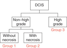
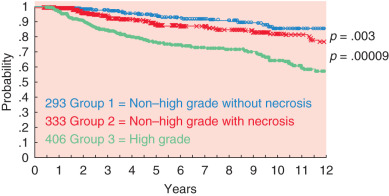
The Van Nuys classification is useful because it divides DCIS patients into three biological groups with different risks of local recurrence after breast conservation therapy ( Fig. 39.2 ). When combined with tumor size, age, and margin status, the Van Nuys Classification is an integral part of the USC/Van Nuys Prognostic Index (USC/VNPI), a system that will be discussed in detail.
Progression to Invasive Breast Cancer
Which DCIS lesions will become invasive, and when will that happen? These are the most important questions in the DCIS field today. Currently, there is intense molecular biological study regarding the progression of genetic changes in normal breast epithelium to DCIS and then to invasive breast cancer. It is now appreciated that DCIS is a stage in the neoplastic continuum in which the majority of genetic and epigenetic changes required for the development of invasive breast cancer are already present, including but not limited to proliferation, evading growth suppression, resisting cell death, replicative immortality, inducing angiogenesis, and activating invasion and metastasis.
Immunohistochemical and Molecular Phenotypes in DCIS
It has been recognized for some time that there is a substantial concordance between the nuclear grade of DCIS and its associated invasive carcinoma, such that low-grade DCIS lesions, regardless of the classification scheme used, are largely associated with lower-grade invasive carcinomas, whereas high-grade DCIS tumors are associated with higher-grade invasive carcinomas. Similarly, the frequency of specific biomarkers in DCIS varies with the grade of the lesion. Estrogen and progesterone receptors are usually expressed in low-grade DCIS but less so in high-grade lesions. In contrast, HER2/neu overexpression and elevated proliferative markers such as ki67 are more often observed in high-grade DCIS and less often in low-grade lesions. More recently, surrogate molecular phenotypes defined by immunohistochemistry have been used to identify DCIS phenotypes corresponding to luminal A, luminal B, HER2, and triple-negative/basal phenotypes in invasive breast cancer. Luminal A and B DCIS phenotypes are more frequent in the low to intermediate nuclear grade lesions, whereas HER2, triple-negative/basal phenotypes are more common among high-grade DCIS.
Microinvasion
The incidence of microinvasion was difficult to quantitate until 1997 because there was no formal or universally accepted definition of what constituted microinvasion. The first official definition of what is now classified as pT1mic disease was published in the 5th edition of the Manual for Cancer Staging read as follows: “Microinvasion is the extension of cancer cells beyond the basement membrane into adjacent tissues with no focus more than 1 mm in greatest dimension. When there are multiple foci of microinvasion the size of only the largest focus is used to classify the microinvasion (do not use the sum of the diameters of all individual foci). The presence of multiple foci of microinvasion should be noted, as it is with multiple larger invasive carcinomas.”
If even the smallest amount of invasive disease is found upon excision or mastectomy in the presence of a large DCIS, the lesion should not be classified as DCIS but as invasive cancer. In concordance with the TNM staging system, if the invasive foci is 1 mm or smaller, the tumor should be defined as a T1mic with an extensive intraductal component (EIC).
Foci of microinvasion that consist of single cells have been shown to have no impact on patient outcome whereas foci comprising cohesive groups of cells have been found to be associated with an increased rate of distant recurrence and death. In contrast to the number of cells within a focus of microinvasion, the number of microinvasive foci in a lesion have a small impact on breast cancer mortality; patients with multiple foci have a comparable outcome to patients with a single focus of microinvasion, 5.8% versus 1%.
Multicentricity and Multifocality of Ductal Carcinoma in Situ
Multicentricity is generally defined as DCIS in a quadrant other than the quadrant in which the original DCIS (index quadrant) was diagnosed. There must be normal breast tissue separating the two foci. Because the definition of multicentricity differs between investigators, the reported incidence of multicentricity also varies. Reported rates vary from 0% to 78%, averaging about 30%, have been reported. Twenty-five years ago, the 30% average rate of multicentricity was used by surgeons as a rationale for mastectomy in patients with DCIS.
In 1990, Holland and colleagues investigated the rate of multicentricity in 82 mastectomy specimens by preparing whole-organ sections every 5 mm, a variation of Egan’s subgrossing technique. Each section was radiographed, and paraffin blocks were made from every radiographically suspicious spot. In addition, an average of 25 blocks were taken from the quadrant containing the index cancer; random samples were taken from all other quadrants, the central subareolar area, and the nipple. The microscopic extension of each lesion was verified on the radiographs. The resulting data demonstrated that most DCIS lesions were larger than expected (50% were greater than 50 mm), involved more than one quadrant by continuous extension (23%), but, most importantly, were unicentric (98.8%). Only 1 of 82 mastectomy specimens (1.2%) had multicentric distribution with separate lesions in a different quadrant separated by normal tissue. This study suggested that complete excision of a DCIS lesion was possible due to unicentricity but might be extremely difficult due to larger than expected size. In an update, Holland reported whole-organ studies in 119 patients, 118 of whom had unicentric disease. This information, when combined with the fact that most local recurrences are at or near the original DCIS, suggests that the problem of multicentricity is not important in the DCIS treatment decision-making process.
Multifocality is defined as separate foci of DCIS within the same ductal system. Studies of both Holland and colleagues and Noguchi and colleagues suggest that a great deal of multifocality may be artifactual, resulting from examining a three-dimensional entity in two dimensions on a glass slide. It would be analogous to saying that the branches of a tree were not connected if the branches were cut through one plane, placed separately on a slide, and viewed in cross section. Multifocality may be due to small gaps of DCIS or skip areas within ducts as described by Faverly and colleagues and is more easily recognized when a serial sequential tissue processing technique as opposed to random sampling is employed.
Detection and Diagnosis
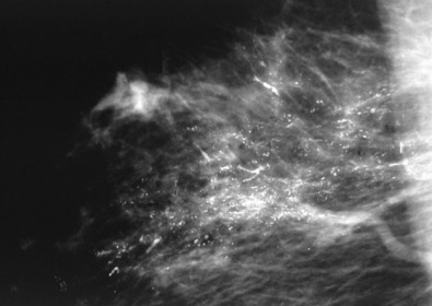
The importance of quality mammography in the identification of DCIS cannot be overemphasized. Currently, more than 90% of patients with DCIS present with a nonpalpable lesion detected by mammography. The most common mammographic finding is microcalcification, frequently clustered and generally without an associated soft tissue abnormality. More than 80% of DCIS patients exhibit microcalcifications on preoperative mammography, the patterns of which may be focal, diffuse, or ductal, with variable size and shape. Patients with comedo DCIS tend to have “casting calcifications” that are linear, branching, and bizarre and are almost pathognomonic for comedo DCIS ( Fig. 39.3 ). However, when noncomedo lesions are calcified, they tend to have fine granular powdery calcifications or crushed stone–like calcifications ( Fig. 39.4 ). It is important to note that some DCIS lesions, even with prominent comedonecrosis, fail to exhibit mammographic microcalcifications; among others, microcalcifications are seen only intermittently. Indeed, 32% of noncomedo lesions in our series did not have mammographic calcifications, making the DCIS more difficult to find and the patients more difficult to follow, if treated conservatively.
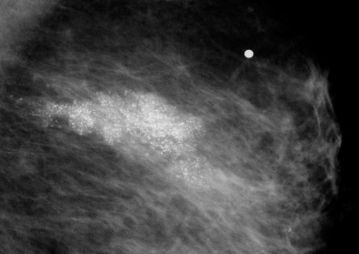
A major problem confronting surgeons relates to the fact that calcifications do not always map out the entire DCIS lesion, particularly those of the noncomedo type. Even if all the calcifications are removed, noncalcified DCIS may be left behind. Conversely, the majority of the calcifications may suggest a lesion larger than the true DCIS lesion. However, calcifications more accurately approximate the size of high-grade and/or comedo lesions than low-grade and/or noncomedo lesions.
If a patient’s mammogram shows any abnormality (i.e., calcifications, architectural distortion, nonpalpable mass), additional radiologic workup needs to be performed. This should include compression and magnification views. Ultrasonography should also be performed on all suspicious calcifications to rule out the presence of a mass that can be biopsied with ultrasound guidance. In addition, magnetic resonance imaging (MRI) has become increasingly popular and is often used to map out the size and shape of biopsy-proven DCIS lesions or invasive breast cancers and to rule out other foci of multifocal, multicentric or contralateral cancer. MRI has the advantage of detecting DCIS that has not undergone calcification.
Biopsy Techniques
If radiologic workup shows an occult lesion that requires biopsy, there are multiple approaches: fine-needle aspiration biopsy (FNAB), core biopsy (i.e., stereotactic, ultrasound guided, MRI guided), and directed surgical biopsy using guide wires or radioactive localization. FNAB is generally of little help for nonpalpable DCIS. Although with FNAB it is possible to obtain cancer cells, there is no architecture. So although cytopathologists can identify the presence of malignant cells, they cannot determine whether the lesion is invasive.
Stereotactic core biopsy became available in the early 1990s and is now widely used. Dedicated digital tables and add-on upright units make this a precise procedure. Large-gauge vacuum-assisted needles are the tools of choice for diagnosing DCIS using these techniques. Ultrasound-guided core biopsy also became popular in the 1990s but is of less value for DCIS because most DCIS lesions do not present with a mass that can be visualized by ultrasound. Nonetheless, all suspicious microcalcifications should be evaluated by ultrasound because a mass will be found in 5% to 15% of patients. Proper pathologic examination of a large-gauge core biopsy for microcalcification requires confirmation of the microcalcification in the core as well as at least serial levels to adequately sample the tissue. Radiographic-pathologic correlation is required to confirm concordance.
Open surgical biopsy should only be used if the lesion cannot be biopsied using minimally invasive techniques. This should be a rare event with current image-guided biopsy techniques and occurs in less than 5% of cases. If excision using needle localization is performed, whether for diagnosis or treatment, intraoperative specimen radiography and correlation with the preoperative mammogram is mandatory. Margins should be inked or dyed and specimens should be serially sectioned and, if necessary, a second x-ray of the slices should be obtained. The tissue sections should be arranged and processed in sequence. Pathologic reporting should include a determination of nuclear grade, an assessment of the presence or absence of necrosis, the measured extent of the lesion (calculated on the basis of the slices prepared), and measurement of all margins, in particular, the closest margin. The major architectural subtypes should also be included in the diagnosis. If the patient is motivated for breast conservation, a multiple wire–directed oncoplastic excision can be planned. This will give the patient her best chance at two opposing goals: clear margins and good cosmesis.
Treatment
For most patients with DCIS, there is no single correct treatment. There will generally be a choice. The choices, although seemingly simple, are not. As the choices increase and become more complicated, frustration increases for both the patient and physician.
Treatment End Points for Patients With Ductal Carcinoma in Situ
When evaluating the results of treatment for patients with breast cancer, a variety of end points must be considered. Important end points include local recurrence (both invasive and DCIS), regional recurrence (such as the axilla), distant recurrence, breast cancer–specific survival, overall survival, and quality of life. No study to date has shown a significant difference in distant disease-free or breast cancer–specific survival in patients with pure DCIS, regardless of any treatment. In our series of 1855 patients with DCIS, the breast cancer–specific mortality rate is 0.7% at 10 years. Numerous other DCIS series also confirm an extremely low mortality rate with DCIS. Consequently, local recurrence has become the most commonly used and important end point when evaluating treatment for patients with DCIS.
Forty to fifty percent of local recurrences after treatment for DCIS are invasive. Approximately 10% to 20% of DCIS patients who develop local invasive recurrences develop distant metastases and die of breast cancer. Long term, this translates into a mortality rate of approximately 0.5% for patients treated with mastectomy, 1% to 2% for conservatively treated patients who receive radiation therapy, and 2% to 3% for patients treated with excision alone.
It is clearly important to prevent local recurrences in patients treated for DCIS. They are demoralizing. They often lead to mastectomy and if they are invasive, they upstage the patient and are a threat to life. However, protecting DCIS patients from local recurrence must be balanced against potential detrimental effects of the treatments given.
Treatment Options
Mastectomy
Mastectomy is, by far, the most effective treatment available for DCIS if the goal is simply to prevent local recurrence. Most mastectomy series reveal local recurrence rates of approximately 1% with mortality rates close to zero. In our series of 576 DCIS patients treated with mastectomy, none of whom received radiation therapy or tamoxifen, we have had 13 local recurrences (9 invasive and 4 DCIS). One of the patients with an invasive local recurrence developed metastatic disease. In addition, two other patients developed metastatic breast cancer without developing a local recurrence. The absolute rate of distant recurrence was 0.5%.
However, mastectomy is an aggressive form of treatment for patients with DCIS. It clearly provides a local recurrence benefit but only a theoretical survival benefit. As well, during an era where breast conservation is increasingly used for treatment of invasive breast carcinoma, it is difficult to justify mastectomy, particularly for otherwise healthy women with screen-detected DCIS. Mastectomy is indicated only in cases of true multicentricity or when a unicentric DCIS lesion is too large to excise with clear margins and an acceptable cosmetic result. In our opinion, no DCIS lesion in too large to excise if the breast is large enough to accommodate an oncoplastic reduction with a good cosmetic result.
Initially it was thought that DCIS was not part of the spectrum of disease related to breast cancer–associated genes BRCA1 and BRCA2 . However, an association between DCIS and these genes is now recognized. Genetic positivity for BRCA1 or BRCA2 is not an absolute contraindication to breast preservation, although many patients who test positive for a deleterious mutation and who develop DCIS seriously consider bilateral mastectomy and salpingo-oophorectomy.
Breast Conservation
Breast conservation for DCIS can take the form of excision alone or excision plus radiation therapy. The most recently available Surveillance Epidemiology and End Results (SEER) data reveal that 70% of patients with DCIS are treated with breast conservation, nearly equally divided between with and without radiation therapy.
Clinical trials have shown that local excision plus radiation therapy in patients with negative margins provides excellent rates of local control. Some cases of DCIS may not recur or progress to invasive carcinoma when treated by excision alone. Although we know that this may be true for many cases of DCIS, it is not true for all cases. Because we are currently unable to determine which DCIS lesions will progress to invasive disease and, if they do, over what period of time, the use of radiation therapy for high-risk DCIS patients should be considered.
Are We Overtreating Ductal Carcinoma in Situ?
Until 2008 the standard treatment for DCIS, recommended by NCCN guidelines, was mastectomy or lumpectomy plus radiation therapy. In 2008, the NCCN modified its recommendations and suggested that selected low-risk DCIS patients could be treated with excision alone. The definition of who was low risk and who could be treated with excision alone was not defined and therefore left to clinical judgment.
During 2015 the media saw numerous lay articles focusing on the issue of whether DCIS was being overtreated. These articles questioned whether excision alone for DCIS was overtreatment.
To address the issue of possible overtreatment, the Low-Risk DCIS (LORIS) Trial was undertaken in the United Kingdom. This trial randomized screen-detected, favorable, low- and intermediate-grade DCIS to standard surgical treatment versus active monitoring (surveillance) (needle biopsy alone with no additional treatment and yearly mammography surveillance). Each group of patients will be followed for 10 years with a yearly mammogram, and the end point will be invasive recurrence.
Given that it will take at minimum of 10 years to gather meaningful results from the LORIS trial, we queried our database for a cohort of patients that could be considered similar to the LORIS Trial’s active monitoring group. We compared them with patients treated using a standard surgical approach. Because NCCN guidelines state that DCIS with surgical margins less than 1 mm are inadequate, we used that definition as a surrogate for the LORIS surveillance arm. In contrast, we considered DCIS patients with surgical margin widths 1 mm or greater as adequately treated and as a surrogate for standard treatment. The patients were subdivided by low nuclear grade versus high.
The 10-year local recurrence probabilities were statistically significant (<0.001) for low grade versus high grade and for narrow margins less than 1 mm versus wide margins 1 mm or greater. When the two factors were combined, excision alone with margins 1 mm or greater yielded a local recurrence rate at 10 years of 13% for low-grade DCIS and 36% for high-grade DCIS ( P < .001). For patients who had inadequate margins of less than 1 mm, excision alone yielded a 10-year local recurrence rate of 51% for low-grade and 67% for high-grade lesions. These data show that margins less than 1 mm lead to local recurrence rates of greater than 50% at 10 years and are inadequate. These data suggest that needle biopsy alone, regardless of grade, will lead to extremely high recurrence rates, half of which will be invasive.
Intraoperative Radiation Therapy for Ductal Carcinoma in Situ
Whole breast radiation therapy (WBRT) is often recommended for women treated for DCIS after breast conserving surgery (BCS) because several prospective randomized trials have demonstrated a 50% to 60% reduction in ipsilateral breast cancer recurrence for DCIS patients treated with WBRT. Local failure in a patient that has received WBRT usually leads to a recommendation for mastectomy, as reirradiation is associated with extremely high levels of toxicity to the breast. As a result, recent advances in radiation therapy have focused on replacing traditional WBRT with shorter hypofractionated regimens or accelerated partial breast irradiation (APBI).
Intraoperative radiation therapy (IORT) is an APBI approach in which all radiation is delivered directly to the lumpectomy site during surgery. Because 60% to 75% of DCIS patients treated with BCS recur at or near the original tumor site, limiting the radiation dose to the tumor bed during lumpectomy allows radiation to be delivered in a single dose to the region where recurrence would most likely happen, eliminating compliance issues, reducing radiation exposure to normal tissues, and reducing radiation-induced toxicity. The simplicity of IORT makes this technique extremely appealing for patients with either invasive or noninvasive breast carcinoma.
The rationale for using IORT in women diagnosed with pure DCIS is supported by the TARGIT-A trial, a prospective randomized IORT-APBI trial that examined the equivalence of IORT compared with standard WBRT treatment for patients with early-stage invasive breast cancer. Half of the patients enrolled in the TARGIT-A trial were found to have concurrent early-stage invasive cancer and DCIS upon pathologic examination. Yet regardless of the presence of a DCIS component, equivalent local recurrence rates were observed among patients treated with WBRT and IORT. Thus data from TARGIT-A demonstrates that IORT is capable of preventing recurrences in both DCIS and early-stage invasive breast carcinomas.
Since publication of the TARGIT-A trial, additional studies have documented the efficacy of APBI in patients with DCIS. In 2011 an update on the American Society for Breast Surgery MammoSite Registry Trial was published, examining a subset of 194 patients with DCIS as the primary pathology. The local recurrence rate for DCIS patients treated with APBI was 3.4%, comparing favorably with the 5-year recurrence rate of 7.5% for WBRT patients reported in the NSABP B-17 trial. In addition, publications from William Beaumont Hospital and Bryn Mawr Hospital studies support the findings of the MammoSite Registry Trial, concluding that that APBI as part of BCS for pure DCIS is associated with excellent local control and survival rates. Other studies treating DCIS patients with IORT reached similar conclusions. Taking these, and other, studies into account there is no reason to conclude that IORT would be less effective in treating DCIS patients than WBRT. Indeed, DCIS is now included as an acceptable histology by the American Brachytherapy Society and American Society for Breast Surgery.
In summary, IORT is a promising new treatment modality that greatly simplifies the delivery of postexcision radiation therapy in patients diagnosed with DCIS. The efficacy of IORT for the treatment of DCIS has been confirmed in numerous trials. IORT makes breast conservation possible for women that could not tolerate or would not be available for 3 to 6 weeks of conventional whole breast radiation therapy.
Reasons to Consider Excision Alone
There clearly are patients with DCIS who require mastectomy. They generally have lesions too large to remove with a cosmetically acceptable result. In addition, some patients are simply more comfortable with mastectomy. However, the majority of patients, more than 70%, are good candidates for breast conservation, and half of these can probably be treated with excision alone, if adequate margins are obtained. Here are a number of reasons to consider excision alone for selected patients with DCIS.
Common Use.
Excision alone is already common in spite of the randomized trial data that suggest that all conservatively treated patients benefit from radiation therapy. SEER Data reflect that excision alone is being used as complete treatment for DCIS in 35% of all DCIS patients. American doctors and patients have embraced the concept of excision alone for DCIS.
Anatomic.
Evaluation of mastectomy specimens using the serial subgross tissue processing technique reveals that most DCIS is unicentric (involves a single breast segment and is radial in its distribution). Using the same technique and evaluating patients with 25 mm or less of disease provided additional support that the majority of image detected DCIS can be adequately excised. This means that in many cases, it is possible to excise the entire lesion with a segment or quadrant resection, possibly curing the patient without additional therapy. Holland and Faverly have shown that if 10-mm margins are achieved in all directions, the likelihood of residual DCIS is less than 10%.
Biological.
DCIS is a heterogeneous group of diseases with different architectures, different nuclear grades, and unpredictable malignant potentials. Some nonaggressive DCIS lesions carry a low potential, about 1% per year, of developing into an invasive tumor. This is only slightly more than lobular carcinoma in situ, a lesion that is routinely treated with careful clinical follow-up.
Pathology Errors.
The differences between atypical ductal hyperplasia and low-grade DCIS may be subtle. It is not uncommon for atypical ductal hyperplasia to be classified as DCIS. Such patients treated with excision and radiation therapy are indeed “cured of potential DCIS” but incur significant risks of morbidities.
Prospective Randomized Data.
Prospective randomized DCIS trials show no difference in breast cancer–specific survival or overall survival, regardless of treatment after excision with or without breast irradiation.
Radiotherapy May Cause Harm.
Numerous studies have shown that WBRT for breast cancer may increase mortality from both lung cancer and cardiovascular disease. As well, radiation fibrosis due to therapy may change the texture of the breast and skin, making mammographic follow-up more difficult and can result in delayed diagnosis if there is a local recurrence. Because there is no proof that breast irradiation for patients with DCIS improves survival and there is proof that radiation therapy may cause harm, it makes perfect sense to spare patients from this potentially dangerous treatment whenever possible.
Socioeconomic.
Radiation therapy is expensive and time consuming (as much at $40,000 and taking 3–7 weeks).
Increased Risk.
Some studies show that there are more invasive recurrences in irradiated patients than nonirradiated patients. In our own series, 44% of excision only patients that recurred did so with invasive disease whereas 55% of irradiated patients who recurred, recurred with invasive cancer ( P < .01). In our series, the median time to recurrence after excision alone was 40 months, whereas after excision and irradiation it was 78 months ( P < .01). All subsets of DCIS show substantial delays in recurrence after irradiation—less in high grade and longest in low grade—and this delay can alter the perceived benefit of irradiation.
Only One Time.
If radiation therapy is given for the initial DCIS, it cannot be given again, at a later time, even if there is a small invasive recurrence. In general, we prefer to withhold radiation in DCIS patients initially and only give it to the few that ultimately recur with invasive disease. The use of radiation therapy with its accompanying skin and vascular changes make skin-sparing mastectomy, if needed in the future, more difficult to perform.
Improved Patient Selection.
The gold standard for local recurrence rates in irradiated patients is a 16% at 12 years as established by the NSABP B-17 trial. A subsequent update showed a 19.8% local recurrence rate in the irradiated arm of B-17 at 15 years. However, by using tools such as the USC/VNPI, it is now possible to select patients that recur at a rate of 8% or less at 12 years without radiation therapy (USC/VNPI scores 4–6).
NCCN Guidelines.
Finally, within the 2008 NCCN guidelines, excision without radiation therapy has been added as an acceptable treatment for selected DCIS patients with low risk of recurrence.
Prospective Randomized Ductal Carcinoma in Situ Trials
The NSABP B-06 protocol is the only prospective randomized trial that has compared mastectomy with breast conservation for patients with DCIS, albeit inadvertently. Although this study investigated invasive disease, during central slide review a subgroup of 78 patients was confirmed to have pure DCIS without any evidence of invasion. There were three treatment arms: total mastectomy, excision plus radiation therapy, and excision alone. Axillary nodes were removed regardless of the treatment assignment. After 83 months of follow-up, the percent of patients with local recurrences were zero for mastectomy, 7% for excision plus radiation therapy, and 43% for excision alone. Despite these large differences in local recurrence, there was no difference among the three treatment groups in breast cancer-specific survival.
Numerous prospective randomized trials have demonstrated a significant reduction in local recurrence for DCIS patients treated with radiation therapy compared with excision alone: the NSABP (protocol B-17) ; the European Organization for Research and Treatment of Cancer (EORTC) protocol 10853 ; the United Kingdom, Australia, New Zealand DCIS Trial (UK/ANZ Trial) ; and the Swedish Trial. However, none of these trials has reported a survival benefit.
In the NSABP B-17 Trial, more than 800 patients with DCIS excised with clear surgical margins were randomized into two groups: excision alone versus excision plus radiation therapy. The main end point was local recurrence, invasive or noninvasive. The definition of a clear margin was nontransection of the DCIS. The results of NSABP B-17 were updated in 1995, 1998, 1999, 2001, and 2011. After 15 years of follow-up, there was a statistically significant, 50% decrease of both invasive and noninvasive local recurrences in patients treated with radiation therapy compared with those treated with excision alone (19.8% and 35%, respectively). There was no difference in distant disease-free or overall survival in either arm. These data led the NSABP to continue recommending postoperative radiation therapy for all patients with DCIS who chose to save their breasts. Clearly, this recommendation was based on the decreased local recurrence rates rather than survival advantages.
Results from the EORTC 10853 trial, designed almost identically to the NSABP B-17 trial, were published in 2000 and updated in 2006. After 10 years of follow-up, 15% of patients treated with excision plus radiation therapy had recurred locally compared with 26% of patients treated with excision alone. As in the NSABP B-17 Trial, there was no difference in distant disease-free or overall survival in either arm of the EORTC Trial. Although in the initial report there was a statistically significant increase in contralateral breast cancer in patients who were randomized to receive radiation therapy, this was not observed when the data were updated.
The UK/ANZ Trial was published in 2003 and updated in 2011. In this trial, 1694 patients that had been excised with clear margins (nontransection of DCIS) were randomized to receive radiotherapy (yes or no) and/or to tamoxifen versus placebo. This yielded four subgroups: excision alone, excision plus radiation therapy, excision plus tamoxifen, and excision plus radiation therapy plus tamoxifen. With a median follow-up of 12.7 years, those who received radiation therapy demonstrated a statistically significant decrease in ipsilateral breast tumor recurrence, similar in magnitude to the NSABP B-17 and EORTC trials. As with the NSABP and the EORTC, there was no difference in survival, regardless of treatment, in any arm of the UK DCIS trial.
The Swedish DCIS Trial randomized 1067 patients into two groups: excision alone versus excision plus radiation therapy. In contrast to the trials discussed earlier, microscopically clear margins were not mandatory. Indeed, 22% of patients had microscopically unknown or involved margins. The cumulative incidence of local recurrence at 10 years was 21.6% for excision only and 10.3% for excision plus radiation therapy with an overall hazard ration of 0.33 ( P < .0001). There were 15 distant metastases and breast cancer related deaths in the excision only arm and 18 in the excision plus radiation therapy ( P = nonsignificant). As in the NSABP B-17, EORTC, and UK/ANZ trials, women treated with radiotherapy in the Swedish DCIS trial exhibited lower recurrence rates. However, the trial found no evidence in the relative risk of invasive and noninvasive recurrences and no difference in distant disease-free or overall survival in either arm.
In 2007 Viani and colleagues published a meta-analysis of the four prospective randomized DCIS trials comparing excision alone with excision plus radiation therapy. Pooled data on 3665 patients revealed a 60% reduction of both invasive and DCIS recurrences ( P < .00001) with the addition of radiation therapy. There was, however, no decrease in distant metastases in those who received radiation therapy, nor was there any survival benefit. Patients with high-grade lesions and involved margins received the most benefit from radiation therapy.
In 2010, the Early Breast Cancer Trialists Collaborative Group (EBCTCG) published an overview of the four randomized DCIS trials, reaching very similar conclusions to Viani and colleagues. The EBCTCG reaffirmed a lower local recurrence rate in all subgroups of patients who received adjuvant radiation therapy but no significant effect on breast cancer or all-cause mortality.
Tamoxifen for Ductal Carcinoma in Situ
Tamoxifen is now considered a standard adjuvant agent for local control in DCIS patients undergoing breast conservation with or without irradiation. This viewpoint is largely due to the initial results of NSABP B-24 and the UK/ANZ Trial, both which claimed a small but significant benefit for ipsilateral local control and contralateral chemoprevention.
Results of the NSABP B-24 trial were first published in 1999 and updated in 2011. In the B-24 protocol, more than 1800 DCIS patients were treated with excision and radiation therapy, and then randomized to receive either tamoxifen or placebo. After 15 years of follow-up, 16.6% of patients treated with placebo had recurred locally, whereas only 13.2% of those treated with tamoxifen had recurred. The difference, although small, was statistically significant for invasive local recurrences but not for DCIS recurrences.
Similar to the results of the NSABP B-24 trial, the UK/ANZ Trial also demonstrated that tamoxifen significantly reduced the incidence of ipsilateral DCIS recurrences but not invasive recurrences. The scale of risk reduction was comparable to those observed in the NSABP B-24 trial. Moreover, tamoxifen provided no additional benefit in those who were irradiated.
However, in 2012 when Allred and colleagues reexamined the rates of ipsilateral recurrences in a subset of 732 patients from NSABP B-24 trial, p values for the differences between tamoxifen versus placebo fell short of statistical significance. Similarly, when Cuzick and colleagues provided an update of the UK trial in 2011, there was no significant difference in either ipsilateral or contralateral events between patients with or without tamoxifen therapy. These findings are exactly opposite those seen in the NSABP B-24 trial. Of interest, upon reanalysis, patients that did not receive radiation showed significant differences in contralateral events related to tamoxifen. Because only the ipsilateral breast undergoes radiation therapy, it is unclear why contralateral events are suppressed by tamoxifen in the nonirradiated group, but not in the irradiated group.
Based on the results of these clinical trials, Warrick and Allred concluded that tamoxifen is probably overused and advocate more selective use of the drug. They particularly note that a major benefit would be seen in patients with estrogen-positive disease who are premenopausal with extensive high-grade disease and/or narrow margins. On the whole, the clinical benefit of tamoxifen intervention based on the randomized trials is meager at best.
Determination of HER2/neu Status and Potential Benefit of Neoadjuvant Trastuzumab
The HER2/neu gene is amplified or overexpressed in approximately 25% to 30% of invasive breast carcinomas. It is now standard of care to treat HER2/neu-positive invasive breast cancers greater than 10 mm with the monoclonal antibody trastuzumab (Herceptin). Indeed, this therapy has had a major impact on relapse in patients with HER2/neu-positive invasive breast cancers. Although approximately 40% of DCIS lesions also exhibit amplification and/or overexpression of HER2/neu, there is a lack of evidence that HER2/neu-positive DCIS will respond to trastuzumab therapy in a manner equivalent to invasive disease.
In 2012, Von Minckwitz and colleagues examined the effect of chemotherapy plus trastuzumab on HER2/neu-positive DCIS adjacent to HER2/neu-positive invasive breast cancer. Treatment reduced the volume of adjacent DCIS suggesting the possibility of a therapeutic impact of chemotherapy plus trastuzumab on the HER2/neu-positive in situ component.
In contrast, in 2011 Kuerer and colleagues described the results of a pilot study in which patients with large areas of HER-2/neu-positive DCIS (mean 5.2 cm) received a single dose of trastuzumab with follow-up surgical excision and reevaluation 14 to 28 days post therapy. No overt histologic response to the biological therapy was recorded; there was no alteration in ki67 or cleaved caspase. However, pretreatment increased antibody-dependent cell-mediated cytotoxicity.
Predicting Local Recurrence in Conservatively Treated Patients With DCIS
There are now sufficient, readily available data that can aid clinicians in differentiating patients who significantly benefit from radiation therapy after excision from those who do not. These same data can identify patients who are better served by mastectomy because recurrence rates with breast conservation even with the addition of radiation therapy are unacceptably high.
Our research and the research of others has shown that various combinations of nuclear grade, the presence of comedo-type necrosis, tumor size, margin width, and age are all important factors that can be used to predict the probability of local recurrence in conservatively treated DCIS patients.
Treatment Selection for Patients With DCIS of the Breast Using the University of Southern California/Van Nuys Prognostic Index
In 2008 the NCCN included excision alone as an acceptable treatment alternative for patients with DCIS, validating an actual practice in the United States in which almost 50% of conservatively treated patients do not receive postexcisional breast irradiation. However, the NCCN did not define the subset of patients in which excision without radiation therapy was appropriate. Researchers have attempted to accomplish this for years but with only marginal success. Multivariate analysis has shown that six factors are independent predictors of local recurrence in patients with DCIS treated with breast conservation: treatment (radiation therapy yields a lower local recurrence rate than excision alone), age (older age is better), size (smaller size is better), nuclear grade (lower grade is better), margin width (wider margins are better), comedonecrosis (no necrosis is better).
In 1995, the Van Nuys Classification was developed that used a combination of nuclear grade and necrosis to predict local recurrence. In 1996, the Van Nuys Prognostic Index added size and margin width to the numerical algorithm and in 2002, the USC/VNPI added age at diagnosis to the algorithm. These studies collected all pathologic features in a prospective fashion but treatment (excision alone vs. excision plus radiation therapy) was not randomized.
The USC/VNPI was devised by combining four statistically significant independent prognostic factors for local tumor recurrence (tumor size, margin width, age, and pathologic classification (determined by nuclear grade and the presence or absence of comedo-type necrosis). Each of the four prognostic predictors was scored 1, 2, or 3, where 1 is the most favorable and 3 the least favorable. Table 39.1 details this scoring system. The individual scores for each of the four prognostic factors were added together to give an overall score ranging from a low of 4 (least likely to recur) to a high of 12 (most likely to recur). Prior published recommendations were excision alone for those who score 4, 5, or 6; excision plus radiation therapy for those who score 7, 8, or 9; and mastectomy for those who scored 10, 11, or 12.

