Abstract
In determining breast cancer prognosis, axillary lymph node (ALN) status has been, to date, the single best prognostic factor. Despite this, the outcome for good-prognosis breast cancer is not uniformly favorable, and as many as 30% of ALN-negative patients die of distant metastases despite adequate local therapy. It is logical to suspect that more intensive surveillance for subclinical regional or systemic disease might uncover this metastatic potential, and for patients with breast cancer, this search has focused on three sites: the ALN, the bone marrow, and the peripheral blood. This chapter focuses on the identification, significance, and surgical management of occult metastases in the ALN recognizing a shifting paradigm regarding both the clinical significance and the appropriate surgical management for women with micrometastatic nodal disease. A large body of retrospective literature consistently reports the identification of occult micrometastases among patients who are ALN negative by routine examination with mixed results regarding the prognostic significance of these findings. More recently, the significance and surgical management of ALN micrometastases have been reevaluated in the era of sentinel lymph node biopsy and routine use of systemic therapy. We now have definitive prospective studies, the American College of Surgeons Oncology Group (ACOSOG) Z0010 and National Surgical Adjuvant Breast and Bowel Project (NSABP) B-32, which show that routine use of immunohistochemical staining is no longer justified for additional evaluation of negative sentinel lymph nodes by standard evaluation, and two randomized trials showing that for many patients with SLN micrometastases, ALND is no longer needed. Looking ahead, genomic profiling for both prognostication and prediction of response to therapy may ultimately trump conventional histopathology, challenging the necessity of lymph node staging altogether.
Keywords
occult metastases, axillary lymph node, sentinel lymph node, breast cancer
Since the days of Halsted, breast cancer has been viewed as a disease that begins locally and is subject to a predictable, orderly, and sequential process of spread, first to regional lymph nodes and then to systemic sites. From the 1970s, Fisher has popularized the alternative hypothesis that most breast cancers are systemic from the outset and that outcome is governed more by the presence of occult systemic metastases than by variations in local treatment. In the Halstedian model, the most aggressive local treatment should be the most curative, and in the “Fisherian” model, greater emphasis is placed on systemic treatment. In fact, neither model by itself can encompass the broad range of clinical behaviors familiar to any physician who treats breast cancer. This broad range has given rise to the most recent paradigm, the spectrum hypothesis, which suggests that the optimal treatment of each cancer must be tailored to its unique (and perhaps separate) propensities for local progression and for systemic metastasis.
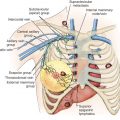
In determining where a given breast cancer lies on the spectrum of potential outcomes, axillary lymph node (ALN) status has been, to date, the single best prognostic factor. Despite this, the outcome for good-prognosis breast cancer is not uniformly favorable, and as many as 30% of ALN-negative patients die of distant metastases despite adequate local therapy. It is logical to suspect that more intensive surveillance for subclinical regional or systemic disease might have uncovered this metastatic potential, and for patients with breast cancer, this search has focused on three sites: the ALN, the bone marrow, and the peripheral blood. This chapter focuses on the identification, significance, and surgical management of occult metastases in the ALN.
Definition and Classification of Axillary Lymph Node Micrometastases
The concept of occult ALN metastasis in breast cancer was first articulated in 1948 by Saphir and Amromin. In radical mastectomy specimens previously found to be ALN negative on routine single-section pathologic examination, they retrospectively performed serial sections (SS), taking an average of 332 sections per paraffin block. They identified ALN metastases in 33% (10/30) of cases and recommended that SS of the ALN in breast cancer should become routine.
The term micrometastasis was first used in 1971 by Huvos and colleagues, who suggested a distinction between ALN macrometastasis (tumor foci ≥2 mm in diameter) and micrometastasis (<2 mm in diameter). This distinction was carried forward to the 1997 (5th) edition of the American Joint Committee on Cancer (AJCC) Staging Manual, in which micrometastases less than or equal to 2 mm were categorized as pN1a disease and regarded as prognostically equivalent to pN0 (node-negative) disease. The AJCC has more recently further subcategorized nodal micrometastases into those less than or equal to 0.2 mm (pN0(i+) or “isolated tumor cells” [ITCs]) and 0.2 to 2 mm (pN1mi or “micrometastases”) in the 2002 (6th) edition, and 2010 (7th) edition. Nodal metastases greater than 2 mm in diameter are categorized as pN1 or “macrometastases.”
Prognostic Significance of Axillary Lymph Node Micrometastases: Retrospective Data
A large body of retrospective data assessing the prognostic significance of micrometastases fits into two categories: (1) studies in which patients classified initially as ALN positive are stratified by size of ALN metastasis and (2) studies in which patients classified initially as ALN negative are found on further study to be ALN positive.
Classification by Size of Axillary Lymph Node Metastasis
Three early studies from Memorial Sloan Kettering Cancer Center found (1) comparable overall survival at 8 years for patients with ALN micrometastases (<2 mm) versus negative ALN ; (2) better overall survival at 14 years for those with micrometastases rather than macrometastases ; and (3) worse disease-free survival (DFS) at 12 years for those with single micrometastasis or single macrometastasis than node-negative disease. In parallel, a contemporaneous report from the National Surgical Adjuvant Breast and Bowel Project (NSABP) B-04 trial demonstrates worse 4-year DFS for patients with axillary micrometastases and macrometastases compared with patients with negative nodes. A subsequent report from Cox and colleagues reaches the same conclusion, finding significantly worse overall survival and DFS for patients with pN1mi versus pN0 disease. Conversely, in a multiinstitutional French retrospective review comprising 8001 patients treated between 1999 and 2008, Houvenaeghel and colleagues found no significant difference in recurrence-free survival or overall survival in patients with nodal micrometastases (either pN0(i+) or pN1mi) compared with pN0 patients.
Classification by Frequency of Occult Axillary Lymph Node Metastases
Dowlatshahi and colleagues reviewed in detail 31 studies (1948–1996) that attempted to define the frequency and significance of occult metastases found on further analysis of ALN originally classified as negative. All are retrospective, span different time periods, and comprise different patient populations. Many contain fewer than 100 patients and lack the statistical power to detect small but significant differences in outcome. All use various combinations of SS and/or immunohistochemistry (IHC) staining for cytokeratins. Despite these caveats, some strong overall trends emerge:
- •
Occult micrometastases are found consistently among patients who are ALN negative by routine examination. All studies but one identify ALN micrometastases in 7% to 42% of patients initially staged as ALN negative.
- •
Occult micrometastases may be prognostically significant. Six of the seven studies with more than 100 patients demonstrate significantly worse overall survival and/or DFS for patients with micrometastases compared with those with negative ALN.
- •
The yield of IHC-detected micrometastases is greater for lobular than for duct carcinomas. All of the studies that distinguish between duct and lobular tumor type demonstrate IHC micrometastases 2 to 10 times more frequently with invasive lobular than with invasive duct cancers.
Table 43.1 summarizes series with more than 100 patients, including (1) eight published before 1997, (2) the second Ludwig study (1999), and (3) six more recent studies (2002–2008) of similar methodology. Of the six recent studies, half find ALN micrometastases to be prognostically significant and half do not. A more recent (2004–2011) Surveillance, Epidemiology, and End Results study of 93,070 patients who were pN0 by routine H&E reports the results of IHC cytokeratin staining; 5% were converted to pN0(i+) and 7% converted to pN1mi. In a multivariate model controlling for demographic and tumor characteristics, compared with pN0 patients, overall survival was significantly worse for pN1mi (hazard ratio [HR] 1.399, p < .0001) but not for pN0(i+) disease (HR 1.071, P = .33).
| Method Author (Year) | No. of Patients | Converted to Node-Positive (%) | Follow-Up (yr) | Disease-Free Survival | Overall Survival |
|---|---|---|---|---|---|
| Serial Sections | |||||
| Wilkinson (1982) | 525 | 17 | 15 | NS | NS |
| Ludwig (1990) | 921 | 9 | 5 | p = .003 | p = .002 |
| Neville (1991) | 921 | 9 | 6 | p = .0008 | p = .0009 |
| Cote (1999) | 736 | 7 | 12 | p = .001 | p = .0005 |
| IHC | |||||
| Trojani (1987) | 122 | 11 | 10 | p < 0.003 | p = .02 |
| de Mascarel (1992) | 129 | 10 | 10 | p = .01 | p = .007 |
| Hainsworth (1993) | 343 | 12 | 6.5 | p < .05 | NS |
| Cote (1999) | 736 | 20 | 12 | p = .09 | NS |
| Umekita (2002) | 148 | 14 | 8 | p = .0009 | p = .0001 |
| Reed (2004) | 385 | 12 | 25 | NS | NS |
| Serial Sections + IHC | |||||
| McGuckin (1996) | 208 | 25 | 5 | p = .007 | p = .02 |
| Nasser (1993) | 159 | 31 | 11 | p = .04 | p = .07 |
| Millis (2002) | 477 | 13 | 19 | — | NS |
| Cummings (2002) | 203 | 25 | 10 | p = .016 | — |
| Kahn (2006) | 214 | 14 | 8 | NS | NS |
| Tan (2008) | 368 | 23 | 18 | p < .001 | p = .02 |
The Ludwig Studies of Axillary Lymph Node Micrometastases
The Ludwig Trial V was designed to test the effect of a single dose of perioperative combination chemotherapy, and it reported 5-year results for ALN-positive and ALN-negative patients in 1988 and 1989, respectively. The authors then examined the lymph node tissue blocks from 921 of the 1275 patients originally staged as pN0 using a meticulous SS methodology and central pathologic review. Occult ALN metastases were found in 9% of patients; at 5 years, both overall survival and DFS were significantly worse.
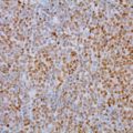
In 1999 Cote and colleagues reported 10-year results for 736 of the original 921 patients, adding IHC (a single section from a single level, stained with the anticytokeratin antibodies AE-1 and CAM 5.2) to the previous SS methodology (two H&E stained sections from each of six levels). Single-section IHC detected micrometastases far more often than SS/H&E (20% vs. 7%), particularly in patients with invasive lobular carcinomas (39% vs. 3%). Although 10-year DFS and overall survival were worse for SS/H&E-detected micrometastases, by 23% and 18%, and for IHC-detected micrometastases, by 8% and 5%, respectively, this margin was significant only for H&E-detected disease. Grouped by menopausal status, both H&E- and IHC-detected micrometastases were highly significant in postmenopausal but not in premenopausal women.
Logistical Hurdles in the Detection of Axillary Lymph Node Micrometastases
Although the preceding results present heterogenous outcomes for patients with micrometastasis, the authors of the first Ludwig study in 1991 present a persuasive case for their conclusion that SS analysis of “negative” ALN should “be considered as part of the routine pathology examination”; few pathology departments then or since have had the resources to do so. The Ludwig investigators examined an average of 12 tissue blocks per patient, and 12 H&E-stained slides per block, in 921 patients to identify occult metastases in 83 individuals, or nearly 1600 slides to identify one additional node-positive patient. The earlier Wilkinson SS study had a strikingly similar yield, examining 1449 slides to identify a single positive ALN. On the basis of these formidable logistics, the pathologic examination of axillary lymph node dissection (ALND) specimens with a single H&E-stained section per node has remained standard care at most institutions worldwide.
The advent of sentinel lymph node (SLN) biopsy has changed this practice. Pioneered in its modern form by Morton and colleagues and first reported for breast cancer by Alex (1993) and Giuliano and colleagues (1994), SLN biopsy has largely replaced ALND for breast cancer staging at many centers in the United States and worldwide. Sixty-nine observational studies and seven randomized trials of SLN biopsy validated by a “backup” ALND confirm that SLN biopsy is feasible, accurate, and safe for patients with stage I to IIIa invasive breast cancer, with less postoperative morbidity than that of ALND. Most importantly, SLN biopsy is a targeted examination of an average of two to three nodes (those most likely to contain metastases) versus the 15 to 20 removed in a standard ALND. SLN biopsy therefore makes enhanced pathologic analysis by SS and IHC logistically feasible and allows the identification of a group of patients whose risk of systemic relapse might otherwise go unrecognized.
Role of Enhanced Pathology in Sentinel Lymph Node Biopsy
Enhanced pathologic techniques using SS and IHC staining have played at least four roles in the evolution of SLN biopsy for breast cancer: (1) improved staging of the axilla; (2) validation of the SLN hypothesis; (3) reduction in the rate of false-negative SLN; and (4) the prediction of non-SLN metastases in patients with positive SLN.
Improved Axillary Lymph Node Staging
Giuliano and colleagues first documented the improved sensitivity of IHC analysis in SLN specimens; comparing patients having SLN biopsy (analyzed both by H&E and IHC) plus completion ALND (analyzed by H&E) with those having conventional ALND (analyzed by H&E), positive ALNs were significantly more frequent in the group having enhanced pathology (42% vs 29%), as was the proportion of micrometastases among the ALN-positive patients (38% vs 10%). A number of subsequent studies have confirmed these findings, which are quite consistent with the data in Table 43.1 ; the yield of positive SLN biopsy is increased 10% to 20% by the addition of IHC to conventional H&E analysis.
Validation of the Sentinel Lymph Node Hypothesis
Two elegant studies have used both SS and IHC to validate the SLN hypothesis. Turner and colleagues prove that a negative SLN is highly predictive of a negative axilla: in 60 patients who had SLN biopsy followed by a completion ALND and whose SLNs proved negative both on H&E and IHC, all of the remaining ALNs were also analyzed by both H&E and IHC, and only one of 1087 nonsentinel nodes, less than 0.1%, contained a metastasis. Conversely, Weaver and colleagues show that the SLN is the node likeliest to be positive; among 431 patients in a multicenter validation trial of SLN biopsy, they found that nodal metastases were far more frequent in the SLN than in non-SLNs (16% vs. 4%). They further demonstrated that occult nodal metastases identified by SS and IHC (in patients initially staged as node negative) were also far more likely to involve the SLN than the non-SLNs (4% vs. 0.35%).
Reduction in the Rate of False-Negative Sentinel Lymph Node Biopsy
The false-negative rate of SLN biopsy (the proportion of node-positive cases in which the SLN is negative) should be as small as possible, ideally 5% or less. Although there is no consensus regarding the optimal method for pathologic examination of SLNs, it is quite clear that the use of IHC increases the sensitivity of SLN biopsy, thereby decreasing the false-negative rate. Liberman has compared the results of 26 SLN validation trials that used H&E staining with seven trials that used IHC and demonstrates increased sensitivity (92% vs 97%) and a decreased false-negative rate (8% vs 3%) for the latter method.
Risk of Nonsentinel Lymph Node Metastasis in Micrometastatic Sentinel Lymph Node–Positive Patients
With the advent of SLN biopsy, it was logical to ask whether ALND should be mandatory for all SLN positive patients, especially those with micrometastases. In a meta-analysis of 25 studies, Cserni and colleagues found that non-SLNs were positive in 20% of patients with SLN micrometastases and in only 9% of those whose SLNs were positive only on IHC. Two other meta-analyses found that in women with pN0(i+) or pN1mi disease, non-SLN metastases were present in 12% to 20% of patients. Other variables (such as larger tumor size or lymphovascular invasion [LVI]) also increase the risk of non-SLN metastases. Van Zee and associates have developed and prospectively validated a multivariate nomogram that estimates the risk of non-SLN metastasis using nine variables, one of which is method of SLN metastasis detection (IHC, SS, or routine H&E); the others are frozen section, tumor size, tumor type/grade, number of positive SLNs, number of negative SLNs, LVI, multifocality, and estrogen receptor status. This tool has been validated by independent data sets worldwide and has allowed a growing number of SLN positive patients in our own practice to avoid ALND altogether.
Stay updated, free articles. Join our Telegram channel

Full access? Get Clinical Tree


