Abstract
Prognostic factors are quantifiable data that provide information regarding the expected outcome for patients with a particular disease prior to therapy. Importantly, though, these prognostic values provide information on a population level and may have only limited application to individuals within that population. In breast cancer, a number of factors are considered truly prognostic; these factors include patient lymph node status, tumor size, histologic grade, age, and race. Additional factors considered both prognostic and predictive of outcomes include steroid receptors (estrogen and progesterone), DNA and proliferative markers, and the epidermal growth factor receptor family. The combination of these prognostic and predictive values provides the clinician with information regarding a specified population of breast cancer patients. This chapter explores these individual factors and how they can aid clinicians in determining the best course of treatment for an individual breast cancer patient.
Keywords
prognostic factors, lymph nodes, histologic grade, tumor size, age, receptors
Nomenclature
Prognostic factors have grown in importance as the options for the treatment of breast cancer have increased. By definition, prognostic factors ( Box 18.1 ) are quantifiable data about the tumor or host that provide information about the expected outcome of a population of patients with similar defining characteristics in the absence of systemic therapy. Several facts that follow from this definition are often overlooked in clinical medicine. The first is that the prognostic value, which may be clearly defined for a population, bears only limited application to any individual within that population. Patients should not be terrorized by membership in a high-risk population, and they should not be made to feel invincible by membership in a favorable risk group. The second fact is that with the broad application of systemic therapy, less and less information will become available about prognostic factors in the absence of such therapy. The best example of a prognostic factor is lymph node status, the degree to which the axillary lymph nodes have been colonized by metastatic breast cancer.
Some parameters of a tumor that were measured and originally described as prognostic factors are now considered primarily predictive factors (see Box 18.1 ). The best example of a predictive factor is estrogen receptor (ER) status. It is of great clinical importance as a predictor of response to hormonal therapy. Certain tumor parameters, such as hormone receptor status, are both prognostic factors and predictive factors and may be considered separately for their contributions to each of these areas.
In the past, prognostic indicators were valued both for their ability to offer a glimpse of risk—desired by both the patient and the physician—and, in a related way, the importance of systemic adjuvant therapy. Prognostic factors can be used to define a population of patients at so little risk of progression or recurrence of breast cancer that systemic therapy may be avoided. This is recognized as increasingly important now that the series of overviews from the Early Breast Cancer Trialists’ Collaborative Group have demonstrated that the relative value of adjuvant therapy applies to all women with breast cancer. Only those with truly minimal risk can be dismissed from consideration because the absolute benefit is so small. On the other hand, studies of the adverse effects of adjuvant therapy that were initially focused on dose-limiting toxicities are beginning to quantify other toxicities, such as neurocognitive dysfunction associated with cytotoxic chemotherapy. With the recognition that an improvement in survival accruing to 2% to 3% of certain subgroups may be achieved at a cost of toxicity accruing to 20% or more of the patients, the need for more precise prognostic factors has grown. Can we divide the most favorable groups of women into those at greater and lesser risk in the future? And of at least as great importance, can we identify predictive factors that will allow us to determine—independent of risk—whether the contemplated therapy will be effective against her tumor?
Clinically established prognostic factors are those that meet the following criteria:
- •
Are reproducibly associated with a better or worse prognosis at a level of clinical utility
- •
Provide independent information not available by more easily measured parameters (this requires multivariate analysis with other established factors)
- •
Are reproducible in multiple clinics or laboratories
- •
Have demonstrated prognostic value in prospective trials
The literature of prognostic and predictive factors is replete with retrospective analysis of data sets. Although these are useful in generating hypotheses, any of multiple parameters may relate to outcome by play of chance in a given data set. If the data set is large, the statistical significance value of such chance associations may appear great. It is only when evaluated prospectively, at best, or in multiple other data sets retrospectively, that prognostic value may be validated. Of such prognostic values, some may be associated with other values, such as nodal status or tumor size. Unless significant additional prognostic information is added, as evaluated with multivariate statistical methods, they lack clinical utility.
The two best established prognostic factors form the basis of clinical and pathologic staging ( Table 18.1 ). Both nodal status and tumor size represent a summation of biologic effects in both the host and tumor that relate to the rate of tumor progression and the time from the initiation of the tumor or the development of its blood supply. Thus a very indolent cancer biologically, long undetected, may present at an identical stage to an extremely biologically aggressive tumor present for a lesser time. Other prognostic factors, such as markers of proliferation, may distinguish between these two scenarios in a specific individual.
| Classification | Definition |
|---|---|
| Primary Tumor (T) | |
| T X | Primary tumor cannot be assessed |
| T 0 | No evidence of primary tumor |
| Tis | Carcinoma in situ |
| Tis (DCIS) | Ductal carcinoma in situ |
| Tis (LCIS) | Lobular carcinoma in situ |
| Tis (Paget) | Paget disease of the nipple with no tumor (Paget disease associated with a tumor is classified according to the size of the tumor) |
| T 1 | Tumor ≤2 cm in greatest dimension |
| T 1 mi | Microinvasion ≤0.1 cm in greatest dimension |
| T 1a | Tumor >0.1 cm but ≤0.5 cm in greatest dimension |
| T 1b | Tumor >0.5 cm but ≤1 cm in greatest dimension |
| T 1c | Tumor >1 cm but ≤2 cm in greatest dimension |
| T 2 | Tumor >2 cm but ≤5 cm in greatest dimension |
| T 3 | Tumor >5 cm in greatest dimension |
| T 4 | Tumor of any size with direct extension to chest wall and/or to the skin (ulcerations or skin nodules) |
| T 4a | Extension to chest wall, not including pectoralis muscle adherence/invasion |
| T 4b | Ulceration and/or ipsilateral satellite nodules and/or edema (including peau d’orange) of the skin, which do not meet the criteria for inflammatory carcinoma |
| T 4c | Both T 4a and T 4b |
| T 4d | Inflammatory carcinoma |
| Regional Lymph Nodes Clinical (N) | |
| N X | Regional lymph nodes cannot be assessed (e.g., previously removed) |
| N 0 | No regional lymph node metastasis |
| N 1 | Metastasis to movable level I, II ipsilateral axillary lymph node(s) |
| N 2 | Metastases in ipsilateral level I, II axillary lymph nodes that are clinically fixed or matted, or in clinically apparent a ipsilateral internal mammary nodes in the absence of clinically evident axillary lymph node metastasis |
| N 2a | Metastasis in ipsilateral level I, II axillary lymph nodes fixed to one another (matted) or to other structures |
| N 2b | Metastasis only in clinically detected a ipsilateral internal mammary nodes and in the absence of clinically evident level I, II axillary lymph node metastasis |
| N 3 | Metastasis in ipsilateral infraclavicular (level III axillary) lymph node(s), with or without level I, II axillary node involvement, or in clinically apparent a ipsilateral internal mammary lymph node(s) and in the presence of clinically evident level I, II axillary lymph node metastasis; or metastasis in ipsilateral supraclavicular lymph node(s), with or without axillary or internal mammary lymph node involvement |
| N 3a | Metastasis in ipsilateral infraclavicular lymph node(s) |
| N 3b | Metastasis in ipsilateral internal mammary lymph node(s) and axillary lymph node(s) |
| N 3c | Metastasis in ipsilateral supraclavicular lymph node(s) |
| Regional Lymph Nodes Pathologic (pN) | |
| pN X | Regional lymph nodes cannot be assessed (e.g., previously removed, or not removed for pathologic study) |
| pN 0 | No regional lymph node metastasis histologically. Note: Isolated tumor cell clusters (ITCs) are defined as small clusters of cells ≤0.2 mm, single tumor cells, or a cluster of <200 cells in a single histologic cross-section; ITCs may be detected by routine histology or by immunohistochemical (IHC) methods; nodes containing only ITCs are excluded from the total positive node count for purposes of N classification but should be included in the total number of nodes evaluated |
| pN 0 (i−) | No regional lymph node metastasis histologically, negative IHC |
| pN 0 (i+) | Malignant cells in regional lymph node(s) ≤0.2 mm (detected by hematoxylin-eosin stain or IHC including ITC) |
| pN 0 (mol−) | No regional lymph node metastasis histologically, negative molecular findings (RT-PCR) |
| pN 0 (mol+) | Positive molecular findings (RT-PCR) but no regional lymph node metastasis detected by histology or IHC |
| pN 1 | Micrometastasis; or metastasis in 1–3 axillary lymph nodes, and/or internal mammary nodes, with metastasis detected by sentinel lymph node dissection but not clinically detected a |
| pN 1mi | Micrometastasis (>0.2 mm and/or >200 cells, but none >2.0 mm) |
| pN 1a | Metastasis in 1–3 axillary lymph nodes (at least 1 metastasis >2.0 mm) |
| pN 1b | Metastasis in internal mammary nodes, with micrometastasis or macrometastasis detected by sentinel lymph node biopsy but not clinically detected a |
| pN 1c | Metastasis in 1–3 axillary lymph nodes and in internal mammary lymph nodes, with micrometastasis or macrometastasis detected by sentinel lymph node biopsy but not clinically detected a |
| pN 2 | Metastasis in 4–9 axillary lymph nodes, or in clinically apparent a internal mammary lymph nodes in the absence of axillary lymph node metastasis |
| pN 2a | Metastases in 4–9 axillary lymph nodes (at least 1 tumor deposit >2.0 mm) |
| pN 2b | Metastases in clinically detected internal mammary lymph nodes in the absence of axillary lymph node metastases |
| pN 3 | Metastases in ≥10 axillary lymph nodes; or in infraclavicular (level III axillary) lymph nodes; or in clinically detected ipsilateral internal mammary lymph nodes in the presence of ≥1 positive level I, II axillary lymph nodes; or in >3 axillary lymph nodes and in internal mammary lymph nodes, with micrometastases or macrometastases detected by sentinel lymph node biopsy but not clinically detected; or in ipsilateral supraclavicular lymph nodes |
| pN 3a | Metastases in ≥10 axillary lymph nodes (at least 1 tumor deposit >2.0 mm); or metastases to the infraclavicular (level III axillary lymph) nodes |
| pN 3b | Metastases in clinically detected ipsilateral internal mammary lymph nodes in the presence of ≥1 positive axillary lymph nodes; or in >3 axillary lymph nodes and in internal mammary lymph nodes, with micrometastases or macrometastases detected by sentinel lymph node biopsy but not clinically detected |
| pN 3c | Metastases in ipsilateral supraclavicular lymph nodes |
| Distant Metastasis (M) | |
| M 0 | No clinical or radiographic evidence of distant metastasis |
| cM 0 (i+) | No clinical or radiographic evidence of distant metastases, but deposits of molecularly or microscopically detected tumor cells in circulating blood, bone marrow, or other nonregional nodal tissue that are no larger than 0.2 mm in a patient without symptoms or signs of metastases |
| M 1 | Distant detectable metastases as determined by classic clinical and radiographic means and/or histologically proven >0.2 mm |
a “Clinically detected” is defined as detected by imaging studies (excluding lymphoscintigraphy) or by clinical examination and having characteristics highly suspicious for malignancy or a presumed pathologic macrometastasis on the basis of fine-needle aspiration biopsy with cytologic examination.
The most powerful adjuvant therapy demonstrated to date—tamoxifen in a premenopausal receptor-positive individual or third-generation aromatase inhibitor in a postmenopausal individual—achieved only a 50% reduction in annual risk of recurrence. Although the ability to identify individuals who lack ERs or progesterone receptors (PRs) and who will consequently not benefit at all from tamoxifen therapy is a great triumph, greater still will be the ability to define predictive factors that will identify the responders from the nonresponders in the receptor-positive population. This is even more true in the case of cytotoxic chemotherapy. Dose-dense therapy is associated with greater population benefit than less intensive chemotherapy in clinical adjuvant trials. Certain data suggest that this benefit accrues from a subpopulation of individuals who require this greater dose density and that many other individuals would do as well with less aggressive chemotherapy. Predictive factors that will reproducibly define these subpopulations are the subject of active investigation.
Prognostic Factors
Axillary Lymph Nodes
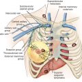
The degree of involvement of axillary lymph nodes by metastatic tumor cells is the dominant prognostic factor for later systemic disease. National Surgical Adjuvant Breast and Bowel Project (NSABP) B-04 and B-06 noted a relationship between survival and the number of nodes involved. There is also a well-established relationship between the size of the primary tumor and the axillary nodal burden. On the basis of this information and more recent studies, oncologists believe that virtually all women with axillary lymph node involvement should receive adjuvant systemic therapy. Other prognostic factors and combinations of such factors have repeatedly been shown to be of equal or greater value in a given retrospective database, but when such factors or a combination of factors have been tested prospectively, axillary lymph node status has been shown to be more predictive. This is understandable because any parameters of the primary tumor are surrogates for the likelihood of metastatic involvement. The potential for metastatic spread also depends on interaction with host resistance. Axillary lymph node status reflects actual end-results data on the interaction between tumor aggressiveness and host defense mechanisms. Therefore it is not surprising that it provides the most important prognostic measure available in clinical decision-making.
In patients with clear axillary metastasis, axillary lymph node dissection (ALND) is often performed providing an absolute number of lymph nodes involved with tumor. However, the extent of surgery is a point of debate in terms of outcomes and prognostic information gained. The early trials of the NSABP failed to show any significant survival benefit based on the extent of axillary surgery. The American College of Surgeons Oncology Group (ACOSOG) Z0011 trial, among patients with limited sentinel lymph node (SLN) metastatic breast cancer treated with breast conservation and systemic therapy, the use of SLN dissection (SLND) alone and ALND did not result in inferior survival. However, when specifically examining patients with a heavy nodal burden, prognosis is poor. A recent examination of the Surveillance, Epidemiology, and End Results Program (SEER) database examining triple-negative breast cancer (TNBC) patients noted N3 patients (undergoing axillary dissection) to have a particularly poor prognosis compared with either lower nodal disease burden or a negative axilla. An additional single institution study of 1711 patients with TNBC also revealed decreased survival for patients with nodal disease, but noted that an additional nodal burden did not further decrease survival in patients with N 2 to N 3 disease.
Clinical staging of axillary lymph nodes is notoriously inaccurate: the difference is 33% in clinical evaluation of axillary nodes, even by experienced clinicians. Cutler and Connelly found that among patients who have clinically negative nodes, 38% had evidence of nodal metastases on pathologic examination, and in those who had clinically suspicious nodes, the nodes were pathologically negative 38% of the time. Fisher and colleagues reported that the false-positive and false-negative clinical evaluation rates for axillary nodes were 24% and 39%, respectively, and the overall error in clinical staging was 32%. Smart, Myers, and Gloeckler reported that 35% of clinically negative lymph nodes had metastases detected on pathologic examination, and 87% of those considered clinically positive contained metastases. The increased utilization of preoperative axillary ultrasound has increased the identification rates of diseased nodes, yet the false-negative rate of ultrasound combined with core needle biopsy is still 25%. Because clinical staging of axillary nodes is so inaccurate and accurate staging is so important, histopathologic axillary lymph node staging remains necessary to stage patients accurately and assign population risks for considering adjuvant therapy. To avoid the consequences of axillary dissection for those with negative axillae, a variety of radiologic and nuclear medicine techniques for diagnosis have been attempted. Even though some of the techniques (e.g., positron emission tomography, magnetic resonance imaging) may surpass clinical examination in accuracy, they still remain less accurate than surgical staging of the axilla.
The use of SLN biopsy to limit axillary dissection in those with nodal metastases has revolutionized axillary lymph node staging. The adoption of SLN biopsy has, however, introduced other areas of controversy in prognostic factor research. The first issue concerns the additional value of the number of involved lymph nodes in planning adjuvant systemic therapy. If a patient has a clinically positive node, the risk of systemic failure is roughly 70% at 10 years. Independent of the question of control of axillary disease is the question of additional prognostic information related to the number of involved lymph nodes. For those patients with a low nodal disease burden, the requisite prognostic information can often be obtained with a minimal amount of surgery. The results of the recent ACSOG Z0011 trial established that patients with T 1 or T 2 tumors undergoing breast conservation surgery followed by radiation and chemotherapy could forgo further axillary staging when two or fewer lymph nodes are involved with cancer. The prognostic role of the axillary lymph nodes in these patients is the presence or absence of disease rather than the absolutely number of involved nodes, as ~27% of patients in the axillary dissection arm were shown to harbor additional nodal disease even when only one or two on the SLNs were positive. At almost 10 years of follow-up, patients in both arms of the trial have similar survival and similarly low rates of axillary recurrence.
The identification of a limited number of SLNs invited a focused pathologic examination of these nodes. This has included multiple histologic sections (vs. one or two), the use of immunohistochemistry (IHC) with cytokeratin stains to identify tiny foci of breast cancer cells that escape notice on hematoxylin and eosin (H&E) staining, and the use of polymerase chain reaction (PCR) to search for “breast cancer RNA” in these lymph nodes. Complicating this question is the pervasive use of core needle biopsy for diagnosis with the introduction of tumor cell clumps into local lymphatics. This has been demonstrated to lead to in transit cell clumps in the subcapsular spaces of axillary lymph nodes. Although this is clearly different from an established metastasis in an axillary lymph node, it may also reflect tumor volume, lack of tumor cellular adhesion, or other factors that may be of prognostic influence.
Micrometastases to axillary lymph nodes, defined as metastases less than 2 mm in diameter, have been found in some studies to have the same prognostic significance as negative nodes. Other authors have suggested a worse prognosis. However, any difference in outcome between the populations is not dramatic and calls into question whether this is additional prognostic information that should influence individual therapeutic decision-making. A large meta-analysis of 58 studies and 299,533 patients attempted to determine the significance of H&E identified micrometastasis. They noted an overall decreased survival in patients with micrometastasis but could not prove the independent prognostic value of those micrometastases. The utilization of IHC to examine for additional isolated tumor cells or IHC-only metastasis has been discouraged. Based on the results of two large randomized trials, there is minimal to no benefit in determining the presence or absence of IHC positive only nodal disease. ACOSOG Z-0010 examined 5538 patients who underwent breast conservation surgery and SLN biopsy. Survival for IHC-only positive patients was 95.1%, similar to the survival of patients who were lymph node (and IHC) negative at 95.8% (adjusted hazard ratio 0.88, 95% confidence interval 0.45–1.71, p = .70). NSABP B-32 similarly examined patients with occult positive nodal disease. There were 5611 patients randomized to SLN versus SLN and immediate axillary dissection. Survival in patients with occult nodal disease had a 5-year survival rate of 94.6% versus 95.8% for patients who were node negative. Although there was a statistically significant decrease in survival in patients harboring occult nodal disease, the authors noted that isolated tumor cells/micrometastasis do not have the same prognostic value as macrometastatic disease deposits within axillary lymph nodes.
Because virtually all women with axillary lymph nodes involved by breast cancer metastases will be offered adjuvant systemic therapy, it is in advising node-negative patients concerning adjuvant therapy decisions that all the other prognostic and predictive factors are considered.
Tumor Size
Tumor size has historically been the most important single, secondary prognostic factor for risk of recurrence and consequent benefit from systemic therapy in axillary node–negative breast cancer. A recent examination of survival and tumor size noted that survival is improved in patients with tumors less than 5 mm versus tumors only slightly larger at 5 to 10 mm. Colzani and coworkers also noted poorer prognosis in patients with primary tumors greater than 20 mm in size.
Tumor size is also related to nodal status, with the increasing tumor diameter related to increased rates of nodal metastases. The relationship between the size of the primary tumor and prognosis in breast cancer was examined by studies in the 1960s. Fisher and colleagues examined a cohort of patients (2578) from the NSABP files and noted that as the size of the tumor increased, so did the number of positive lymph nodes. They also noted a commensurate drop in survival. However, the authors pointed out that it was not a direct relationship and that both tumor size and nodal positivity had prognostic value individually. In fact, outcomes with a similar number of positive lymph nodes will have decreasing survival with increasing tumor size. Rosen and coworkers noted that tumor size 1 cm or smaller was associated with a very favorable prognosis. Carter and colleagues noted that as tumors increased in size (from 1–2 to 5 cm), survival dropped from 68% to approximately 48% in patients with N 2 + disease. Modern series have echoed these earlier conclusions. Axillary nodes were involved in 15% of patients with tumors smaller than 1.1 cm in diameter and in 60% of those with tumors 5.5 cm in diameter or larger. Small tumors associated with positive nodes had a better prognosis than large tumors with positive nodes. Survival decreased with increasing tumor size in all node categories. In examining 1894 patients with tumor ranging in size from 0.1 cm to 5 cm, Narod noted that tumor size was a strong predictor of survival in all groups, but the strongest correlation was noted in node-positive patients.
More recent studies have examined the different subtypes of breast cancer, in terms of hormone receptor status and tumor size as prognostic indicators. When examining ER-negative and HER2-positive tumor, there remains a strong association between tumor size and survival. However, the prognostic value of tumor size and axillary metastases, for that matter, is somewhat altered in patients with triple-negative disease. Although larger tumor size still equates to poorer overall prognosis in TNBC patients, its relationship with node positivity is different. A retrospective examination of TNBC patients noted that the rate of node positivity did not correlate with tumor size in the same manner that non-TNBC patients did. Interestingly, in another study, TNBC patients with smaller tumors and a heavy nodal disease burden (T 1a N 2 +) actually exhibited lower breast cancer–specific survival than those with larger primary tumors (T 1b N 2 +). Some point to these findings as evidence that tumor size is not as prognostically important as nodal status in TNBC.
Subsequent studies in the chemotherapeutic era have reinforced the importance of a prognostic break at 1 cm for node-negative tumors with 98% to 99% distant disease-free outcomes. Node-negative patients with tumors 1 cm or smaller should receive adjuvant systemic therapy only on investigative protocols based on receptor status. Patients with tumors larger than 2 cm benefit significantly from adjuvant therapy, and those with tumors measuring 1 to 2 cm should be evaluated for risks and benefits based on careful examination of other prognostic factors.
Histologic Factors
Histopathologic analysis is based on individual characteristics, such as nuclear grade, gland formation (e.g., tumor grading), or the clustering of various cytologic and histologic features into special types of breast carcinoma. Several histologic grading systems have been described and have prognostic value in the evaluation of breast carcinoma. Two commonly used grading systems were those of Scarff, Blume, Richardson, and Fisher and coworkers. Both evaluated architectural arrangement of cells or tubule formation, degree of nuclear differentiation, and mitotic rate, although each system used distinct and differently weighted histologic criteria. These grading systems have been shown to be poorly reproducible and to have marked interobserver variation. Today the Nottingham combined histologic grade is recommended. Nuclear grading is also subjective, but there is more concordance on grade I of III and grade III of III. Recent examinations of the relationship between Nottingham histologic grade and survival in breast cancer have helped to further solidify its prognostic value. In an examination of 2219 patients with more than 111 months of follow-up, Rakha and colleagues noted improved breast cancer–specific survival and disease-free survival prognostication. In fact, increasing grade from grade I through grade III was noted to lead to decreased breast cancer–specific survival. On multivariate analysis, grade persisted as a strong independent predictor of outcomes. There are indications that the grade of the tumor may reflect the molecular composition of the tumor, portending a much better prognosis with lower grade tumors when combined with other favorable prognostic factors. In fact, regardless of the grading system used, grade I, or its equivalent, identifies a subset of axillary node–negative patients at very low risk of recurrence and death from breast cancer. Grade I cancers up to 2 cm in diameter have a systemic failure rate of only 2% at 5 years.
In addition to nuclear grade, another important indicator of favorable prognosis is histologic tumor type. A number of classifications are aimed at grouping breast cancer according to the histologic growth pattern and structural characteristics. Breast cancers generally arise from the two major functional units of the breast: lobules and ducts. Invasive ductal and invasive lobular histologies behave similarly, and the differentiation has no particular prognostic significance. They are further classified as noninvasive or cancers in situ if the malignant cells fail to traverse the basement membrane and as infiltrating or invasive if the malignant cells do invade the basement membrane.
Certain histologic types of breast cancer, even though they are invasive, have a more favorable prognosis. Approximately 20% to 30% of all breast cancers are classified as special, and their frequency has increased as a result of mammographic detection of smaller carcinomas. The histologic features that define special types of carcinomas are present homogeneously throughout more than 90% of the lesion; however, when these features are present throughout only 75% to 90% of the carcinoma, the prognosis may be only slightly better. The three special types of invasive breast cancer are tubular, mucinous (or colloid), and medullary.
Tubular carcinoma has an excellent prognosis. It accounts for some 3% to 5% of all breast cancers but may be the most prevalent of the special breast cancers. It is associated with a favorable prognosis when it occurs in its pure form and meets the histologic criteria. Invasive cribriform carcinoma is very similar, both histologically and biologically, to tubular carcinoma.
Colloid carcinoma is a glandular papillary or glandular cystic tumor that demonstrates a high degree of maturity and prominent mucin surrounding the cellular aggregates. It has also been called mucinous or gelatinous carcinoma. A favorable prognosis is associated with colloid carcinoma only when it occurs in the pure form. It accounts for 2% to 4% of all invasive breast cancers and usually affects older women. Women who have pure mucinous carcinomas have a 10-year survival rate of approximately 90%. It is more often seen in the mixed form and, in that context, does not have the favorable prognosis. Generally, special-type carcinomas are low grade. An exception is medullary carcinoma.
Medullary carcinoma is a parenchyma-rich tumor with little stroma that shows a marked lymphoid infiltrate. These tumors have a favorable prognosis despite a high degree of cellular pleomorphism and a high mitotic rate. Generally, the tumors are well circumscribed and may be large, but size does not seem to affect prognosis adversely. Medullary carcinomas account for 5% to 7% of all breast cancers. Bloom, Richardson, and Field, in a 20-year follow-up, reported a 74% survival rate for patients with medullary carcinoma, compared with 14% for those who had other types. Typical medullary breast carcinoma is a favorable histologic type of breast carcinoma with very good prognosis for pathologically node-negative patients.
Pure infiltrating papillary carcinoma is rare, accounting for only 0.3% to 1.5% of all breast cancers. Intraductal papillary growth is a common component of breast cancer of many other histologic types, and like colloid carcinoma, unless the papillary carcinoma is present in the pure form, it is not associated with a more favorable prognosis.
Adverse histologic features such as lymphatic vessel or blood vessel invasion may be noted at the time of diagnosis. These findings are strongly related to the presence of lymph node metastases and are consequently of moderate prognostic significance. There have been studies suggesting lymphovascular invasion (LVI) as a prognostic marker, even calling for its inclusion in the staging of breast cancers. Mirza and coworkers performed a meta-analysis examining prognostic factors for early-stage, node-negative tumors and noted lymphovascular invasion to be significantly associated with survival. A more recent examination placed patients into high- and low-risk categories based on tumor size, age, tumor grade, and receptor status. Although a significant association between LVI and survival was noted for high-risk tumors, there was no association noted for low-risk tumors. The authors concluded that although LVI could provide prognostic information for some subgroups, its role as an independent prognostic factor for all breast cancer patients was not supported. Despite the association with increased risk, LVI is not of independent significance sufficient for them to influence clinical decision-making regarding such things as systemic therapy.
Age and Race
Age at diagnosis has proved to be an important prognostic factor. Younger age is a major risk factor for bad outcome in breast cancer. A multivariate analysis of more than 4000 women younger than 50 years of age demonstrated that the hazard ratio set at 1 for women 40 to 44 years of age and 45 to 49 years of age, was 1.8 for those younger than 30 years of age, 1.7 for those 30 to 34, and 1.5 for those 35 to 39. These differences were highly statistically significant. There have been numerous reviews of population studies examining race as an independent prognostic marker in breast cancer. Using SEER data, Eley noted a higher mortality for African Americans, but this was not statistically significant when other variables were controlled. Simon and Severson did not identify race as an independent predictor of survival in their study. Joslyn and West also used SEER data to examine race and breast cancer survival. Race was an independent predictor of survival in this study and others. This epidemiologic observation that race is associated with mortality almost certainly reflects differences in the molecular biology of breast cancer in these populations.
Stay updated, free articles. Join our Telegram channel

Full access? Get Clinical Tree


