(1)
State University of New York at Buffalo Kaleida Health, Buffalo, NY, USA
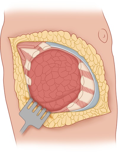
The incision used to resect a tumor in the chest wall is determined by the shape of the tumor and the orientation of its longitudinal axis. If the tumor is elongated in any direction, the incision is made along the longitudinal axis of the ellipse outlined by the tumor protuberance. Most tumors tend to be somewhat spherical in shape, however, and then the preferred incision is an oblique one over the middle of the mass, extending well proximal and distal to the mass along the direction of the ribs. An ellipse is made to circumscribe the site of a prior biopsy of the mass, which then can be removed en bloc with the underlying tumor (Fig. 19.1). Flaps are developed well beyond the palpable extent of the tumor. The investing fascia is incised above and below the area of the tumor (Fig. 19.2). Depending on whether the tumor is located on the posterior or anterior chest wall, the latissimus dorsi and/or the serratus anterior becomes exposed.
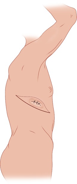
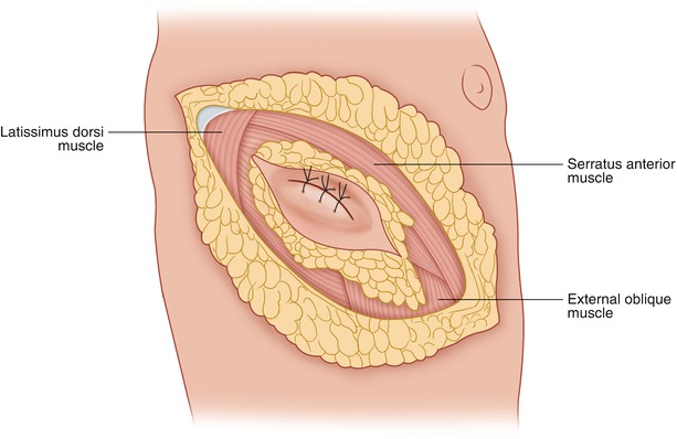

Fig. 19.1
An elliptical incision is made around the previous biopsy incision so that the biopsy track may be removed en bloc with the underlying tumor

Fig. 19.2
Flaps are raised superior and inferior to the tumor
To determine the number and specific ribs which will have to be resected with the tumor, the ribs are exposed anterior and posterior to the tumor (Fig. 19.3). Starting with the rib that most clearly goes through or under the center of this chest wall mass, an inch or so of the rib is removed and the intercostal muscles above and below it are divided, enabling one to insert a finger in the pleural cavity and palpate the extent of the tumor, both from the external surface of the tumor and from inside the thoracic cavity. One should then be able to determine exactly which ribs should be sacrificed and removed en bloc with the chest wall tumor. On the basis of these clinical findings, the involved ribs are divided above and below the central rib that was already divided, being careful to ascertain not only whether a rib needs to be sacrificed but also the appropriate length of the rib that must be removed en bloc with the tumor. The intercostal vessels and nerve running at the inferior border of each rib are carefully ligated and divided.
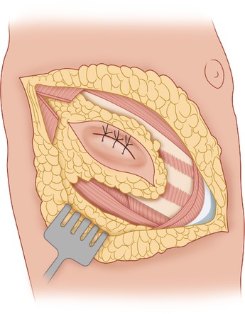

Fig. 19.3
The deep fascia and underlying muscles are divided in the direction of the ribs at a sufficient distance from the tumor mass, exposing the ribs above and below the area of tumor involvement
Early on, as the dissection around the tumor mass proceeds, it is determined whether the underlying lung is adherent to and possibly infiltrated by the tumor. If the lung is adherent to the undersurface of these rib segments, the dissection around the chest wall mass incorporating the involved ribs is completed first. One then can decide whether it is appropriate to wedge out the involved piece of lung, to be removed en bloc with the rest of the specimen, or whether a more formal resection of the lung should take place. In the latter case, the incision is continued anteriorly and/or posteriorly sufficiently in the appropriate intercostal space to provide the room to perform an en bloc lobectomy. As the finger dissection and palpation of the inner surface of the chest wall area to be resected with the overlying tumor proceeds, one should be careful not to break any adhesions between the chest wall mass and the underlying lung, which may contain tumor. Therefore, one should use blunt dissection sparingly, in favor of sharp dissection; it is preferable to remove a piece of the lung in order to make sure that no microscopic tumor residual remains.
The length of the portion to be removed from each rib will not be the same for all the resected ribs, as one follows at the desired distance the shape of the tumor mass on the ribs above and below its location. An additional soft tissue margin may be obtained by dissecting the periosteum off the ribs above and below the area of the tumor that are apparently free of involvement. In this fashion, one obtains a wider soft tissue margin with the tumor to be resected (Fig. 19.4). A mesh or patch may then be placed in the defect and sutured with a running monofilament nylon suture, which is used in several interrupted segments (Fig. 19.5). The edge of the mesh is folded as one goes around the curvature of the defect, in order to keep it taut at all times. The result will be a taut piece of mesh without wrinkles. Suture bites are taken through the intercostal muscles and, where appropriate, around a rib, to provide a firm and reliable closure. A chest tube is placed in the pleural cavity, with suction drains on top of the mesh (Fig. 19.6) to promote adherence of the flaps to the mesh. To avoid a pneumothorax, the suction drains are removed postoperatively at least 1 day earlier than the chest tube.
