Breast Cancer during Pregnancy and Subsequent Pregnancy in Breast Cancer Survivors


Breast Cancer during Pregnancy and Subsequent Pregnancy in Breast Cancer Survivors


Jennifer K. Litton

Richard L. Theriault


INTRODUCTION

Pregnancy and fertility issues have become a significant concern for younger breast cancer patients. Young breast cancer patients can be faced not only with the diagnosis and treatment of their breast cancer, but also with concerns regarding fertility, future pregnancies and, for some, breast cancer diagnosis and treatment during pregnancy. Gestational or pregnancy-associated breast cancer (PABC) is defined as breast cancer that is either diagnosed during pregnancy or within 1 year postpartum. Since women seem to be delaying childbirth to later ages than in previous generations, the incidence of breast cancer and pregnancy, as well as the importance of future pregnancies subsequent to successful treatment for breast cancer, must be considered as part of the informed decision process when discussing treatment of breast cancer with younger women. Given this special situation and the amount of reports in the literature, national societies such as the American Society of Clinical Oncology (ASCO) and National Comprehensive Cancer Network (NCCN) have both issued guidelines on how to approach the treatment of the pregnant cancer patient. We will review the diagnosis and treatment of breast cancer and concurrent pregnancy, the prognosis for those treated during pregnancy, effects of treatment for children exposed to systemic therapies in utero and, for women with successfully treated primary breast cancer, the breast cancer prognosis in relation to future pregnancies.


EPIDEMIOLOGY: BREAST CANCER AND CONCURRENT PREGNANCY

Breast cancer and cervical cancer are the most commonly diagnosed malignancies during pregnancy. In a large retrospective population-based study in California between 1991 and 1997, there were 1.3 cases of breast cancer per 10,000 live births (1). In women under the age of 50 who are diagnosed with breast cancer, approximately 0.2% to 3.8% are diagnosed during pregnancy (2, 3). As women delay childbearing, the incidence of breast cancer coinciding with pregnancy may increase since the frequency of breast cancer diagnosis increases with age (4). In a population-based cohort study based on the Healthcare Cost and Utilization Project—Nationwide Inpatient Sample (HCUP-NIS), Abenhaim et al. reported 573 breast cancers identified in 8,826,137 births over a 10-year interval. The 10-year incidence was noted to be 6.5 cases per 100,000 births. The diagnosis of breast cancer was more common among women over 35 years of age (5).

In patients with breast cancer with a recent past pregnancy, some retrospective analyses have shown a worse prognosis. In a multi-institutional retrospective case-control study, Guinee et al. (6) examined the impact of recent prior pregnancy on breast cancer outcome in a group of 407 women, aged 20 to 29, with breast cancer. The women were matched for age and stage of disease and had never been pregnant. For each 1-year increment in the time between the latest previous pregnancy and breast cancer diagnosis, the risk of dying decreased by 15% (relative risk 0.85, p = .011) (6). In a study of 540 patients from Memorial Sloan-Kettering Cancer Center, patients with previous childbirth within 2 years of the diagnosis of breast cancer also were shown to have a worse prognosis with an adjusted relative risk (RR) of dying from the cancer of 3.1 (7). Kroman et al. from Denmark noted an increased RR of dying from breast cancer for those women who had childbirth within 2 years of diagnosis. After adjusting for age, cancer characteristics and stage, a breast cancer diagnosis within 2 years of
childbirth was significantly associated with death (RR = 1.58, 95% confidence interval 1.24-2.02) compared to patients who gave birth more than 5 years before their breast cancer diagnosis (8). These studies were not able to control for delay in diagnosis, treatment, or treatment modalities of the breast cancer. Future translational research may help identify whether or not there is a true biologic difference in breast cancers diagnosed soon after pregnancy to account for these differences. A recent case-control study concludes that current or recent pregnancy is associated with adverse pathologic features but breast cancer survival is not impaired (9).


BRCA1 and BRCA2 Mutation Carriers and Pregnancy

Women who are more susceptible to breast cancers at younger ages, such as those with deleterious mutations in the BRCA1 or BRCA2 genes may be overrepresented in the population of pregnant breast cancer patients. Few studies, with small numbers of patients, have evaluated the potential increased risk in hereditary breast cancer patients (10, 11). Women with genetic predispositions to cancer, such as those with BRCA1 and BRCA2 deleterious mutations, tend to develop breast cancer at earlier ages and therefore may be more likely to have cancer diagnosed during childbearing years.

Potentially, the relationship of number of pregnancies and age of parity may be significant in BRCA1 and BRCA2 mutation carriers. Antoniou et al. evaluated 457 mutation carriers who developed breast cancer and 332 mutation carriers without a history of cancer. Parous BRCA1 and BRCA2 mutation carriers had a lower risk of developing cancer, but only among carriers who were older than 40 years of age (hazard ratio 0.54, 95% CI 0.37-0.81). Patients with an increased age at first parity had an increased breast cancer risk if a BRCA2 mutation carrier, but not if they were a BRCA1 mutation carrier (12). Kotsopoulos et al. performed a matched case-control study on 1,816 pairs of BRCA1 and BRCA2 mutation carriers, and showed that age at first parity did not influence the development of breast cancer in mutation carriers. Also they did not show a difference between BRCA1 and BRCA2 mutation carriers in univariate or multivariable models (13). An Icelandic study examining 100 BRCA2 mutation carriers did not show a decrease in risk of breast cancer in BRCA2 mutation carriers with increased number of births as has been seen in nonmutation carriers (14). Not only was the protective effect of parity modulated in these patients, but also, in a large matched case-control study comparing 1,260 pairs of women with known BRCA mutations, increasing parity was associated with an increased risk of breast cancer in BRCA2 mutation carriers compared to nulliparous women (OR 1.53, 95% CI 1.01-2.32, p = .05). BRCA2 mutation carriers who were under the age of 50, when compared to nulliparous BRCA2 mutation carriers, had a 17% increase in adjusted risk of breast cancer with each additional birth (OR 1.17, 85% CI 1.01-1.36, p = .03) (15).

Given the risk of a deleterious mutation in women who develop breast cancer at an early age, genetic counseling should be part of the clinical discussion and evaluation for all such patients. Having children at an early age does not appear to provide the same protective effects in patients with deleterious BRCA1 or BRCA2 gene mutations as for those without mutations. In fact, recent parity may increase the risk of being diagnosed with breast cancer potentially more notably in those with deleterious BRCA2 mutations.


EVALUATION OF BREAST MASSES DURING PREGNANCY


Imaging of the Breast during Pregnancy

Mammography: Mammography should be ordered in pregnancy with abdominal shielding. With mammography the fetal radiation exposure is estimated to be 0.4 mrad (17). This level is substantially below the level of 5 rad, a level at which multiple studies have shown no known increase in congenital malformations or growth retardation (18).

Ultrasonography: Ultrasound (US) can distinguish between cystic and solid breast masses in approximately 97% of cases with no radiation exposure. One study diagnosed 100% of the breast masses as well as axillary metastases in 18 of 20 women (19). Ultrasound was also shown to be effective for evaluating response to preoperative chemotherapy in the pregnant breast (19).

Breast MRI: Magnetic resonance imaging (MRI) has not been studied for the diagnosis of a breast mass in pregnant or lactating women. Gadolinium-enhanced MRI may be more sensitive than conventional mammography; however, data regarding the safety of gadolinium during pregnancy are limited. Gadolinium has been shown to cross the placenta and be associated with fetal abnormalities in animal models (20, 21). Animal studies have shown diverse fetal effects and gadolinium is considered a pregnancy category C drug (Table 65-1). There have been no controlled human studies to date, however; several studies have observed no significant toxicity when gadolinium has been given during human pregnancy. But there does remain some controversy as to the safety of gadolinium during pregnancy and therefore should be used with caution.


Staging and Diagnosis of Breast Cancer during Pregnancy


Biopsy

Any clinically suspicious breast mass requires biopsy, even if the ultrasound and mammogram are equivocal or nondiagnostic. Fine-needle aspirate (FNA) in the pregnant breast is well established. While FNA may provide cytologic confirmation of cancer, core biopsy is preferred because it can confirm invasive disease and provide tissue for biologic
marker evaluation. Core or excisional biopsies can be performed safely with local anesthesia, with only one found report of the development of a milk fistula after a core needle biopsy (22).


TABLE 65-1 U.S. FDA Pregnancy Category Definitions


A


Controlled studies in women fail to demonstrate a risk to the fetus in the first trimester, and the possibility of fetal harm appears remote.


B


Animal studies do not indicate a risk to the fetus and there are no controlled human studies or animal studies to show an adverse effect on the fetus, but well-controlled studies in pregnant women have failed to demonstrate a risk to the fetus.


C


Studies have shown that the drug exerts animal teratogenic or embryocidal effects, but there are no controlled studies in women, or no studies are available in either animals or women.


D


Positive evidence of human fetal risk exists, but benefits in certain situations (e.g., life-threatening situations or serious diseases for which safer drugs cannot be used or are ineffective) may make use of the drug acceptable despite its risks.


X


Studies in animal or humans have demonstrated fetal abnormalities or there is evidence of fetal risk based on human experience, or both, and the risk clearly outweighs any positive benefit.


Pathology of Breast Cancer Diagnosed during Pregnancy

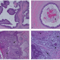
The majority of breast cancer cases are infiltrating ductal adenocarcinomas with one prospective cohort showing 84% with poorly differentiated tumors (23). When compared to nonpregnant premenopausal women, pregnant patients are reported to have tumors with a lower frequency of ER and/or PR expression (23, 24, 25 and 26). Amplification of HER2/neu is seen in approximately 20% to 30% of breast cancers. In some series, HER2/neu amplification has been reported to be disproportionately amplified in pregnant patients (up to 58% vs. 16 % in nonpregnant counterparts) (27); however, another series show similar HER2/neu amplification (28%) in pregnant and nonpregnant patients (23).


Staging Evaluations during Pregnancy

The American Joint Committee on Cancer (AJCC) TNM staging system is used and, along with tumor biologic characteristics, forms the basis for treatment decisions. Staging procedures may need to be modified for the pregnant breast cancer patient with safety considerations for both the patient and the fetus. Staging is important, however, for better understanding of the full extent of disease, informing treatment recommendations, considering the potential influence of the treatment on cancer outcome and the potential impact of cancer treatment on the fetus and pregnancy. Radiation exposure for the fetus and outcomes regarding different imaging modalities are available (28).

Guidelines have been established regarding imaging and staging during pregnancy (29, 30). A recent practice patterns analysis recommends an algorithm that pregnant patients with breast cancer be considered for chest CT scan if pulmonary metastases are suspected and that MRI of the abdomen and pelvis along with a bone scan may be considered if metastases at these sites are suspect (31). However, staging procedures need to be tailored to minimize fetal radiation exposure, provide adequate information to determine disease stage, and provide sufficient knowledge for informed decisions for clinicians and patients. Recommended initial staging should include the following: complete history and physical examination with special attention to breast and nodal basin examinations; comprehensive metabolic panel; and complete blood count with differential. It is important to note that pregnant patients may have anemia due to the increase in circulating plasma volume. They may have increases in serum alkaline phosphatase level that can be doubled or tripled due to the pregnancy itself. The physical examination of the breast and nodal basins needs to include tumor measurements when possible and an assessment of extent of clinical nodal involvement. Given that the sites of breast cancer metastases are most commonly bone, liver, and lung, these areas should be evaluated in the pregnant breast cancer patient who has a clinical stage II or higher breast cancer as is done in nonpregnant patients. These staging evaluations may include chest x-ray with abdominal shielding; echocardiogram prior to the use of an anthracycline-based chemotherapy regimen; ultrasound of the liver; and a screening noncontrast MRI of the thoracic and lumbar spine to exclude bone metastases. If there are concerns of liver metastases after ultrasound examination, an abdominal non-gadolinium-enhanced MRI may be considered, especially since the liver may tend to have fattyreplacement during pregnancy. Noncontrast MRI has been used routinely and safely in the pregnant patient for staging. CT scans and bone scans are not recommended for routine use due to concerns of excessive fetal radiation exposures (29). Radionuclide scanning, including bone scanning, has very limited safety data in the pregnant patient, and should be considered only if absolutely necessary with aggressive hydration and frequent voiding.


Locoregional Therapy


Surgery and Anesthesia

Breast surgery can be safely performed in all trimesters of pregnancy; however, patients and surgeons may choose to wait until after the 12th week of gestation when the risk of spontaneous abortion may be lower (29). Multiple studies evaluating the risks of anesthesia during pregnancy have not shown an increase in fetal abnormalities. Mazze and Kallen reported on a registry of 5,405 pregnant patients who had any kind of surgery during pregnancy. They observed no difference in the risk of fetal malformation when they compared the pregnant group to 720,000 women who did not have a surgical procedure during pregnancy. There was no difference in outcomes in women who had their surgeries in
the first trimester. There was, however, an increase in the frequency of low- and very-low-birth-weight infants. This was attributed to the underlying illness or trauma necessitating the surgery (32). In a Canadian report of 2,565 pregnant women who underwent surgery, Duncan et al. reported no increase in fetal abnormalities compared to a control population of pregnant women who did not have an operation (33). A recent review of surgery in the pregnant patient recommends that the preferred timing for surgical intervention is 16 to 20 weeks of gestation (34).

Although in the majority of published reports patients opt for mastectomies for breast cancer treatment due to concerns regarding radiation therapy, breast-conserving surgery is an option, especially in women in the third trimester of pregnancy who can receive radiation therapy after delivery. With the potential of preoperative chemotherapy during pregnancy, breast-conserving surgery can be done later in the pregnancy or after delivery (35).

Safety and efficacy of sentinel lymph node biopsies is currently an area of clinical interest. The sensitivity and specificity of sentinel lymph node biopsies in the pregnant woman with breast cancer has not been well established. Estimated radiation exposure to the fetus is low and calculated to a maximum of 4.3 mGy (36). However, isosulfan blue dye mapping is not recommended due to concerns of unknown effects for the fetus as well as risk of anaphylaxis for the patient. Sensitivity of sentinel lymph node mapping may be significantly decreased without using isosulfan blue. The concern with sentinel node procedure in the pregnant patient is not the technical aspects of the procedure but the accuracy of the diagnostic information obtained as a result of the procedure. Sentinel node excision has not been subjected to the same rigorous study in the pregnant population as in the nonpregnant population (37). Therefore prudent and careful clinical evaluation prior to the procedure is warranted.

Jul 9, 2016 | Posted by in ONCOLOGY | Comments Off on Breast Cancer during Pregnancy and Subsequent Pregnancy in Breast Cancer Survivors

Full access? Get Clinical Tree

Get Clinical Tree app for offline access