ADVANTAGES AND DISADVANTAGES OF IMAGING TECHNIQUES
Traditionally, the first imaging examination in the evaluation of the pituitary-sellar region was a skull radiograph series.1 Plain radiographs offer the advantage of being noninvasive and inexpensive; they yield a certain amount of limited information about the adjacent bony structures (Fig. 20-1). However, potentially important secondary soft-tissue changes can only be inferred, although soft-tissue calcifications can be seen. Thus, in general, routine radiographs provide only minimal information related to the sella, and hence are generally not obtained.
 FIGURE 20-1. Lateral radiograph of sella. Labeled structures include planum sphenoidale (curved solid white arrows), tuberculum sella (short black arrow), lamina terminalis (open white arrow), sphenoid sinus (black arrowheads), anterior clinoid (long thin black arrow), and posterior clinoid (open curved white arrow). This patient has a relatively well-pneumatized sphenoid sinus. The chiasmatic sulcus is not well marginated. |
Computed tomography (CT) was the first imaging modality to directly visualize the pituitary gland, hypothalamus, and optic chiasm.2 The bony structures in this region can be well evaluated with CT (Fig. 20-2). It is more sensitive than either plain radiographs or magnetic resonance imaging (MRI) in the detection of calcifications within soft tissues. However, intravenous contrast agents frequently are necessary to improve the image contrast and to enhance the vasculature, and CT involves radiation exposure.
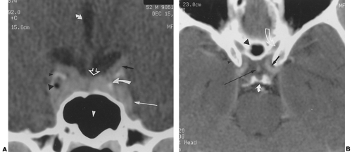 FIGURE 20-2. Normal sellar and suprasellar CT. A, Coronal CT scan obtained with contrast enhancement through the anterior third ventricle (short curved solid white arrow), supraclinoid carotid artery (thick black arrow), calcified cavernous carotid artery (long curved solid white arrow), pituitary gland (open white arrow), anterior clinoid (small black arrowhead), cavernous sinus (long white arrow), incidental fat in the cavernous sinus (large black arrowhead), and sphenoid sinus (white arrowhead). B, Axial CT scan made with contrast enhancement through the suprasellar cistern. The labeled structures include anterior clinoids (small black arrowhead), posterior clinoids (curved solid white arrow), optic canal (curved open white arrow), pneumatized planum sphenoidale (large black arrowhead), pituitary stalk (long thin black arrow), and supraclinoid carotid artery (short black arrow).
Related posts:Stay updated, free articles. Join our Telegram channel
Full access? Get Clinical Tree
 Get Clinical Tree app for offline access

|
