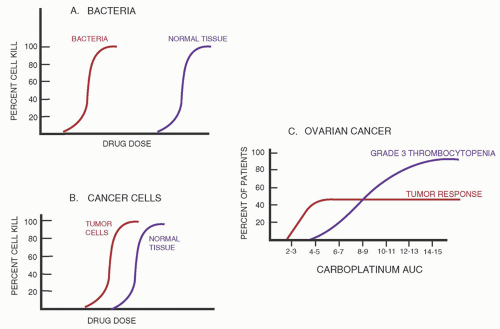
Principles and Pharmacology of Chemotherapy

Principles and Pharmacology of Chemotherapy
Kenneth R. Hande
PRINCIPLES OF CHEMOTHERAPY
To optimally treat cancer patients, clinicians should understand the principles of cancer chemotherapy as well as the pharmacology of the drugs they are administering. The first section of this chapter reviews our understanding of (a) cancer biology, (b) mechanisms of antineoplastic drug action, (c) mechanisms of drug resistance, (d) drug toxicities, (e) pharmacologic principles (common and unique), and (f) potentials and limitations of therapy for individual patients. Specific drugs used to treat hematologic malignancies, their mechanism of action, and their toxicities are summarized in the second section of this chapter.
Cancer Biology
Most cancers are detected when there are 104 to 1011 neoplastic cells present.1 The large number of cells present at diagnosis usually requires multiple courses of treatment or a long duration of therapy to result in a cure. Even if a drug, or a combination of drugs, is quite effective in killing cancer cells, such that it kills 999 of every 1,000 cells present (99.9% cell kill), one treatment will reduce a tumor population from 1011 to 108 cells. At least four treatments are therefore necessary to eliminate the final cancer cell, assuming there is no tumor cell growth during the treatment period. In fact, because of toxicity to normal tissues and the time needed for normal tissue recovery, cancer chemotherapy must be spaced out in treatment cycles. During the period between cycles, tumor regrowth occurs. Thus, most curative treatment regimens require prolonged therapy, often using multiple courses of chemotherapy, with the number of courses depending on the tumor mass at the time of diagnosis and the sensitivity of the tumor to the drugs. The fraction cell kill (e.g., 90%, 99%, 99.99% cell kill) depends on the sensitivity of cells to the antineoplastic drug and the dose of drug administered. Rapidly dividing cells are generally more sensitive to antineoplastic agents while nondividing cells are generally resistant.
Most cancers do not grow at a fixed rate throughout their course. Initial growth rates may be exponential. Gompertzian growth is sigmoid. In the Gompertzian model, the growth fraction of the tumor reduces as the tumor enlarges.2 Because a significant proportion of cells in a large tumor mass are often in a resting or dormant phase, presumably as a result of lack of adequate nutrients or oxygen, they may not be affected by chemotherapy. The best opportunity for cell kill by chemotherapy is in the early portion of the growth curve, when all cells are dividing.
Recurrence of cancer after “complete remission” (disappearance of all detectable disease) can be explained by the inability of current staging methods to detect fewer than 103 to 104 tumor cells in the body even by the most sensitive means. Chemotherapy that kills several logs of tumor cells (e.g., from 1011 to 104) eliminates all visible evidence of cancer and prolongs survival, but the malignancy will recur at a later time. Examples of possible clinical courses associated with cancer chemotherapy are illustrated in Figure 68.1.
The growth fraction of a cancer represents the percentage of cells progressing actively through the cell cycle. A cell cycle is marked by two observable events. In S-phase (synthesis), DNA replication occurs; during M-phase (mitosis), cellular division into two daughter cells is noted.1 G1 (gap) is the time between the end of mitosis and the start of the next S-phase. G2 is the time between the completion of S-phase and the start of M-phase. Cells that have ceased to proliferate for prolonged periods have entered a G0 phase (or resting phase) of the cell cycle (Fig. 68.2). Some drugs are cytotoxic when exposed to cancer cells in any phase of the cell cycle. Other drugs are phase-specific; that is, they are cytotoxic to cells only in a particular phase of the cell cycle. Cytarabine, for example, is an S-phase-specific agent, whereas vincristine is M-phase-specific. For cells to be killed by cytarabine, the drug must be present when the cell is synthesizing DNA.
Mechanisms of Antineoplastic Drug Action
Antineoplastic agents have been discovered by many methods. Exposing cancer cells in culture to natural or synthetic compounds to screen for materials with antineoplastic activity has identified a number of active antineoplastic agents (drugs such as paclitaxel). Since DNA is important in cell division, compounds blocking DNA synthesis have been developed. Over the past 20 years, genes and their protein products that regulate cell division have been identified. The molecular pathways by which these oncogenes cause cellular proliferation have been determined.3 Knowledge of molecular pathways critical to the growth of cancer cells has led to the targeted development of molecules altering specific pathways (e.g., tyrosine kinase inhibitors, angiogenesis inhibitors). During the past two decades, the development of cancer drugs has shifted from a screening process where active agents are identified and their mechanism of action later defined, to first identifying a targetable pathway, finding a compound to block that pathway, and then testing for antineoplastic activity.
No matter how an antineoplastic has been developed, these drugs interfere with an essential step required for cell growth or division. The initial target of antineoplastic drugs varies widely, from direct attack on the DNA molecule to inhibition of signal transduction molecules. However, all antineoplastic agents cause a disruption in a cellular process so significant to the cancer cell that it requires the cell to either quickly repair the damage or to initiate the process of apoptosis (programmed cell death). Essentially, all antineoplastic agents result in cancer cell death through initiation of apoptosis.4 Apoptosis is the normal physiologic process of cellular suicide, which occurs in all living organisms to eliminate unwanted, functionally abnormal, or harmful cells. In apoptosis, in contrast to cellular necrosis, the cell shrinks and condenses, fragmenting into multiple membrane-bound bodies (apoptotic bodies) that are engulfed by surrounding cells without inflammation or damage to the surrounding tissues. Biochemically, apoptosis is characterized by fragmentation of nuclear DNA, demonstrated by a typical ladder pattern on agarose gel electrophoresis.
To understand how cytotoxic agents initiate apoptosis, an understanding of the events that occur during the normal cell cycle is important. As previously mentioned, DNA synthesis is not continuous from one mitosis to the next but takes place in only a specific period of the cell cycle, the S-phase. Similarly, mitosis or the M-phase takes up only a small part of the cell cycle.1 In most, if not all cells, the cell cycle is temporarily halted during the G1-S-phase checkpoint and at the G2-M-phase checkpoint. At these times, cells determine whether to continue into the S-phase, initiate the process of apoptosis, or undergo DNA repair. Passage into a new phase of the cell cycle requires activation of a series of enzymes called cyclin-dependent kinases, which activate another group of enzymes (the cyclins).5 If cells are damaged by chemotherapeutic agents and are unable to repair the damage, apoptosis is initiated at the G1-S or G2-M checkpoint, provided the mechanisms for apoptosis are in place. Several critical factors are needed for apoptosis to occur such as p53, p63, and p21. P53 is a nuclear phosphoprotein capable of binding to specific DNA sequences and activating selected target genes triggering apoptosis. Cells with a mutated P53 gene have a relative resistance to cancer chemotherapeutic agents and are unable to initiate apoptosis properly.6 P21 is a protein induced by p53 that binds to and inactivates the cyclins required for progression of cells into the S-phase. Other factors are known to affect the apoptotic pathway. A protein originally found in B cell lymphomas, bcl-2, blocks apoptosis. Caspases are the key machinery causing the breakdown of DNA and proteins. Caspases are specialized proteases existing as proenzymes, which can be rapidly activated by a number of factors. The Fas ligand and tumor necrosis factor stimulate the protease cascade. It is clear that antineoplastic agents provide the initial trigger for beginning the pathway to programmed cell death (Fig. 68.3). However, the presence, or absence, of apoptotic proteins is as important as the initial interaction between a cytotoxic drug and its effecter in determining whether tumor cell kill occurs.4
FIGURE 68.1. Schematic representation of clinical course of two patients with Hodgkin disease. Both patients A and B are diagnosed with a clinically detected tumor mass (1011 cells). Both patients are treated with ABVD (doxorubicin, bleomycin, vincristine, and DTIC) every 2 weeks. One course of therapy for patient A results in a 2-log tumor cell kill. Patient B’s therapy results in only a 1-log tumor cell kill. Some tumor growth occurs while waiting for normal tissue recovery before initiation of the next treatment. Three months into therapy, neither patient A nor B has clinically detectable disease. Treatment is stopped after 6 months of therapy. Patient A is cured, whereas disease recurs in patient B 3 months after stopping therapy.
FIGURE 68.2. The cell cycle. The cell cycle is marked by two observable events. During S-phase (synthesis), DNA replication occurs; and during M-phase (mitosis), cells divide. G1 and G2 (gap) are times between completion of M-phase and start of S-phase and between completion of S-phase and start of M-phase, respectively. During G1 and G2, the cell prepares for the S- and M-phases of the cell cycle. Cells may temporarily cease to divide and enter a resting or G0 phase of the cell cycle. The G1-S checkpoint is a critical phase in the cell cycle, when directions for entering S-phase or committing to apoptosis (programmed cell death) are given.
FIGURE 68.3. Potential pathways involved in cytotoxicity induced by chemotherapy. Chemotherapeutic drugs or growth factor deprivation damages cells. Cells are arrested at the G1-S checkpoint. If the damage is sublethal, it may be repaired and the cell proceeds to S-phase. If significant DNA damage is present, the process of programmed cell death is initiated. Critical factors, such as p53 and p21 gene products, are required for the cell to undergo apoptosis.
Drug Toxicity
As opposed to many other classes of drugs, the therapeutic window for chemotherapeutic agents is narrow (Fig. 68.5). The dose of drug needed to achieve adequate tumor cell kill often causes toxicity to normal tissues. For many antineoplastic agents, a sigmoidal curve defining the relationship between toxicity and the dose of drug administered (or the area under the plasma drug concentration [AUC] versus time curve) has been demonstrated. At low drug concentrations, no cytotoxicity is observed. With increasing concentration, cell kill is proportional to dose. At higher concentrations, the effect plateaus. Similar relationships correlating the dose of drug and the antitumor response have also been demonstrated.14 A positive relationship between the drug dose administered and the tumor response rate has been found for many tumors, including the lymphomas. Higher response rates and an increased chance for cure are achieved with higher doses of selected agents.15, 16 For this reason, clinicians generally try to push doses of agents to toxicity to maximize the chance for cure. The optimal dose or AUC for an individual patient with a particular cancer is unknown.
Chemotherapeutic agents cause a wide variety of toxicities (Table 68.1). Myelosuppression and nausea are seen, to some degree, with many anticancer drugs. Nausea and vomiting, although generally self-limited and not life-threatening, are very distressing to patients. The emetogenic potential of antineoplastic agents varies from drug to drug and is dose-dependent17 (Table 68.2). Serotonin receptors, located within the vagal and splanchnic nerves of the gastrointestinal system and in the brain, are critical in the initiation of nausea by chemotherapeutic agents. The development of specific type 3 serotonin (5-hydroxytryptamine or 5HT3) receptor blockers, such as ondansetron, dolasetron, and granisetron, and NK-1 inhibitors, such as aprepitant, has resulted in major improvement in control of chemotherapy-induced emesis.17, 18
FIGURE 68.5. Schematic representation curves relating drug concentrations to response. Antibacterial agents (A) have a wide therapeutic window, with a significant difference in the dose that produces bacterial cell kill and the dose that causes toxicity to normal tissues. For antineoplastic drugs (B), the therapeutic window is narrow (e.g., doses adequate to produce tumor cell kill usually produce toxicity to normal tissues). For a few cancers, such as ovarian (C), the shape of the dose-response curve has been defined.14 For most tumors, however, a specific dose-response (or AUC-response curve) has not been defined.
Myelosuppression is the most common dose-limiting antineoplastic agent toxicity. The suppression of hematopoietic cell lines is determined by the kinetics of the specific cell line in the peripheral compartment. Anemia occurs as a late effect because of the long half-life of red blood cells (120 days). Thrombocytopenia occurs in an intermediate time frame (platelet half-life 5 to 7 days), whereas granulocyte suppression occurs earliest. Granulocytopenia is a more frequent occurrence than thrombocytopenia or anemia. The white blood cell count usually drops 5 to 14 days after drug administration, with recovery by 7 to 21 days. Several exceptions are recognized, such as a predominant thrombocytopenia seen with carboplatin and delayed myelosuppression with busulfan and the nitrosoureas (occurring 4 to 5 weeks posttherapy). The degree and duration of bone marrow suppression are related directly to the dose of drug administered. High-dose chemotherapy regimens, used with stem cell transplantation, generally result in 10 to 25 days with neutrophils <500/mm3 and thrombocytopenia lasting for a more extended period. The length of cytopenias depend upon the stem cell source with peripheral blood shorter (10 to 14 days) than bone marrow (20 to 25 days). An increased risk of infectious complications occurs that is related directly to the degree and duration of granulocytopenia.19 The development of recombinant hematopoietic colony-stimulating factors (CSFs), such as erythropoietin, thrombopoietin, granulocyte-macrophage CSF, and granulocyte-CSF, has shortened the duration of bone marrow suppression but not eliminated it. CSFs are now commonly used prophylactically with regimens expected to produce an incidence of febrile neutropenia >20%. Guidelines for the use of these important, yet expensive, factors are available and should be followed.20
The range of toxicities associated with antineoplastic agents is too great to review in detail in this chapter. Readers are referred to reviews on endocrine,21 oral,22 renal,23 neurologic,24 hepatic,25 pulmonary,26 cardiac,27 and cutaneous28 toxicities of antineoplastic agents. Physicians must also remember that most antineoplastic agents have teratogenic and mutagenic potential. Alkylating agents are the most damaging to testicular and ovarian function. Damage is dependent on drug dose, the age of the patient (older patients are more likely to experience toxicity), and sex (males more than females).29 Second malignancies (primarily acute leukemia) have been associated with alkylating agents, epipodophyllotoxins, nitrosoureas, and anthracyclines.30 Concomitant radiation therapy increases the risk of second malignancies (such as sarcomas or breast cancer) 5 to 20 years following therapy.
General Pharmacologic Principles
Antitumor activity of an antineoplastic agent is best correlated with the concentration of drug that reaches the site of drug action within the cancer cell (Fig. 68.5A). Several factors may affect what happens to a drug following administration until it reaches its site of action (Fig. 68.6). Pharmacokinetics describes what happens to a drug following administration (what the body does to the drug). Pharmacokinetics represents an attempt to predict quantitatively how a patient will handle a given dose of drug.31 Important pharmacokinetic parameters include (a) bioavailability or absorption, (b) volume of distribution, (c) clearance or drug elimination, and (d) drug half-life (Fig. 68.7). Pharmacodynamics describes what effect the drug has on a particular tissue (what the drug does to the body).
TABLE 68.1 ANTINEOPLASTIC DRUG TOXICITY

Nausea/Vomiting

Bone Marrow

Mucositis

Alopecia

Neurologic

Diarrhea

Hepatic

Vesicant

Pulmonary

Renal

Cardiac

Hypersensitivity

Actinomycin

Actinomycina

Actinomycina

Actinomycin

Ara-Ca

Arsenic trioxide

Asparaginase

Daunorubicin

Arsenic trioxide

5-Azacytidine

Arsenic trioxide

Asparaginasea

Asparaginase

Alemtuzumab

Ara-C

Amsacrine

Asparaginase

Asparaginase

Azathioprine

Doxorubicin

ATRAa

Bevacizumab

Cyclophosphamide

Bleomycin

Arsenic trioxide

Alkylating agents

Busulfan

Ara-C

Bortezomiba

Ara-C

Ara-C

DTIC

Ara-Ca

Carboplatin

Desatinib

Denileukin diftitoxa

Ara-C

Asparaginase

Bleomycin

Bleomycin

Busultan

Denileuken diftitox

BCNU

Epirubicin

Azathioprine

Cisplatin

Daunorubicin

Docetaxel

Azacytidine

Ara-Ca

Capcitibine

Busulfan

Carboplatin

Dasatinib

Busulfan

Idarubicina

Bleomycina

Cladribine

Doxorubicin

Etoposide

Bortezomib

Azacytidine

Cyclophosphamide

Cyclophosphamide

Cisplatina

Doxorubicin

Clofarabine

Mechlorethamine

Busulfan

Fludarabinea

Epirubicin

Monoclonal antibodies

Carboplatin

Bendamustine

Daunorubicina

Cytarabine

Cladribine

Fludarabine

Cytarabine

Mitomycina

Chlorambucil

Gemcitabine

Fluorouracil

HMM

Carmustinea

Bortezamib

Docetaxel

Daunorubicin

Fludarabine

Fluorouracila

Dactinomycin

Mitoxantronea

Cyclophosphamide

Ifosfamidea

Idarubicina

Paclitaxel

Cisplatina

Busulfana

Doxorubicina

Docetaxel

Fluorouracila

HMM

DTIC

Vinblastinea

Dasatinib

Interferon

Mitoxantrone

Teniposide

Cyclophosphamidea

Carboplatina

Epirubicin

DTIC

Gemcitabine

Idarubicin

Gemcitabine

Vincristinea

Etoposide

Methotrexate

Trastuzumab

Daunorubicina

Chlorambucila

Etoposide

Doxorubicin

HMM

Imatinib

Interferon

Vinorelbinea

Fludarabine

Mitomycin

Decitibine

Cladribine

Fludarabinea

Etoposide

Ifosfamidea

Irinotecana

Interleukin-2

Gemcitabine

Nitrosoureas

Denileuken- diftitox

Clotarabine

Hydroxyurea

Fluorouracil

Interferon

Methotrexate

L-Asparaginasea

Ifosfamide

Pentostatin

Doxorubicina

Cyclophosphamidea

Fluorouracil

Hydroxyurea

L-Asparaginasea

Thioguanine

Imatinib

Irinotecan

Sorafenib

DTICa

Daunorubicina

Idarubicin

Idarubicin

Lenalidomide

Topotecana

Interferona

Lenalidomide

Streptozocina

Epirubicin

Decitibine

Ifosfamide

Ifosfamide

Methotrexate

Mercaptopurine

Mercaptopurine

Sunitinib

Etoposide

Docetaxela

Methotrexatea

Irinotecan

Nitrosoureas

Methotrexatea

Methotrexatea

Carboplatin

Fludarabine

Doxorubicina

Mercaptopurine

Methotrexate

Nelarabine

Paclitaxel

Melphalan

Cisplatin

Fluorouracil

DTICa

Mitomycin

Mitoxantrone

Oxaliplatina

Mitomycin

Mitomycina

Cladribine

Gemcitabine

Etoposidea

Mitoxantrone

Mustard

Paclitaxela

Nitrosoureas

Nitrosoureasa

Fludarabinea

HMMa

Fludarabinea

Nitrosoureas

Nitrosoureas

Pentostatin

Sunitinib

Oxaliplatin

Gemcitabine

Imatinib mesylate

Hydroxyureaa

Paclitaxel

Paclitaxel

Procarbazine

Thioguanine

Procarbazine

Hydroxyurea

Ibitumomab

Procarbazine

Procarbazine

Rituzimab

Tretinoin

Paclitaxel

Idarubicin

Idarubicina

Taxotere

Vinblastine

Thalidomidea

Thalidomide

Ifosfamide

Ifosfamidea

Thioguanine

Vincristine

Tretinoin

Vinca alkaloids

Interferon

Interferon

Thiotepa (high-dose)

Vinorelbine

Vinblastinea

Irinotecan

Irinotecan

Vinblastine

Vincristinea

Mechlorethaminea

Lenalidomide

Vincristine

Vinorelbinea

Methotrexate

L-PAMa

Mitomycin

Methotrexate

Mitoxantrone

Mercapto purinea

Mustarda

Mitomycina

Nitrosoureas

Mitoxantronea

Irinotecan

Mustarda

Pentostatin

Nelarabine

Procarbazine

Nitrosoureasa

Romidespin

Paclitaxela

Streptozocina

Pentostatin

Thioguanine

Procarbazine

Thiotepa

Romidespin

Topotecan

Teniposidea

Vorinostat

Thioguanine

Thiotepa

Tositumomab

Vinblastine

Vinorelbine

Vorinostat

Ara-C, cytosine arabinoside; ATRA, all-trans retinoic acid; DTIC, decarbazine; HMM, hexamethylmelamine; l-PAM, L-phenylalanine mustard.

a Major toxicity.

TABLE 68.2 EMETIC POTENTIAL OF SELECTED ANTINEOPLASTIC AGENTS

High (>90% risk)

Carmustine

Dactinomycin

Cisplatin

Mechlorethamine

Cyclophosphamide (>1.5 mg/m2)

Streptozocin

Dacarbazine

Moderate (30-90% risk)

Alemtuzumab

Doxorubicin

Azacytidine

Epirubicin

Bendamustine

Idarubicin

Carbazitaxel

Ifosfamide

Carboplatin

Irinotecan

Clofaribine

Methrotrexate (>250 mg/m2)

Cyclophosphamide (>1.5 g/m2)

Oxaliplatin

Cytarabine (>1 g/m2)

Pralatrexate

Daunorubicin

Temozolomide

Low (10-30% risk)

Bortezomib

Mitomycin

Cytarabine (<1 g/m2)

Mitoxantrone

Docetaxel

Paclitaxel

Eribulin

Panitumumab

Etoposide

Pegylated liposomal doxorubicin

5 Fluorouracil

Pemetrexed

Chlorambucil

Romidesin

Gemcitabine

Temsirolimus

Ixabepilone

Topotecan

Hydroxyurea

Trastuzumab

Melphalan

Vorinostat

Methotrexate (<250 mg/m2)

Minimal (<10% risk)

Bevacizumab

2-Chlorodeoxyadenosine

Bleomycin

Fludarabine

Busultan

Rituximab

Cetuximab

Vinca alkaloids

Bioavailability, the percentage of a dose of drug that reaches the plasma compartment, defines drug absorption (Fig. 68.7). Drugs given intravenously have, by definition, 100% bioavailability. Factors that decrease the bioavailability of orally administered drugs include poor solubility in aqueous solutions or metabolism of the drug by the intestine or liver before entry into the systemic circulation (first-pass effect). Bioavailability of poorly soluble drugs, such as etoposide, decreases at high oral drug doses.32 Bioavailability of orally administered 6-mercaptopurine (6-MP) and 5-fluorouracil is low because of extensive first-pass hepatic drug metabolism.33, 34 With poor or widely variable bioavailability of a particular agent, an intravenous route of administration is preferred. The most important factor regarding bioavailability is the variation from patient to patient in the amount of oral drug absorbed (coefficient of variation). If bioavailability of an oral anticancer drug were 50% in all patients, simply doubling the drug dose would produce an equivalent response to intravenously administered drug. However, the variation from patient to patient in the AUC achieved is usually greater with oral than with intravenously administered drug.35 Therefore, variation in toxicity is also greater.
After a drug reaches the bloodstream, it is distributed into tissues. Figure 68.8 illustrates a plasma concentration-versustime curve for a drug with a typical two-phase (distribution and elimination) decline in plasma concentration. Distribution may be affected by drug binding to plasma proteins (usually albumin or α1-acid glycoprotein). Only free drug is biologically active. Decreasing the amount of bound drug by having a low serum albumin concentration or displacing drug from its binding site may increase toxicity (as seen with etoposide).36 Distribution of drug into a “third space,” such as ascites or a pleural effusion, may slow clearance and increase toxicity, as is noted with MTX.37
Clearance is expressed as the volume of biologic fluid (usually plasma) from which a drug can be removed per unit of time. Clearance of most drugs is constant over a range of plasma concentrations, which means that the mechanism for elimination is not saturated. However, some drugs, such as paclitaxel, demonstrate saturable elimination at high plasma concentrations.38 This means that a greater proportional drug effect is noted when high drug doses are employed. Clearance measurements allow physicians to maintain a particular plasma concentration because the dosage rate = clearance × desired plasma concentration.
Removal of drugs from the circulation occurs primarily by metabolism, renal elimination, or hepatic excretion. If urinary excretion is an important route of elimination, renal failure results in slower removal of the drug from the body. Administration of the usual dosage of a drug cleared by the kidney to a patient with renal insufficiency leads to greater drug accumulation and an increased likelihood of toxicity. In such cases, drug dosage needs to be modified to prevent excess toxicity. Using available data on drug clearance, the appropriate dose in renal insufficiency may be calculated as follows: Dose in renal insufficiency = normal dose × (clearance in renal insufficiency ÷ normal clearance)
In contrast to a predictable decline in renal drug clearance when glomerular filtration is reduced, it is not possible to make a general prediction of the effect of liver disease on hepatic biotransformation of drugs. Even in advanced hepatocellular disease, the magnitude of impairment in drug clearance usually is only two- to fivefold. The extent of such changes cannot be predicted by common tests of liver function. Consequently, even when it is suspected that drug elimination is altered in liver disease, there is no quantitative base on which to adjust the dosage regimen other than assessment of clinical response. Antineoplastic drugs for which dose modifications are indicated for renal or hepatic insufficiency are listed in Table 68.3.
The hepatic metabolism of drugs may be altered by genetic deficiency of a metabolizing enzyme or by inhibition of metabolism by another drug. Pharmacogenomics is the study of how a particular patient’s genetic makeup affects drug metabolism. It is now recognized that some patients inherit a mutated gene which produces an inactive or much less active enzyme important in clearing a drug from the body.39 Such patients have a markedly lower drug clearance and a higher incidence of toxicity. Examples include the inherited deficiencies of dihydropyrimidine dehydrogenase, the enzyme that degrades 5-fluorouracil40; UGT1A1, the enzyme that inactivates irinotecan41; and thiopurine methyltransferase, the enzyme that degrades azathioprine and 6-MP.42
FIGURE 68.6. Schematic representation of drug disposition in the body. For a drug to function, it must be taken into the body (1) and the drug must avoid being cleared from the body by metabolism (2) or excretion (5). It must reach its site of action in active form (3), without being inactivated by protein binding (4).
Certain antineoplastic drugs are metabolized by the hepatic P-450 microsomal enzyme system (vincas, epipodophyllotoxins, cyclophosphamide, tyrosine kinase inhibitors, and the taxanes). The activity of the microsomal enzyme systems may be increased with concomitant use of phenobarbital, phenytoin, or other drugs. Use of anticonvulsants has been shown to increase the catabolism of teniposide, a drug eliminated from the body through hepatic microsomal metabolism.43 Clearance of drugs metabolized by the hepatic P-450 enzyme system, such as paclitaxel or Taxotere, can be decreased with concomitant use of P-450 inhibitors, such as ketoconazole or selected antiretroviral agents.
It is important to recognize the unpredictable variation in the way chemotherapeutic drugs are handled by the body. Mean values for bioavailability, clearance, and volume of distribution of anticancer agents have standard deviations of 20%, 50%, and 30%, respectively. This means that target drug concentrations may vary widely from patient to patient, even those whose renal and hepatic function appears similar. This is particularly important for drugs with a low therapeutic index and necessitates that all patients receiving chemotherapy must be carefully monitored.
FIGURE 68.7. Pathway of a drug administered to a patient. Pharmacokinetics describes what the patient’s body does to a drug. Pharmacodynamics describes what the drug does to the patient’s tissues or cancer cells.
Approach to the Patient with Cancer
Before initiating cancer chemotherapy, a physician should (a) verify the accuracy of the diagnosis, (b) understand the natural history of the illness, and (c) identify, with the patient, the goals of therapy. Verification of the diagnosis, in nearly every case, requires histologic documentation of cancer. Once the diagnosis is established, the physician and patient must decide whether cure is possible or palliation is the optimal goal. If cure is the goal, the patient and physician may be willing to tolerate more severe toxicity. The patient must be a partner in such decisions. In many cases, several options may be reasonable and an informed patient can direct the physician as to whether intensive, potentially toxic therapy should be tried for a relatively small chance of cure.44, 45 Fortunately, curative therapy, even for disseminated disease, is available for many hematologic malignancies, including Hodgkin and non-Hodgkin lymphomas, and acute and chronic leukemia. In some illnesses, such as chronic lymphocytic leukemia and low-grade lymphomas, curative therapy may not be available, but the disease is often indolent. A discussion of these illnesses and their natural histories can reassure patients and help them understand why chemotherapy is not being immediately initiated. If high-dose, aggressive curative chemotherapy is planned, patients and their families need to be aware of anticipated and potential toxicities.
FIGURE 68.8. Plasma concentrations of a typical drug at various times following intravenous administration. Following an early distribution phase, drug concentrations decrease in a log-linear manner. Cp0 is the hypothetical drug plasma concentration at time zero if equilibrium were achieved instantaneously. The drug’s half-life is the time required for its concentration to decrease by half.
TABLE 68.3 DRUGS REQUIRING DOSE ALTERATIONS FOR ORGAN TOXICITY

Nephrotoxicity

Hepatic Toxicity

Arsenic trioxide

Bortezomib

Bleomycin

Cytarabine

Capecitabine

Daunorubicina

Carboplatina

Doxorubicina

Cisplatina

Docetaxela

Cladribine

Epirubicina

Cyclophosphamide (if CrCl < 20 ml/min)

Etoposide

Daunorubicina

Everolimus

Deoxycoformycin

5 Fluorouracil

Etoposide

Idarubicin

Fludarabine

Imatinib

Hydroxyurea

Irinotecana

Ifosfamide

Ixabepilone

Irinotecan

Nilotinib

Lenalidomide

Paclitaxela

Methotrexatea

Procarbazine

Mithramycin

Temsirolimus

Mitomycina

Vinblastinea

Nitrosoureasa

Vincristinea

Oxaliplatin (if CrCl < 20 ml/min)

Vinorelbinea

Pemetrexede

Pentostatin

Sorafenib

Streptozocin

Topotecana

Vandetanib

a Major dose adjustment.

The use of chemotherapy for treatment of an individual requires a detailed knowledge of the patient, including his or her medical and psychological status, specific knowledge of the drugs to be used, and the availability of appropriate laboratory and hospital support services. Combination chemotherapy (use of several drugs simultaneously) is usually employed and multiple cycles of drugs administered to achieve adequate tumor cell kill without life-threatening toxicity or the development of tumor cell resistance. Certain patient selection factors are important in planning treatment. Age alone is seldom a reason to exclude patients from chemotherapy.46 However, age-related changes in organ function, including reduced marrow reserve, abnormal liver function tests, and decreased renal function, are commonly seen and may increase the risk of toxicity. The performance status of a patient (either Karnofsky or American Joint Committee on Cancer scale) usually correlates with response and tolerance to chemotherapy (Table 68.4).
The nutritional state of a patient is important. Malnourished patients with hypoalbuminemia may have increased toxicity when highly protein-bound drugs are used.36
Guidelines for dosing of chemotherapy drugs in obese patients have been developed.47 Doses based on actual body weight are generally recommended, as myelosuppression is the same or less pronounced in the obese than the non-obese cancer patient given full-weight-based doses. Fixed dosing of carboplatin, vincristine, and bleomycin is appropriate. Altered organ function may eliminate the opportunity to use certain drugs (e.g., doxorubicin in patients with heart failure or bleomycin in patients with severe pulmonary toxicity). Drug doses may require modification for decreases in blood counts (Table 68.5) and also for changes in renal and hepatic function (see General Pharmacologic Principles).
It is common to re-evaluate patients after two to four cycles of chemotherapy to determine treatment effectiveness. If a response is seen, therapy is usually continued for a set number of courses or two cycles past a complete response. If tumor progression is noted, therapy should generally be discontinued. For patients with stable disease, an assessment of drug toxicity is important. If therapy is tolerable, a decision to continue treatment is reasonable, with the understanding that disease progression will eventually occur. Physicians administering potentially toxic chemotherapy must make certain that they have appropriate monitoring and support facilities available for their patients in the event of untoward toxicity.
DRUGS USED TO TREAT CANCER
A comprehensive review of the pharmacology of all chemotherapeutic agents is beyond the scope of this chapter. This section focuses on those agents most commonly used in the therapy of hematologic malignancies. Important information necessary for the optimal use of these drugs requires knowledge of their (a) mechanism of action; (b) pharmacology, including bioavailability, routes of elimination, and important drug interactions; and (c) toxicities. Table 68.6 summarizes this information for the majority of antineoplastic agents. A more detailed review of the agents commonly used to treat hematologic malignancies follows.
TABLE 68.4 KARNOFSKY AND AMERICAN JOINT COMMITTEE ON CANCER (AJCC) PERFORMANCE STATUS SCALES

Karnofsky Description

Karnofsky Scale (%)

AJCC Scale

AJCC or ECOG Description

Normal; no complaints; no evidence of disease

100

0

Normal activity

Able to carry on normal activity; minor signs or symptoms of disease

90

Normal activity with effort; some signs or symptoms of disease

80

1

Symptomatic and ambulatory; cares for self

Cares for self; unable to carry on normal activity or to do active work

70

Requires occasional assistance but is able to care for most of own needs

60

2

Ambulatory >50% of time; occasionally needs assistance

Requires considerable assistance and frequent medical care

50

3

Ambulatory 50% or less of time; nursing care needed less of time

Disabled; requires special care and assistance

40

Severely disabled; hospitalization indicated although death not imminent

30

Very sick; hospitalization necessary; active supportive treatment necessary

20

4

Bedridden; may need hospitalization

Moribund, fatal processes progressing rapidly

10

Dead

0

Alkylating Agents (Actinomycin-D, Bendamustine, Busulfan, Chlorambucil, Cyclophosphamide, Dacarbazine, Hexamethylmelamine, Ifosfamide, Melphalan, Mechlorethamine, Mitomycin C, Nitrosoureas (BCNU, CCNU), Procarbazine, Streptozocin, Temozolomide, Thiotepa)
Mechanism of Action
Alkylating agents covalently bind alkyl groups (one or more saturated carbon atoms) to cellular molecules, including DNA, RNA, and proteins. Alkylating agents form reactive carbonyl groups in plasma and within tissues. Attack at electron-rich sites on adenine or guanine (SN 1 agents) in DNA is the primary mechanism for cytotoxicity.48 The pattern of DNA lesions generated by an alkylating agent depends on the number of reactive sites within the alkylating agent, its particular reactivity, the type of alkyl group addition (methyl or chloroethyl) and the DNA substrate (double- or single-strand). Many alkylating agents (bendamustine, chlorambucil, cyclophosphamide, ifosfamide, mustard, melphalan) contain two reactive nitrochlorethyl groups, which allow them to react with both strands of DNA, forming cross-linkage (Fig. 68.9). Other agents (procarbazine, dacarbazine, temozolomide) produce single-strand alkylation. Most alkylators preferentially attack the N7 position of guanine. Nitrosoureas, procarbazine, and dacarbazine (DTIC) attack the O6 position. Depending on the site of DNA binding, the base adducts produced by alkylating agents block DNA replication and transcription, leading to cell death. However, alkylating agents may also compromise genome integrity, inducing mutagenesis. Alkylating agents are cell cycle-nonspecific.
TABLE 68.5 GENERALIZED DOSE ADJUSTMENT GUIDELINES FOR HEMATOLOGIC TOXICITYa

100% Dose

75% Dose

50% Dose

Omit

Granulocyte

>2,000

1,500-1,999

1,000-1,499

<1,000

White blood cell

>3,500

3,000-3,500

2,500-2,999

<2,500

Platelet

>100,000

75,000-100,000

50,000-75,000

<50,000

aIn selected circumstances, such as the treatment of leukemia, these guidelines do not apply. Specific guidelines accompanying individual protocols should be sought.

There are multiple enzymes that have been identified that can repair the genomic damage caused by alkylating agents.48 An example is the repair of alkylation sites on DNA by O6 methyl transferase; the overexpression of this enzyme in a cancer cell produces resistance to nitrosoureas but not to nitrogen mustard or cyclophosphamide.49 An imbalance of any one repair enzyme can increase or decrease the alkylation sensitivity of a cell. Inhibitors of DNA-repair enzymes (such as Poly ADP ribose polymerase or PARP) may increase cell sensitivity to alkylating agents.
The most widely used alkylating agents in hematology are the nitrogen mustards: mechlorethamine, cyclophosphamide, ifosfamide, melphalan, and chlorambucil. Thiotepa is an aziridine that is closely related to the mustards. Busulfan is an alkyl sulfonate that has a poorly understood selective toxicity for myeloid precursors. Procarbazine and DTIC are metabolized to reactive intermediates that decompose to produce methyl diazonium, which covalently binds DNA. The basis of cytotoxicity of these nonfunctional alkylating agents is probably the formation of DNA strand breaks and inhibition of DNA polymerase.50 Bendamustine, a recently FDA-approved alkylating agent for treatment of chronic lymphocytic leukemia and lymphomas, regulates different genes compared to other alkylating agents.51 It is not cross resistant with other alkylators and is effective against both quiescent and dividing cells. It produces more durable and extensive DNA double-stand breaks than do cyclophosphamide or melphalan.
Cellular thiols can provide nucleophilic targets, which bind alkylating agents before they reach their DNA target. Increasing or decreasing the concentration of thiols can decrease or increase antineoplastic drug activity and/or toxicity. Buthione sulfonamide (BSO) decreases glutathione synthesis (a naturally occurring thiol) and increases alkylating agent cytotoxicity.52 Amifostine (WR2721) provides an exogenous nucleophilic thiol that can decrease alkylating agent toxicity.53
TABLE 68.6 CHEMOTHERAPEUTIC AGENTS (EXCLUDING HORMONAL AGENTS)

Name (Synonym)

Drug Class

Action

Clearance Routea

Major Toxicity

Alemtuzumab (Campath®)

Radioactive monoclonal antibody

Binds to CD52 to target radioactivity

Radioactive extinction

Myelosuppression Hypersensitivity reaction Infection

Altretamine (Hexalen®, hexamethylmelamine)

Nonclassical alkylating agent

Unknown, may alkylate DNA

Hepatic metabolism

Hypersensitivity reaction, deficient synthesis of key proteins (clotting factors, insulin), CNS depression, pancreatitis

Anagrelide (Agrylin®)

Phospholipase inhibitor

Prevents megakaryocytes from maturing

Metabolism via CYP/A2

Palpitations, headache, nausea, abdominal pain, dizziness

Arsenic trioxide (Trisenox®, ATO)

Targeted drug

Degrades PML-RAR fusion protein

Hepatic metabolism

APL differentiation syndrome, Q-T prolongation, nausea, fatigue

Asparaginase (Elspar®, Oncaspar®, pegasparaginase)

Enzyme

Breaks down the amino acid asparagine; sensitive lymphocytes lack ability to synthesize asparagine

Hepatic metabolism

N&V, neurotoxicity, myelosuppression, diarrhea, diabetes, anticoagulation

Azacitidine (Vidaza®)

Hypomethylating agent

Inhibitor of DNA methylation

Hepatic metabolism

Myelosuppression

Bexarotene (Targretin®)

Retinoid

Binds to the retinoid X receptor to induce cellular differentiation

Oxidative hepatic metabolism

Hepatotoxicity, hyperlipidemia, hypothyroidism, photosensitivity, teratogenicity

Bendamustine (Trenda®)

Alkylating agent

Forms DNA cross-links

Hydrolysis in plasma to inactive metabolites

Nausea, fatigue, myelosuppression, fever

Bevacizumab (Avastin®)

Monoclonal antibody to VEGF

Decreases angiogenesis

Protein degradation

Hypertension, headache, bleeding, thrombosis, proteinurea

Bleomycin (Blenoxane®)

Antibiotic

Single-strand DNA breaks

Renal

Hypersensitivity reaction, pulmonary fibrosis, skin and mucocutaneous reactions, fevers

Bortezomib (Velcade®)

Proteosome inhibitor (targeted agent)

Inhibits protein destruction blocking NFK-β

Oxidative hepatic metabolism

Nausea, fatigue, diarrhea, peripheral neuropathy, thrombocytopenia

Brentuximab vedotin

Monoclonal antibody

Binds with CD30 antigen with toxin then internalized

Hepatic

Hypersensitivity reaction, neuropathy, fatigue, fever, diarrhea, neutropenia

Busulfan (Myleran®, Busulfex®)

Alkylating agent

Forms DNA cross-links.

Metabolism

Myelosuppression, hepatotoxicity (venoocclusive disease), pulmonary fibrosis

Capecitibine (Xeloda®)

Antimetabolite

A 5-FU prodrug

Hepatic metabolism

Diarrhea, myelosuppession, palmar-plantar erythrodysethesia

Carboplatin (CBDCA, Paraplatin®)

Platinum complex

Produces DNA cross-links

Renal

Thrombocytopenia, leukopenia, nephrotoxicity, ototoxicity, neuropathy, N&V

Carmustine (BCNU)

Nitrosourea

Alkylates DNA at O6 position of guanine

Hepatic metabolism

Delayed (4-6 wk) myelosuppression, pulmonary toxicity, hepatotoxicity

Cetuximab (Erbitux®)

Monoclonal antibody

Binds to the epidermal growth factor receptor

Binding of antibody to receptor

Anaphylactic reaction, skin rash, fevers

Chlorambucil (Leukeran)

Alkylating agent

Cross-links DNA

Metabolism

Myelosuppression, pulmonary toxicity, hepatotoxicity

Cisplatin (CDDP) (Platinol®)

Platinum complex

Produces DNA cross-links

Protein binding

Nephrotoxicity, N&V, ototoxicity, alopecia, neuropathy

Cladribine (LeustatinTM) (2-chlorodeoxy adenosine)

Antimetabolite (purine analog)

Incorporation into DNA; NAD consumption

Renal

Myelosuppression, fever, renal toxicity (high-dose)

Clofarabine (Clolar®)

Antimetabolite

Incorporates into DNA; inhibits DNA polymerase

Renal

Nausea, hepatotoxicity, palmar-plantar erythrodysesthesia

Cyclophosphamide (Cytoxan®, Neosar®)

Alkylating agent

Cross-links DNA strands

Hepatic metabolism (renal)

Myelosuppression, N&V, cystitis, cardiac (high-dose)

Cytarabine (Cytosar®, ara-C, cytosine arabinoside, DepoCytTM)

Antimetabolite (pyrmidine analog)

Incorporates into DNA; inhibits DNA polymerase

Hepatic metabolism

Myelosuppression, N&V, mucositis, ocular, hepatic

Dacarbazine (DTIC)

Nonclassical alkylating agent

DNA methylation

Renal (hepatic metabolism)

Vesicant, myelosuppression, N&V, hepatic

Dactinomycin (Cosmegen®) (actinomycin-D)

Antibiotic

DNA intercalation

Biliary

Myelosuppression, N&V, vesicant, mucositis

Dasatinib (SprycelTM)

Targeted agent, signal transduction inhibitor

Inhibits the tyrosine kinase of several growth factor receptors including bcr-abl

Hepatic metabolism (CYP 3A4) and biliary excretion

Fluid retention, N&V, diarrhea, myelosuppression, hypothyroidism

Daunorubicin (Cerubidine®, Dauno Xome®)

Antibiotic (anthracycline)

Topoisomerase inhibition, DNA intercalation, free-radical formation

Biliary excretion, hepatic metabolism

Myelosuppression, N&V, cardiomyopathy, vesicant, red urine, mucositis

Decitabine (Dacogen®)

Hypomethylating agent

Allows activation of tumor suppressor genes

Hepatic deamination

Myelosuppression, fatigue, nausea, teratogen

Denileukin diftitox (Ontak®)

Toxin-fusion protein

Binds to the IL-2 receptor, where the diphtheria toxin is internalized

Proteolytic degradation

Infusion reactions (fever, hypotension, myalgias), skin rash, transaminitis, vascular leak syndrome, hypothyroidism

Docetaxel (Taxotere®)

Tubulin binder

Mitotic spindle inhibitor

Hepatic metabolism, biliary excretion

Myelosuppression, hypersensitivity (steroids needed), fluid retention, neuropathy

Doxorubicin (Adriamycin®, Rubex®, MyocetTM, Doxil®)

Topoisomerase inhibitor (Anthracycline)

Topoisomerase inhibition, free-radical formation

Biliary excretion, hepatic metabolism

Myelosuppression, N&V, cardiomyopathy, vesicant, red urine, mucositis

Epirubicin (EllenceTM)

Topoisomerase inhibitor (Anthracycline)

Inhibits topoisomerase II

Hepatic metabolism and excretion

Nausea, vomiting, myelosuppression, cardiac toxicity

Erlotinib (Tarceva®)

Targeted agent

Inhibits the tyrosine kinase of the epidermal growth factor receptor

Hepatic oxidative metabolism

Skin rash, diarrhea

Etoposide (VePesid®, VP-16, Etopophos®, Toposar®, etoposide phosphate)

Topoisomerase inhibitor

Inhibits topoisomerase II

Renal (hepatic metabolism)

Myelosuppression, mucositis, hypersensitivity reaction

Everolimus (Affinitor®)

Targeted therapy

Blocks oncogenic pathway through m-TOR inhibition

Hepatic metabolism through CYP3A4

Edema, rash, stomatitis, diarrhea, myelosuppression, infection

Fludarabine (fludarabine phosphate, Fludara®)

Antimetabolite (purine analog)

Inhibits DNA polymerase, incorporation into DNA and RNA, NAD depletion

Renal

Myelosuppression, mucositis, hypersensitivity reaction, neurologic

Fluorouracil (5-FU, Adrucil®, FUDR®)

Antimetabolite (pyrimidine analog)

Inhibits thymidylate synthetase, incorporated into DNA and RNA

Hepatic metabolism

Myelosuppression (more with bolus), diarrhea & mucositis (more with continuous infusion), stomatitis, cardiac ischemia, CNS (cerebellar ataxia)

Gefitinib (Iressa®)

Targeted therapy

Block the tyrosine kinase of EGFR

Hepatic metabolism via CYP3A4

Rash, diarrhea

Gemcitabine (Gemzar®)

Antimetabolite

Inhibits ribonucleotide reductase, incorporated into DNA as false nucleotide

Metabolism

Myelosuppression, nausea, diarrhea, hepatic, fever

Hydroxyurea (Hydrea®, DroxiaTM, MylocelTM)

Antimetabolite

Inhibits ribonucleotide reductase

Hepatic metabolism, renal

Myelosuppression, mucositis

Idarubicin (Idamycin®)

Topoisomerase inhibitor (anthracycline)

Similar to doxorubicin

Hepatic

Similar to doxorubicin

Ifosfamide (Ifex®)

Alkylating agent

Cross-links DNA strands through alkyl groups

Hepatic metabolism, renal excretion.

Myelosuppression, N&V, neurologic, alopecia, cystitis (must be given with MESNA)

Imatinib mesylate (GleevecTM, STI-575)

Targeted agent

Inhibits the tyrosine kinase of the bcr-abl and c-kit oncogenes

Hepatic metabolism

Nausea, diarrhea, fluid retention, abnormal LFTs, hypothyroidism

Interferon-α (INF-α, Intron A®, Roferon®)

Biologic

Degradation of messenger RNAs, modulation of oncogene expression, increase in NK cells and other immunoregulatory elements

Renal metabolism

Fever, chills, myalgias, headache, fatigue, anorexia, myelosuppression, hepatic, CNS, depression

Ibritumomab (ZevalinTM)

Monoclonal antibody

Antibody to CD20 coupled to Y90

Radioactive decay

Myelosuppression, allergic reactions, hypothyroidism

Irinotecan (Camptosar®, CPT-11)

Topoisomerase I inhibitor

Inhibits topoisomerase I

Metabolism, biliary excretion

Myelosuppression, diarrhea, pneumonitis, stomatitis

Lenalidomide (Revlimid®)

Immunomodulator

Uncertain—possible TNF-α inhibitor; inhibits angiogenesis

Renal

Teratogenicity, myelosuppression, DVTs, diarrhea, fatigue

Lomustine (CeeNU®, CCNU)

Alkylating nitrosourea

Same as carmustine

Same as carmustine

Same as carmustine

Mechlorethamine (nitrogen mustard, Mustargen®)

Alkylating agent

Cross-links DNA via alkylation

Tissue binding

Vesicant, ototoxicity, myelosuppression, N&V

Melphelan (Alkeran®, L-PAM, phenylalanine mustard)

Alkylating agent

Cross-links DNA strands via alkylation

Spontaneous degradation, protein binding

Myelosuppression, pulmonary fibrosis (rare), N&V (high-dose)

Mercaptopurine (6-MP, Purinethol®)

Antimetabolite (purine analog)

Incorporation into DNA

Hepatic metabolism

Myelosuppression, hepatotoxicity

Methotrexate (MTX)

Antimetabolite (folic acid analog)

Inhibits dihydrofolate reductase with decreased thymidylate and protein synthesis

Renal excretion

Myelosuppression, mucositis, hepatotoxicity (chronic low-dose), renal (high-dose), pulmonary

Mitomycin (Mutamycin®)

Antibiotic

Cross-links DNA strands

Hepatic metabolism

Myelosuppression, N&V, vesicant, pulmonary, hepatic, renal

Mitoxantrone (Novantrone®, DHAD)

Anthraquinone

Similar to doxorubicin

Hepatic metabolism

Similar to doxorubicin, blue-green urine

Nelarabine (Arranon®)

Antimetabolite (purine analog)

Incorporated into DNA and blocks DNA replication

Hepatic demethylation

Neurotoxicity including somnolence, fatigue, dizziness, headache, myelosuppression

Nilotinib (Tasigna®)

Targeted therapy

Inhibits the tyrosine kinase of BCR/ABL

Hepatic metabolism via CYP3A4

Myelosuppression, QT prolongation, N & V, hepatic toxicity, edema

Ofatumumab (Arzerra®)

Monoclonal antibody

Binds to CD20

Proteolytic degradation

Infusion reaction, infection, myelosuppression, HBV reactivation

Oxaliplatin (Eloxatin®)

Platinum complex

Produces DNA cross-links

Renal and tissue binding

Hypersensitivity reaction, neuropathy, hepatitis, pulmonary fibrosis

Paclitaxel (Taxol®, Abraxane®)

Plant alkaloid

Mitotic spindle inhibitor, stabilizes microtubulin.

Hepatic metabolism, biliary excretion

Myelosuppression, hypersensitivity syndrome (use with steroids and antihistamines), mucositis, neuropathy, myalgia

Panitumumab (VectibixTM)

Monoclonal antibody

Binds to EGFR

Proteolytic degradation

Rash, infusion reaction, diarrhea

Pazopanib (Votrient®)

Targeted agent

Inhibits the TKI of VEGF receptors

Hepatic metabolism via CYP3A4

Hypertension, hair color change, diarrhea, myelosuppression, QT prolongation, hepatotoxicity

Pemetrexed (Alimta®)

Antimetabolite

An antifolate that inhibits dihydrofolate reductase, and thymidylate synthetase

Renal

Myelosuppression, fatigue, N&V

Pentostatin (Nipent®, 2-deoxycoformycin)

Antimetabolite (purine analog)

Adenosine deaminase inhibitor.

Renal

Myelosuppression, fever, rash, hepatotoxicity, pulmonary, CNS

Prelatrexate (Folotyn®)

Antimetabolite

Inhibits DHFR (see methotrexate)

Renal

Myelosuppression, mucositis

Procarbazine (Matulane)

Nonclassical alkylating agent

Alkylates DNA; DNA strand breaks

Hepatic metabolism

Myelosuppression, N&V, CNS (confusion, depression), MAO inhibition, hepatic, pulmonary

Rituximab (Rituxan®)

Monoclonal antibody

Binds to CD20 on lymphocytes and initiates complementmediated cytotoxicity

Proteolytic degradation

Fevers, hypersensitivity reaction, Hepatitis B reactivation, infection

Sorafenib (Nexavar®)

Targeted therapy

Inhibits the tyrosine kinase of VEGER, PDGFR, c-kit, and FLT-3

Heptic metabolism: Oxidation and glucuronidation

Fatigue, palmar-plantar erythrodysethesia, hypertension, hyperphosphatemia, rash proteinuria

Streptozocin (Zanosar®)

Alkylating nitrosourea

Methylation of O6-guanine of DNA

Renal

Myelosuppression, N&V, renal, diabetes, vesicant

Sunitinib (Sutent®)

Targeted therapy

Inhibits the tyrosine kinases of VEGFR, PDGFR, c-kit; inhibits angiogenesis

Hepatic oxidative metabolism

Hypertension, bleeding, diarrhea, mucositis, fatigue

Temozolomide (Temodar®)

Atypical alkylating agent

Methylates DNA guanine resulting in strand breaks

Metabolism by hydrolysis

Myelosuppression, N & V, fatigue

Teniposide (VM-26, Vumon®)

Microtubulin inhibitor

Binds to topoisomerase II, causing DNA strand breaks

Hepatic metabolism

Myelosuppression, hypersensitivity reactions

Temsirolimus (Torisel®)

Targeted agent

Inhibits m-TOR

Hepatic metabolism and biliary excretion

Edema, hyperlipidemia, myelosuppression, hepatic toxicity, hyperglycemia

Thalidomide (Thalomid®)

Immunomodulatory agent

Suppresses TNF, blocks angiogenesis, increases IL2 and interferon

Nonenzymatic hydrolysis

Birth defects, thrombosis, fatigue, somnolence, neuropathy

Thioguanine (6-thioguanine, 6-TG, Tabloid®)

Antimetabolite (purine analog)

Incorporates into DNA as fraudulent nucleotide

Hepatic metabolism

Myelosuppression, hepatic venoocclusive disease

Thiotepa (Thioplex®)

Alkylating agent

Trifunctional alkylating agent, cross-links DNA

Metabolism

Myelosuppression, stomatitis

Topotecan (Hycamtin®)

Topoisomerase I inhibitor

Inhibits the enzyme topoisomerase I, causing DNA stand breaks

Renal

Myelosuppression, N&V

Tositumomab (Bexxar®)

Monclonal antibody

Radioactive drug that binds to CD20

Proteolytic degradation

Myelosuppression, fever, nausea, hypersensitivity reaction

Trastuzumab (Herceptin®)

Monoclonal antibody

Binds to the Her2neu oncogene, resulting in apoptosis

Protein binding and proleotytic degradation

Hypersensitivity reaction, cardiomyopathy, fever, diarrhea

Tretinoin (ATRA, Vesanoid®)

Targeted retinoid therapy

Induces maturation of promyelocytes

Hepatic oxidative metabolism and glucuronidation

Fever, dyspnea, pulmonary infiltrates (retinoic acid symdrome), headache, fever, neurologic, hepatic

Venmurafanib (Zelboraf®)

Targeted agent

Inhibits the tyrosine kinase of the BRAF oncogene

Hepatic metabolism

Rash, erythema, squamous cell skin cancers, photosensitivity, abnormal LFTs, fatigue

Vinblastine (Velban®, VLB)

Microtubulin inhibitor

Binds to tubulin, prevents formation of mitotic spindle

Hepatic

Myelosuppression, vesicant, neurotoxin

Vincristine (Oncovin®, Vincasar®, VCR)

Microtubulin inhibitor

Binds to tubulin, prevents formation of mitotic spindle

Hepatic

Neurotoxin, vesicant, CNS

Vinorelbine (Navelbine®)

Microtubulin inhibitor

Binds to tubulin, prevents formation of mitotic spindle

Hepatic

Myelosuppression, vesicant, neuropathy

Vismodegib (Erivedge®)

Targeted agent

Inhibits the Hedgehog signaling pathway

Hepatic metabolism and excretion

Teratogenic, fatigue, muscle spasms, diarrhea, alopecia

Vorinostat (Zolinza®)

Histone deacytylase inhibitor

Inhibits histone deacytelase to unmask DNA methylation

Hepatic

Fatigue, diarrhea, N&V pulmonary emboli

Only gold members can continue reading. Log In or Register to continue

Stay updated, free articles. Join our Telegram channel

Oct 21, 2016 | Posted by in HEMATOLOGY | Comments Off on Principles and Pharmacology of Chemotherapy

Full access? Get Clinical Tree

Get Clinical Tree app for offline access