INTRODUCTION
SUMMARY
The care of a patient with a suspected hematologic abnormality begins with a systematic attempt to determine the nature of the illness by eliciting an in-depth medical history and performing a thorough physical examination. The physician should identify the patient’s symptoms systematically and obtain as much relevant information as possible about their origin and evolution and about the general health of the patient by appropriate questions designed to explore the patient’s recent and remote experience. Reviewing previous records may add important data for understanding the onset or progression of illness. Hereditary and environmental factors should be carefully sought and evaluated. The use of drugs and medications, nutritional patterns, and sexual behavior should be considered. The physician follows the medical history with a physical examination to obtain evidence for tissue and organ abnormalities that can be assessed through bedside observation to permit a careful search for signs of the illnesses suggested by the history. Skin changes and hepatic, splenic, or lymph nodal enlargement are a few findings that may be of considerable help in pointing toward a diagnosis. Additional history is obtained during the physical examination, as findings suggest an additional or alternative consideration. Thus, the history and physical examination should be considered as a unit, providing the basic information with which further diagnostic information is integrated: blood and marrow studies, imaging studies, and biopsies.
Primary hematologic diseases are common in the aggregate, but hematologic manifestations secondary to other diseases occur even more frequently. For example, the signs and symptoms of anemia and the presence of enlarged lymph nodes are common clinical findings that may be related to a hematologic disease but occur frequently as secondary manifestations of disorders not considered primarily hematologic. A wide variety of diseases may produce signs or symptoms of hematologic illness. Thus, in patients with a connective tissue disease, all the signs and symptoms of anemia may be elicited and lymphadenopathy may be notable, but additional findings are usually present that indicate primary involvement of some system besides the hematopoietic (marrow) or lymphopoietic (lymph nodes or other lymphatic sites). In this discussion, emphasis is placed on the clinical findings resulting from either primary hematologic disease or the complications of hematologic disorders so as to avoid presenting an extensive catalog of signs and symptoms encountered in general clinical medicine.
In each discussion of specific diseases in subsequent chapters, the signs and symptoms that accompany the particular disorder are presented, and the clinical findings are covered in detail. In this chapter, a more general systematic approach is taken.
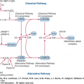
Acronyms and Abbreviations:
Ig, immunoglobulin; IL, interleukin; POEMS, polyneuropathy, organomegaly, endocrinopathy, monoclonal gammopathy, and skin changes; PS, performance status.
THE HEMATOLOGY CONSULTATION
Table 1–1 lists the major abnormalities that result in the evaluation of the patient by the hematologist. The signs indicated in Table 1–1 may reflect a primary or secondary hematologic problem. For example, immature granulocytes in the blood may be signs of myeloid diseases such as myelogenous leukemia, or, depending on the frequency of these cells and the level of immaturity, the dislodgment of cells resulting from marrow metastases of a carcinoma. Nucleated red cells in the blood may reflect the breakdown in the marrow–blood interface seen in primary myelofibrosis or the hypoxia of congestive heart failure. Certain disorders have a propensity for secondary hematologic abnormalities; renal, liver, and connective tissue diseases are prominent among such abnormalities. Chronic alcoholism, nutritional fetishes, and the use of certain medications may be causal factors in blood cell or coagulation protein disorders. Pregnant women and persons of older age are prone to certain hematologic disorders: anemia, thrombocytopenia, or intravascular coagulation in the former case, and hematologic malignancies, pernicious anemia and the anemia of aging in the latter. The history and physical examination can provide vital clues to the possible diagnosis and also to the rationale choice of laboratory tests.
| Decreased hemoglobin concentration (anemia) |
| Increased hemoglobin concentration (polycythemia) |
| Elevated serum ferritin level |
| Leukopenia or neutropenia |
| Immature granulocytes or nucleated red cells in the blood Pancytopenia |
| Granulocytosis: neutrophilia, eosinophilia, basophilia, or mastocytosis |
| Monocytosis |
| Lymphocytosis |
| Lymphadenopathy |
| Splenomegaly |
| Hypergammaglobulinemia: monoclonal or polyclonal |
| Purpura |
| Thrombocytopenia |
| Thrombocytosis |
| Exaggerated bleeding: spontaneous or trauma related |
| Prolonged partial thromboplastin or prothrombin coagulation times |
| Venous thromboembolism |
| Thrombophilia |
| Obstetrical adverse events (e.g., recurrent fetal loss, stillbirth, and HELLP syndrome) |
THE HISTORY
In today’s technology- and procedure-driven medical environment, the importance of carefully gathering information from patient inquiry and examination is at risk of losing its primacy. The history (and physical examination) remains the vital starting point for the evaluation of any clinical problem.1,2,3
Performance status (PS) is used to establish semiquantitatively the extent of a patient’s disability. This status is important in evaluating patient comparability in clinical trials, in determining the likely tolerance to cytotoxic therapy, and in evaluating the effects of therapy. Table 1–2 presents a well-founded set of criteria for measuring PS.4 An abbreviated version sometimes is used, as proposed by the Eastern Cooperative Oncology Group (Table 1–3).5
| Able to carry on normal activity; no special care is needed. | |
| 100% | Normal; no complaints, no evidence of disease |
| 90% | Able to carry on normal activity; minor signs or symptoms of disease |
| 80% | Normal activity with effort; some signs or symptoms of disease |
| Unable to work; able to live at home, care for most personal needs; a varying amount of assistance is needed. | |
| 70% | Cares for self; unable to carry on normal activity or to do active work |
| 60% | Requires occasional assistance but is able to care for most personal needs |
| 50% | Requires considerable assistance and frequent medical care |
| Unable to care for self; requires equivalent of institutional or hospital care; disease may be progressing rapidly. | |
| 40% | Disabled; requires special care and assistance |
| 30% | Severely disabled; hospitalization is indicated though death not imminent |
| 20% | Very sick; hospitalization necessary; active supportive treatment necessary |
| 10% | Moribund; fatal processes progressing rapidly |
| 0% | Dead |
| Grade | Activity |
|---|---|
| 0 | Fully active, able to carry on all predisease performance without restriction |
| 1 | Restricted in physically strenuous activity but ambulatory and able to carry out work of a light or sedentary nature, e.g., light housework, office work |
| 2 | Ambulatory and capable of all self-care but unable to carry out any work activities; up and about more than 50% of waking hours |
| 3 | Capable of only limited self-care, confined to bed or chair more than 50% of waking hours |
| 4 | Completely disabled; cannot carry on any self-care; totally confined to bed or chair |
| 5 | Dead |
Weight loss is a frequent accompaniment of many serious diseases, including primary hematologic malignancies, but it is not a prominent accompaniment of most hematologic diseases. Many “wasting” diseases, such as disseminated carcinoma and tuberculosis, cause anemia, and pronounced emaciation should suggest one of these diseases rather than anemia as the primary disorder.
Fever is a common early manifestation of the aggressive lymphomas or acute leukemias as a result of pyrogenic cytokines (e.g., interleukin [IL]-1, IL-6, and IL-8) released as a reflection of the disease itself. After chemotherapy-induced cytopenias or in the face of accompanying immunodeficiency, infection is usually the cause of fever. In patients with “fever of unknown origin,” lymphoma, particularly Hodgkin lymphoma, should be considered. Occasionally, primary myelofibrosis, acute leukemia, advanced myelodysplastic syndrome, and other lymphomas may also cause fever. In rare patients with severe pernicious anemia or hemolytic anemia, fever may be present. Chills may accompany severe hemolytic processes and the bacteremia that may complicate the immunocompromised or neutropenic patient. Night sweats suggest the presence of low-grade fever and may occur in patients with lymphoma or leukemia.
Fatigue, malaise, and lassitude are such common accompaniments of both physical and emotional disorders that their evaluation is complex and often difficult. In patients with serious disease, these symptoms may be readily explained by fever, muscle wasting, or other associated findings. Patients with moderate or severe anemia frequently complain of fatigue, malaise, or lassitude and these symptoms may accompany the hematologic malignancies. Fatigue or lassitude may occur also with iron deficiency even in the absence of sufficient anemia to account for the symptom. In slowly developing chronic anemias, the patient may not recognize reduced exercise tolerance, or other loss of physical capabilities except in retrospect, after a remission or a cure has been induced by appropriate therapy. Anemia may be responsible for more symptoms than has been traditionally recognized, as suggested by the remarkable improvement in quality of life of most uremic patients treated with erythropoietin.
Weakness may accompany anemia or the wasting of malignant processes, in which cases it is manifest as a general loss of strength or reduced capacity for exercise. The weakness may be localized as a result of neurologic complications of hematologic disease. In vitamin B12 deficiency (e.g., pernicious anemia), there may be weakness of the lower extremities, accompanied by numbness, tingling, and unsteadiness of gait. Peripheral neuropathy also occurs with monoclonal immunoglobulinemias. Weakness of one or more extremities in patients with leukemia, myeloma, or lymphoma may signify central or peripheral nervous system invasion or compression as a result of vertebral collapse, a paraneoplastic syndrome (e.g., encephalitis), or brain or meningeal involvement. Myopathy secondary to malignancy occurs with the hematologic malignancies and is usually manifest as weakness of proximal muscle groups. Foot drop or wrist drop may occur in lead poisoning, amyloidosis, systemic autoimmune diseases, or as a complication of vincristine therapy. Paralysis may occur in acute intermittent porphyria.
Headache may be the result of a number of causes related to hematologic diseases. Anemia or polycythemia may cause mild to severe headache. Invasion or compression of the brain by leukemia or lymphoma, or opportunistic infection of the central nervous system by Cryptococcus or Mycobacterium species, may also cause headache in patients with hematologic malignancies. Hemorrhage into the brain or subarachnoid space in patients with thrombocytopenia or other bleeding disorders may cause sudden, severe headache.
Paresthesias may occur because of peripheral neuropathy in pernicious anemia or secondary to hematologic malignancy or amyloidosis. They may also result from therapy with vincristine.
Confusion may accompany malignant or infectious processes involving the brain, sometimes as a result of the accompanying fever. Confusion may also occur with severe anemia, hypercalcemia (e.g., myeloma), thrombotic thrombocytopenic purpura, or high-dose glucocorticoid therapy. Confusion or apparent senility may be a manifestation of pernicious anemia. Frank psychosis may develop in acute intermittent porphyria or with high-dose glucocorticoid therapy.
Impairment of consciousness may be a result of increased intracranial pressure secondary to hemorrhage or leukemia or lymphoma in the central nervous system. It may also accompany severe anemia, polycythemia, hyperviscosity secondary, usually, to an immunoglobulin (Ig) M monoclonal protein (uncommonly IgA or IgG) in the plasma, or a leukemic hyperleukocytosis syndrome, especially in chronic myelogenous leukemia.
Conjunctival plethora is a feature of polycythemia and pallor a result of anemia. Occasionally blindness may result from retinal hemorrhages secondary to severe anemia and thrombocytopenia or blurred vision resulting from severe hyperviscosity resulting from macroglobulinemia or extreme hyperleukocytosis of leukemia. Partial or complete visual loss can stem from retinal vein or artery thrombosis. Diplopia or disturbances of ocular movement may occur with orbital tumors or paralysis of the third, fourth, or sixth cranial nerves because of compression by tumor, especially extranodal lymphoma, extramedullary myeloma, or myeloid (granulocytic) sarcoma.
Vertigo, tinnitus, and “roaring” in the ears may occur with marked anemia, polycythemia, hyperleukocytic leukemia, or macroglobulinemia-induced hyperviscosity. Ménière disease was first described in a patient with acute leukemia and inner ear hemorrhage.
Epistaxis may occur in patients with thrombocytopenia, acquired or inherited platelet function disorders, and von Willebrand disease. Anosmia or olfactory hallucinations occur in pernicious anemia. The nasopharynx may be invaded by a granulocytic sarcoma or extranodal lymphoma; the symptoms are dependent on the structures invaded. The paranasal sinuses may be involved by opportunistic organisms, such as fungus in patients with severe, prolonged neutropenia. Pain or tingling in the tongue occurs in pernicious anemia and may accompany severe iron deficiency or vitamin deficiencies. Macroglossia occurs in amyloidosis. Bleeding gums may occur with bleeding disorders. Infiltration of the gingiva with leukemic cells occurs notably in acute monocytic leukemia. Ulceration of the tongue or oral mucosa may be severe in the acute leukemias or in patients with severe neutropenia. Dryness of the mouth may be caused by hypercalcemia, secondary, for example, to myeloma. Dysphagia may be seen in patients with severe mucous membrane atrophy associated with chronic iron-deficiency anemia.
Painless swelling
Related posts:


Stay updated, free articles. Join our Telegram channel

Full access? Get Clinical Tree


