Small Cell Lesions
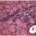
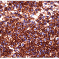
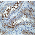
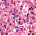
Small cell carcinomas and lymphomas constitute a common source of diagnostic error, often misclassified as anaplastic carcinomas [1, 2, 3, 4] (Figs. 13.1, 13.2, 13.3, e-Figs. 13.1, 13.2, 13.3, 13.4, 13.5).
MEDULLARY CARCINOMA AND OTHER NEUROENDOCRINE CARCINOMAS
A variant of medullary carcinoma composed of poorly differentiated cells resembles small cell carcinoma of lung or neuroblastoma [5]. Immunohistochemical staining identifies these lesions as medullary carcinoma when they contain calcitonin and carcinoembryonic antigen (CEA). Difficulty arises when the lesion is negative for those markers, but the possibility of a primary nonmedullary neuroendocrine carcinoma of thyroid exists [6]. Metastatic disease is more likely and metastases have been described from lung [7], bladder [8], cervix [9], and other sites. Unfortunately, TTF-1 is not a helpful marker in this distinction, since it is also positive in small cell tumors of lung and other primary sites [10]. The role of Pax-8 in this setting has not been reported, but one case reported expression of the TSH-receptor as proof of thyroid origin [6].
Stay updated, free articles. Join our Telegram channel

Full access? Get Clinical Tree


