Figure 18.1
40× magnification. CK7 positive staining of the cytoplasm
Diagnosis
Cutaneous metastatic carcinoma, the source is most likely a primary lung cancer.
Microscopic Features
Metastases are typically categorized broadly as adenocarcinoma, squamous cell carcinoma or undifferentiated carcinomas [1]. The neoplastic cells are usually found in the mid to deep dermis but may also be found in the subcutaneous tissue (Fig. 18.2). The cells tend to demonstrate patterns that resemble the primary source of the tumor. Some patterns that are commonly observed include: nodular growths of tumor cells with scanty intervening stroma, strands or rows of cells infiltrating a fibrotic dermis, dense sheets of cells, or malignant glandular clusters of eosinophilic cells containing mucin or glycogen. Some of the cells may demonstrate epidermotropism by abutting the epidermis. Lymphatic invasion and diffuse, intralymphatic tumor emboli may be seen [1, 2].
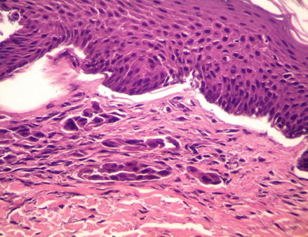
Figure 18.2
H&E, 40× magnification. Multiple nests and single cells are found just under the epidermis. These cells show marked variability with irregularly-shaped nuclei
Discussion
Cutaneous metastases are not commonly seen in dermatology offices and occur in 0.6–10.4 % of all patients with cancer [3]. Therefore, if a patient does not provide a history of cancer, it is easy for this diagnosis to be missed. Cutaneous manifestations of metastases can present in various ways, some of which may resemble other common dermatological diagnoses. One study showed that in 45 % of cases of biopsied cutaneous metastases, the diagnosis was missed altogether [2].
There are cases reported in the literature of cutaneous metastases presenting as a rash, melanoma, basal cell carcinoma, keratoacanthoma, subcutaneous nodules, hidradenitis suppurativa, herpes zoster, vascular tumors, and epidermal inclusion cysts. The sites most commonly involved in descending order include the upper trunk, abdomen, head (especially the scalp), and the neck. Metastatic lung cancers were noted to present most commonly on the head, neck and upper trunk. Metastases to the extremities are extremely uncommon [2–5].
A retrospective review performed by Sariya and colleagues found that 86 % of patients who presented with cutaneous findings were found to have Stage 4 cancer. Of those patients, 76 % succumbed to the disease in an average of 9.4 months. Therefore, cutaneous metastases in most often a late finding in advanced disease states. The same retrospective study showed that the average time between diagnosis of primary cancer and the development of cutaneous findings was approximately 36 months. By that point, the majority of patients had disease progression to Stage 3 or greater [2].
Related posts:
 Amelanotic Malignant Melanoma of the Toe Presenting as an Ulcer: Management and Biopsy Guidelines
Metastatic Cutaneous Adenocarcinoma
Adenocystic Carcinoma
Balloon Cell Nevi and Balloon Cell Melanomas: What Are They?
The Keystone Design Perforator Island Flap: An Easy Option for the Lower Limb, But How Does It Actually Work?
Rotation Flaps of the Scalp: Study of the Design, Planning and Biomechanics of Single, Double and Triple Pedicle Flaps
Amelanotic Malignant Melanoma of the Toe Presenting as an Ulcer: Management and Biopsy Guidelines
Metastatic Cutaneous Adenocarcinoma
Adenocystic Carcinoma
Balloon Cell Nevi and Balloon Cell Melanomas: What Are They?
The Keystone Design Perforator Island Flap: An Easy Option for the Lower Limb, But How Does It Actually Work?
Rotation Flaps of the Scalp: Study of the Design, Planning and Biomechanics of Single, Double and Triple Pedicle Flaps
Stay updated, free articles. Join our Telegram channel

Full access? Get Clinical Tree


