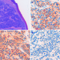
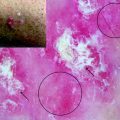
Fig. 15.1
Clinical (a), dermoscopic (b), and HFUS/HRUS (c) images of a retroauricular basal cell carcinoma (BCC)
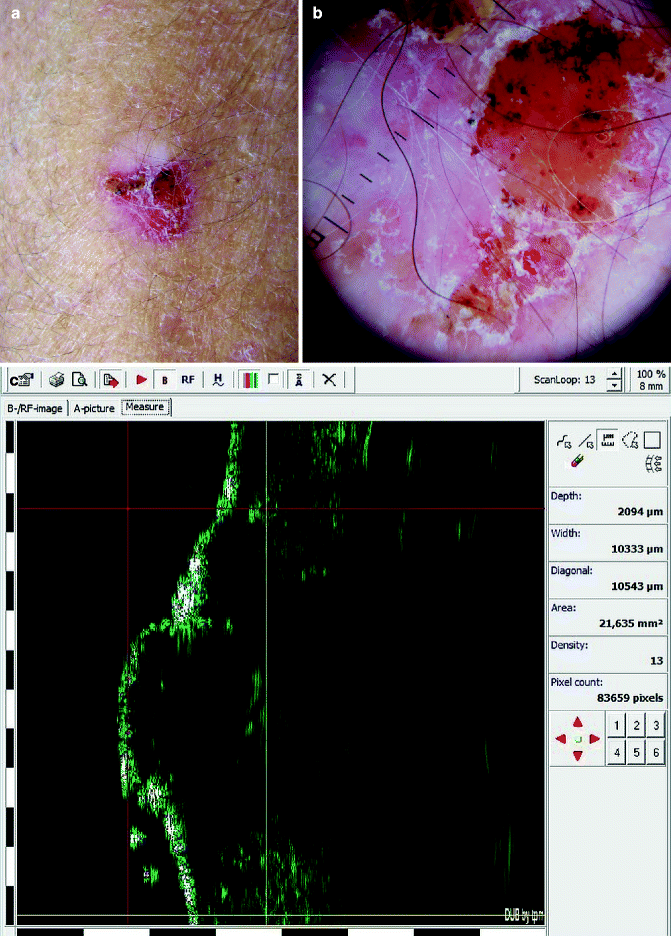
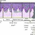
Fig. 15.2
Clinical (a), dermoscopic (b), and HFUS/HRUS (c) images of a squamous cell carcinoma (SCC) in the arm
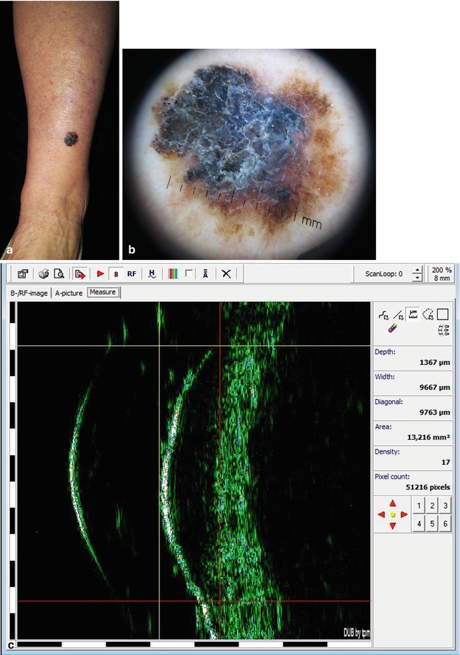
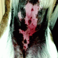
Fig. 15.3
Clinical (a), dermoscopic (b), and HFUS/HRUS (c) images of a malignant melanoma (MM) in the leg
One limitation of the HFUS/HRUS is the lack of sensitivity to detect lesions localized to the epidermis or extremely thin (<0.1 mm) lesions.
HFUS/HRUS is not meant to replace histologic evaluation of BCC but may be a useful adjunct in surgical planning [3] since it allows knowing the exact measures of the lesion including the depth.
The previous knowledge of the tumor dimensions allow to choose the best treatment modality (surgical excision, cryosurgery, curettage, PDT, laser).
Determination of Volume/Depth/Length of the Tumor
The first important consideration is to know if the image obtained with the HFUS equipment corresponds to the real tumor [9–12]. In a study done by the authors of this chapter (Pasquali P, Camacho E, Fortuny, A) and using a 22 MHz HFUS (Taberna Pro Medicum), it was determined that the length obtained by HFUS/HRUS in 57/60 BCC was larger than the one obtained by measuring the histological specimen, the average increase being 60.1 %. There was a strong correlation (R 2 = 0.66) among the length obtained by HFUS/HRUS and the one obtained by histology. For larger tumors, the percentage increase of the HFUS/HRUS length vs. histology length was smaller, with a medium correlation of R 2 =0.46 (Fig. 15.4).
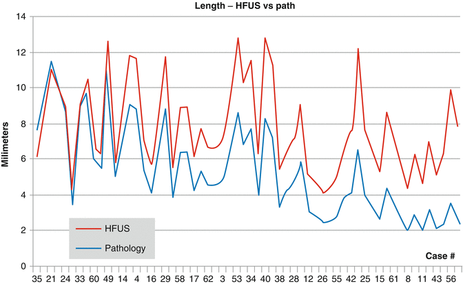

Fig. 15.4
Comparison between the length obtained by HFUS/HRUS (22 MHz Taberna Pro Medicum®) and the length obtained by measuring the histological specimen in 60 cases of BCC
As far as the depth in 49/61 BCC, the depth obtained by HFUS/HRUS was larger than the one obtained by measuring the histological specimen, the average increase being 27 %. There was a strong correlation (R 2 = 0.65) among the depth obtained by HFUS/HRUS and the one obtained by histology. For larger tumors, the percentage increase of the HFUS/HRUS depth vs. histology depth was smaller, with a small correlation of R 2 = 0.29 (Fig. 15.5).
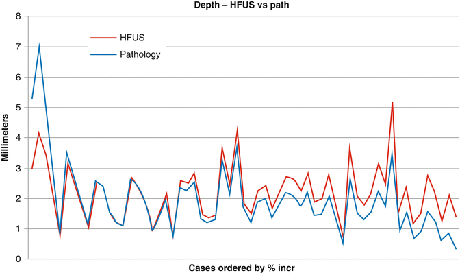

Fig. 15.5
Comparison between the depth obtained by HFUS/HRUS (22 MHz Taberna Pro Medicum®) and the length obtained by measuring the histological specimen in 61 cases of BCC
There is excellent correlation between dermoscopy, HFUS/HRUS, and histology of the BCCs. Dermoscopy is essential in achieving the diagnosis of a malignant tumor. It allows defining margins, and the length of the tumor obtained by dermoscopy correlates very closely with the HFUS/HRUS length. We compared the length obtained by dermoscopy (DMS), only for those cases with less than 12 mm (the largest length obtained through HFUS/HRUS). We found a strong correlation between the DMS length and the one from HFUS/HRUS (R 2 = 0.66) and also with the histology length (R 2 = 0.75) (Fig. 15.6).
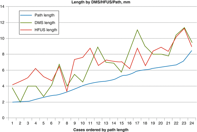

Fig. 15.6
Comparison of the length obtained from HFUS/HRUS and dermoscopy in relation to the length obtained by measuring the histological specimen in 24 cases of BCC
From this information, the surgeon can feel confident to plan a treatment for a BCC knowing the information obtained from the dermoscopy and HFUS/HRUS.
Control of Freezing Fronts During a Cryosurgical Procedure
Monitoring the freezing front with HFUS/HRUS helps the cryosurgeon to visualize the extent of the freezing front and the development of the cryoinjury [13].
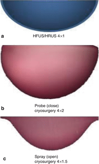
From the measurements mentioned above, we observed that the ratio between length and depth (L/D) for BCC was 4 × 1 mm (Fig. 15.7). In probe (close) cryosurgery, the iceball has an L/D ratio of 4 × 2 mm. In spray (open) cryosurgery, the ratio is 4 × 1.5 mm (Fig. 15.8). The radial distribution of the isotherms allows the surgeon to know that the skin surface temperature at a given distance from the application point of LN (blue arrow in Fig. 15.9) will be the same at the depth of the tumor (Fig. 15.9).
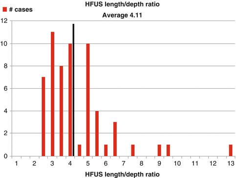

Fig. 15.7
The ratio between HFUS/HRUS length and depth from the measured BCC was 4 × 1
