Fig. 41.1
(a) Focal airway infantile hemangioma of the left subglottis. (i) Initial presentation (ii) and (iii) after two CO2 laser procedures. (b) Focal airway infantile hemangioma of right subglottis. (i) Initial presentation. (ii) After 6 months of oral propranolol therapy (Photos courtesy of Nancy Bauman, MD, Children’s National Medical Center)
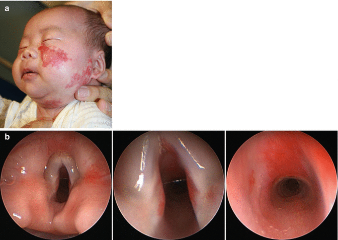
Fig. 41.2
(a) Child with a unilateral maxillary (V2) and mandibular (V3) segmental infantile hemangioma. (b) Direct laryngoscopy/bronchoscopy of the same child from Fig. 41.2a. (i) Note supraglottic staining of midline and right false vocal fold. (ii) Medial edges of bilateral true vocal folds are involved. (iii) Anterior tracheal wall staining toward carina
Presentation
The clinical presentation is determined by the type of lesion. Focal lesions are solitary and not usually associated with cutaneous hemangiomas. Patients may present with stridor in the first 3 months of life, and diagnosis is made during laryngoscopy.
Segmental airway lesions will always have a cutaneous manifestation; depending on the study, 29–63 % of mandibular/beard or V3 segmental hemangiomas have laryngeal involvement [1, 2], while all patients with airway staining will have cutaneous involvement. Thus, it is essential that these patients be evaluated by an otorhinolaryngologist when the diagnosis of facial segmental IH is made.
Management
The first-line treatment of airway infantile hemangioma is medical. Oral propranolol is used for both focal and segmental lesions and can often prevent tracheotomy [3–6].
Corticosteroids may be used as adjuvant therapy in cases of added airway edema, especially during surgical procedures.
Concerning focal lesions, intralesional corticosteroid injection (triamcinolone 40 mg/mL (latent effect) and betamethasone 6 mg/mL (fast acting), 1:1) is still popular among some otolaryngologists and may be performed at the time of laryngoscopy [7].
CO2 laser may also be used to ablate and debulk IH. Complications such as subglottic stenosis from overaggressive laser treatment have been reported. As always, judicious use of the laser is still very helpful. Although the pulsed dye laser has a large role in the treatment of cutaneous IH, it does not have a role in the treatment of mucosal IH.
Surgical excision is possible for focal subglottic lesions. A direct translaryngeal approach was popular prior to the current propranolol era [3, 8, 9]. This method is still used by some and has its benefits as well as associated morbidity. In rare cases, tracheotomy is used as a temporary measure to bypass any subglottic obstruction.
Segmental IH are usually treated with propranolol. Because of the longer proliferative phase in segmental disease (2–3 years versus 1 year), longer treatment should be anticipated. In cases of poor response and significant obstruction, CO2 laser ablation may be useful. In general, these lesions are followed with serial laryngoscopy. The staining may involve all levels of the endolarynx: supraglottis, glottis, and subglottis.
Airway Lymphatic Malformations
A large percentage (up to 73 %) of patients with head and neck LMs will have upper airway involvement. Areas most commonly involved are the oral cavity (tongue), oropharynx (base of tongue), and supraglottis. Notably, the disease may extend to the supraglottis and not involve structures inferiorly such as the glottis, subglottis, and trachea [10]. Approximately, 30 % of patients will require tracheotomy for massive cervicofacial disease with associated airway involvement. Airway disease is often microcystic (less than 1–2 cm in diameter).
Presentation
The most common clinical presentation shows lymphatic malformation vesicles on the tongue surface which may be mucosal colored or hemorrhagic (Fig. 41.3). There is often accompanied swelling, pain, and malodor. The base of the tongue is often enlarged, and the epiglottis is omega shaped with or without LM involvement. Glossoptosis may be caused by either macroglossia or floor of the mouth disease.
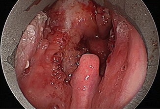
Fig. 41.3
Lymphatic malformation, mucosal and hemorrhagic vesicles involving bilateral tonsils, soft palate, and tongue
Management
Patients who present with extensive facial LM or with oral cavity LM should undergo an airway survey. MR imaging will delineate the extent of disease. Bilateral disease increases the risk of airway obstruction.
The treatment of LM as with all vascular anomalies is multidisciplinary.
Medical treatment is not curative and is used to control symptoms during acute exacerbations and remissions of lymphangitic episodes. High-dose corticosteroids and antibiotics are used with good effect.
Surgical intervention is staged and may incorporate various modalities of treatment.
CO2 laser ablation is used to treat superficial mucosal disease (Figs. 41.4 and 41.5) [15, 16]. One must be cognizant of the risk of stenosis. Generally, one side at a time is treated.
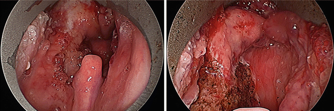
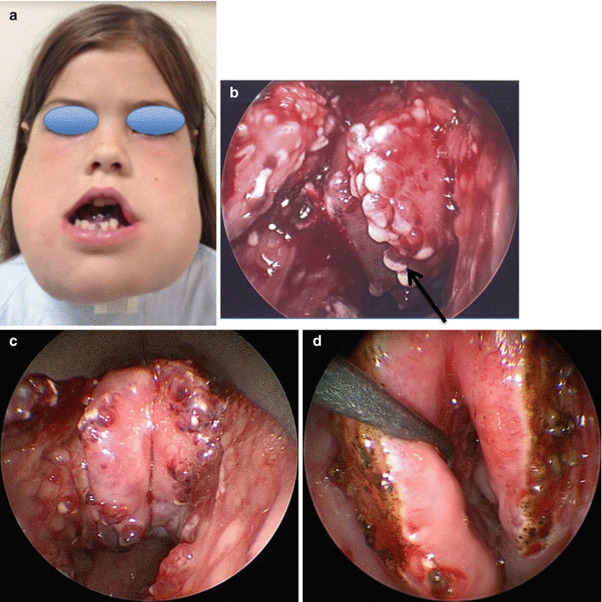
Fig. 41.4
Pre- and intraoperative CO2 laser ablation of LM vesicles on mucosal surface or oropharynx
Fig. 41.5
(a) Child with bilateral massive cervicofacial LM. Note tracheotomy and anterior open mouth deformity. Patient is unable to vocalize. (b) Laryngoscopy at presentation. Epiglottis is omega shaped and collapsed with LM hemorrhagic vesicles and edema. Black arrow delineates midline of epiglottic opening. (c) 6 weeks after 1st laser treatment. Profile of epiglottis more defined. (d) Intraoperative view after CO2 laser ablation of bilateral lingual surface of epiglottis. Midline is not treated
Macroglossia is treated with surgical debulking of the tongue. A midline wedge excision decreases the width (Fig. 41.6). This may be followed by a second stage to address the vertical height of the tongue. Floor of the mouth disease may be treated with excision, with or without sclerotherapy.
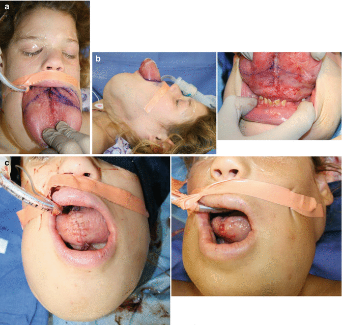
Fig. 41.6
(a) Child with macroglossia and glossoptosis secondary to tongue LM. Intraoperative wedge excision is marked. (b) Lateral view, note significant glossoptosis. (c) Ventral surface marking-distance allows for tongue protrusion. (d) Postoperatively, tongue is now inside the mouth. (e) Intraoperative second stage excision in horizontal vector addresses vertical dimension
Patients may require tracheotomy for impending obstruction or as a temporary measure during sclerotherapy of the airway.
Sclerotherapy is used to treat micro- or macrocystic LM. Traditionally, microcystic disease is not as responsive as macrocystic disease to sclerotherapy. However, recent advances in sclerosants (bleomycin) and techniques with a combined direct laryngoscopy with transmucosal bleomycin injection have offered excellent results and at times obviated the need for tracheotomy (Figs. 41.7 and 41.8).
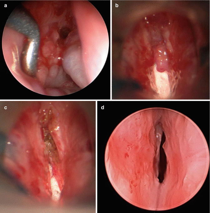
Fig. 41.7


Endolarynx of same patient as Fig. 41.5. (a) Left and right false vocal fold disease. (b) Patient with decreased bulk after one bleomycin injection to bilateral false vocal folds. (c) Intraoperative view after CO2 laser ablation of left false vocal fold disease. (d) 6 weeks post-op. Note resolution of supraglottic disease and opening of airway aperture
Related posts:

Stay updated, free articles. Join our Telegram channel
Full access? Get Clinical Tree
