Fig. 24.1
(a–e) (Infantile) Hemangioma for differential diagnosis with vascular malformation (a) depicts a typical clinical finding of (infantile) hemangioma along the left cheek, appeared suddenly as a small lump month after the birth; soon the tumor started to grow exponentially reaching to the size shown in (b) within a few months. Following initial explosive growth through proliferative stage, the lesion shown in (a) started to regress within first year of age to reach remarkably shrunken condition shown in (b) as unique characteristic of the hemangioma. Before reaching to the age of 4 years, the lesion was almost completely regressed as shown in (c). In such situation, careful history and physical examination are more than enough for correct diagnosis as a hemangioma. However, occasionally simple tests like Duplex ultrasonography or further additional MRI might be needed to confirm the clinical impression for differential diagnosis between vascular malformation and hemangioma; as shown in (d), hemangioma in proliferative stage depicts hypervascular condition, compatible to the findings shown in MRI (e)
In other words, both CVM and hemangioma are two entirely different conditions with different management and prognosis. The CVMs are “self-perpetuating” slowly progressing embryonic tissue remnant as a birth defect. On the contrary, the hemangioma is a “self-limited” vascular (Fig. 24.1) tumor starting in early neonatal period as a rapid growing lesion in its majority but soon followed by steady regression before reaching to the age of 7. Therefore, the differentiation between the CVM and (infantile/neonatal) hemangioma will be the first most important step for the proper diagnosis of the CVMs for correct management [10–12].
The second most important aspect of the CVM is that “CVM is NOT a single vascular disorder;” it represents a group of various vascular defects with much different characteristics and clinical behaviors. The CVM is the outcome of developmental arrest during various stages of the embryogenesis, depending upon the circulation systems affected by this inborn error – artery, vein, lymphatic, and capillary – and also the embryonic stages, early or late, when it developed; the CVM will become a venous, arterial, lymphatic, as well as capillary malformation either as an “extratruncular” or “truncular” form with entirely different clinical significance and prognosis [13–15] (Fig. 24.2).
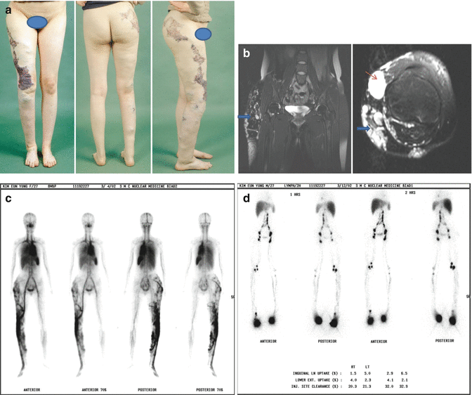
Fig. 24.2
(a–d) Combined form of vascular malformation: (a) represents a clinical condition of diffusely swollen right lower extremity by combined condition of three different vascular malformations: venous, lymphatic and capillary malformations (CM). (b) shows the MRI findings to confirm two different types of venous malformation: extratruncular (thick blue arrow) and truncular (thin red arrow) venous malformation (VM) lesions while (c) confirms abnormal blood pool by two different types of the VM lesions altogether throughout right lower extremity, detected by Whole Body Blood Pool Scintigraphy (WBBPS). (d) illustrates overall lymphatic transport condition of both lower extremities assessed with radionuclide lymphoscintigraphy (LSG); right lower extremity shows reduced isotope uptake at the proximal/inguinal lymph nodes together with delayed clearance with mild dermal backflow as well as collateral routes suggesting substantial lymphatic dysfunction by truncular lymphatic malformation (LM) known as ‘primary’ lymphedema [28]
Therefore, proper differentiation among various CVMs is extremely important following the differentiation with the hemangioma. Although the CVM is often called AV malformation (AVM), the AVM is only one of the CVMs, and AVM should not represent the entire group of the CVMs [16–18].
The CVM, especially of the venous nature, named the venous malformation (VM) is often called the cystic/cavernous hemangioma. But again, this is the wrong term based on the old concept/classification misguiding the clinicians, and its misuse should be stopped [19–21].
The name-based eponyms (e.g., Klippel-Trenaunay syndrome, Parkes Weber syndrome), which have been used for a century, should also be used with proper discretion. In the past era when the clinical findings were the only criteria for the diagnosis, the CVMs had to be classified only with these clinical findings and named after the physicians who first described it. Such old classifications are not able to provide the mandatory information on the etiology, anatomy, embryology, and pathophysiology including the hemodynamic status for the contemporary concept of the CVMs [22–24] (Fig. 24.3 and 24.4).
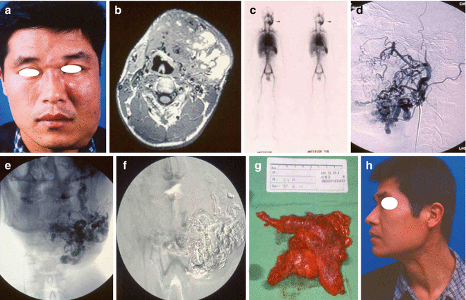
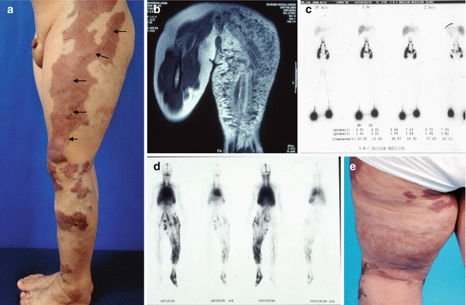
Fig. 24.3
(a–h) AV malformation (a) portrays rapidly expanding swelling along entire left face following minor blunt trauma, which was further extended to the left upper neck across the chin line and also to reach to periorbital region within a year. As shown in MRI (b), the lesion was confirmed as AV malformation (AVM) in infiltrating nature, compatible to Duplex ultrasonographic finding as a high flow lesion. As shown in (c), the lesion was confined only left face and neck and no other lesions were depicted on WBBPS to make it manageable by surgical excision. (d) represents arteriographic finding of the AVM as infiltrating extratruncular lesion, and the lesion was filled with n-BCA glue done preoperatively as shown in (e); this glue filled lesion depicted in (f) became a road map for subsequent surgical excision to allow minimal risk of bleeding and collateral tissue damage. Whole lesions were safely resected in total, shown in surgical specimen (g), and follow up clinical finding in 5 years was excellent shown in (h). Such contemporary approach of surgical and endovascular therapy combined can provide much improved outcome of the management in comparison to two procedures done separately [32]
Fig. 24.4
(a–e) Hemolymphatic Malformation (a) renders typical limb swelling involved to complicated condition of hemolymphatic malformation consisted of VM, LM, and CM, also known as Klippel Trenaunay Syndrome. This photo (4A) was taken soon after the marginal vein (MV) was resected -see the outline of surgical incision by black arrows- due to the lymphatic leakage from upper buttock. Preoperative MRI finding, shown in (b), was lymphedematous soft tissue swelling by truncular LM; further assessment of lymph transporting function with LSG shown in (c) confirmed reduced isotope uptake at inguinal lymph nodes as well as delayed clearance, which is compatible with clinical findings as primary lymphedema but there was no clear evidence for extratruncular LM/ lymphangioma was involved. Based on the WBBPS findings (d) to confirm extratruncular VM and truncular VM both as the major cause of deteriorating venous symptoms (e.g. venous stasis ulcer), surgical excision of a truncular VM confirmed as MV in this patient was performed with priority. But within 2 weeks following MV resection, the lymph started to leak from posterior aspect of left buttock shown in (e) precipitating the local as well as general sepsis to require intense antibiotic therapy, etc. It took more than 6 months to be relieved from recurrent bouts of massive leakage
Hamburg classification, named after the consensus workshop held in Hamburg in 1988, is capable to clear all these confusions caused by old nomenclatures/classifications and became a new guideline for the contemporary management of the CVMs [25–27] (Table 23.1A and B).
Table 24.1
(A & B)
A. Hamburg classificationa of congenital vascular malformations (CVMs) – species |
Arterial malformation |
Venous malformation |
Arterio-venous malformation |
Lymphatic malformation |
Capillary malformation |
Combined vascular malformation |
B. Hamburg classification of CVMsb: forms – embryological subtypes |
1. Extratruncular forms |
Infiltrating, diffuse |
Limited, localized |
2. Truncular forms |
Obstruction or stenosis |
Aplasia; hypoplasia; hyperplasia |
Stenosis; membrane; congenital spur |
Dilatation |
Localized (aneurysm) |
Diffuse (ectasia) |
Much advanced technology through the century is now able to provide accurate diagnosis of such complicated CVM condition; often non- to minimally invasive tests are more than adequate for the diagnosis per se in its majority, and further invasive tests can be reserved as a road map for the group later when the treatment is indicated [28–30].
NOT every CVM would need the treatment; only the CVM lesions with appropriate indications should be selected as a candidate for the treatment. Decision on whether the treatment is indicated or not should be based on the consensus among the multidisciplinary team members; proper evaluation on the benefit versus the risk is essential since the majority of the conventional treatment accompanies high morbidity [31–33].
The VM [34–36] and LM (lymphatic malformation) [37–39] are generally not a life- or limb-threatening condition in its majority on the contrary to the AVMs [40–42]; AVM, VM, and LM are therefore mandated for different treatment principles since their clinical behavior and the prognosis are so different. Identical treatment principle cannot be implemented indiscriminately to all these different types of the CVMs; the treatment principle/strategy for the VM and LM should be different from those for the AVM [43–45].
Selection of the treatment modalities in addition to the basic conservative treatment regimen such as surgical or nonsurgical (e.g., sclerotherapy) should be made by a multidisciplinary team [46–48].
Accurate and thorough hemodynamic assessment of the deep venous system of the lower extremity is absolutely required for the safe management of the CVMs located in the lower extremity, especially for the hemolymphatic malformation (HLM) consisted of the LM and VM combined [49–51]. Priority for treatment among multiple lesion types should be based on the relative degree, extent, and severity of the specific lesion.
Every treatment strategy should be set accordingly based on the multidisciplinary approach with full integration of open surgery and endovascular surgery to improve the treatment outcome.
The endovascular surgery with various modalities of embolotherapy and sclerotherapy should be considered with priority as an independent therapy to the “surgically inaccessible/difficult” lesion. Even the “surgically accessible” lesion should be treated by the combined approach of preoperative endovascular therapy and subsequent open excisional surgery whenever possible to reduce the complication and morbidity [52–54] (Fig. 24.3).
Active incorporation of the pre- and/or postoperative endovascular therapy with embolotherapy/sclerotherapy allows substantial expansion of the traditional role of surgical therapy especially for the “infiltrating extratruncular” form of CVM while maintaining acceptable range of surgical risk. Such perioperative, especially preoperative, therapy could reduce surgical morbidity as well as complications drastically so that such benefit can exceed beyond the boundary for the traditional surgical approach [55–57] (Fig. 24.3).
Such approach with fully integrated endovascular therapy and the traditional open surgical therapy can achieve acceptable goal for the contemporary management of the CVMs as a new strategy. And the treatment strategy should be reviewed periodically based on the response to the initial and subsequent therapy by the multidisciplinary team and should be readjusted accordingly [52–57].
However, not every CVM lesion is amenable to the treatment. Furthermore, not every CVM lesion should be treated. Its mere presence often makes the practitioner feel obligated to treat. The only lesion assessed by the multidisciplinary team with justified indications such as severe symptoms or complications of the lesion (e.g., leakage, sepsis) should be considered for treatment. Although extratruncular CVM lesions are more serious than truncular lesions with much poorer long-term outcome, an overzealous approach sometimes does more harm than good [40, 58, 59].
“Cure” with devastating complication and morbidity is NOT better than limited “control” with less complication and morbidity even for the AVM. Observation sometimes remains the best approach yet until figuring out the exact condition of the lesion; “not to intervene” is sometimes a wiser choice than to casually intervene without a full understanding of the biology and natural history of the CVM lesion.
A “controlled” aggressive approach is therefore favored especially for the less vicious CVM lesion like LM, where every effort is made to minimize collateral damage during treatment. The decision to initiate treatment should be based on the accepted indications.
When the benefit of treatment outweighs the risk of complications and morbidity, less risky treatment options (e.g., foam/liquid sclerotherapy) should be the first-line therapy. “No treatment is the best option if feasible.” In contrast to the treatment of AVMs, all the VM and LM lesions can be treated using a less aggressive approach.
The traditional conservative approach to the young pediatric patient with a VM and LM is still valid, especially for the common CVMs as far as there is NO evidence of bony involvement (e.g., leg length discrepancy). It is usually safe to delay treatment until the child reaches to the age of 2 or more years before beginning diagnostic procedures and treatment.
Nevertheless, CVM lesions producing the vascular-bone syndrome (e.g., angio-osteohypotrophy/hypertrophy) are best treated early in order to prevent long bone growth discrepancy [60–62].
Also, for the VM or LM lesion at a life- or limb-threatening anatomic location, an earlier treatment approach is preferred over a more conservative one (e.g., surgical decompression); for the lesion complicated with a life- or limb-threatening condition (e.g., hemorrhage, sepsis, pulmonary embolism), the treatment should be started expeditiously despite the risk of the associated morbidity [63–65].
Related posts:

Stay updated, free articles. Join our Telegram channel

Full access? Get Clinical Tree


