Fig. 7.1
Early hemangioma in a newborn (a). Hemangioma in rapid proliferative phase in the same patient, who presented with associated PHACE syndrome (b)
The territory of the IH is delineated by the premonitory mark, yet in some cases only a part of the area proliferates [3, 4] (Fig. 7.2). They are characterized by a wide clinical heterogeneity (depth of cutaneous involvement, size).
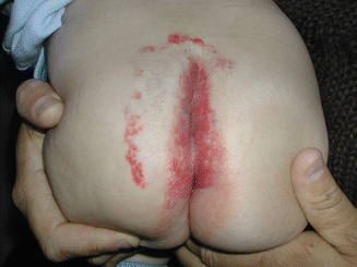

Fig. 7.2
Hemangioma with minimal or arrested growth, fine telangiectasias, and a proliferative component in the periphery in lumbosacral area. The patient has spinal dysraphism
During the last years, IH have been classified according to the level of the skin affected and into three groups: superficial, deep, and mixed hemangiomas [5, 6]. Superficial hemangiomas involve the superficial dermis and appear as bright red lesions (Fig. 7.3). These lesions may be plaque-like or more rounded papules or nodules. Deep hemangiomas involve the deep dermis and subcutis and present as bluish to skin-colored nodules (Fig. 7.4). Mixed hemangiomas have both superficial and deep components and therefore have features of both (Fig. 7.5). The skin and subcutis appear to be most commonly affected, even factoring in more obvious presentation, whereas deep skeletal muscle is spared.
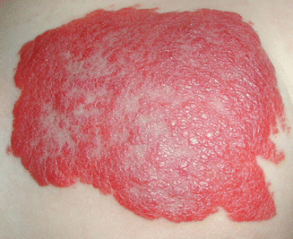
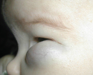
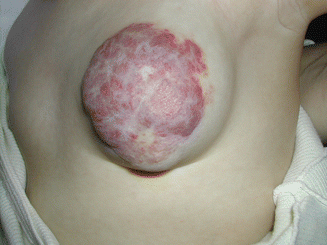

Fig. 7.3
Superficial hemangioma

Fig. 7.4
Deep hemangiomas

Fig. 7.5
Mixed hemangioma
Chiller et al. proposed a new clinical classification, in which they were divided into four groups: localized, segmental, indeterminate, and multifocal hemangiomas. “Segmental hemangiomas” were those hemangiomas or clusters of hemangiomas with a configuration corresponding to known embryologic prominences, derived from the neuroectoderm. They have a geographic shape, have large size and involve an anatomic region, and can be reticular or telangiectatic or flat cherry-red patches [6–8] (Fig. 7.6).
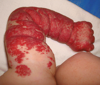

Fig. 7.6
Segmental hemangioma
Four segments or patterns of involvement occur in facial area regions. Segment 1 involved the lateral forehead and temporal and lateral frontal scalp. Segments 2 and 3 correspond to the maxillary and mandibular, and Segment 4 involved the nose, philtrum, and medial frontal scalp. These segmental hemangiomas tend to be greater than 5 cm in diameter and most often span one side of the face and neck (Fig. 7.7).
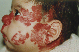

Fig. 7.7
Facial segmental hemangioma associated with PHACE syndrome (the four facial segments are partially involved)
“Localized hemangiomas” were defined as those hemangiomas that seem to grow from a single focal point or were localized to an area without any apparent linear or developmental configuration. They are usually oval or round.
“Indeterminate hemangiomas” were those that were not readily classified as either localized or segmental, and “multifocal hemangiomas” were defined as more than ten cutaneous hemangiomas [6].
Although approximately 60 % of hemangiomas occur on the head and neck, they can also occur in any region of the body on the trunk (25 %), extremities, and genitals (15 %) [7, 8]. Most present as solitary cutaneous and/or subcutaneous lesions (80–90 %), and a significant percentage of patients (15 %) have multiple lesions [7, 8] (Fig. 7.8).
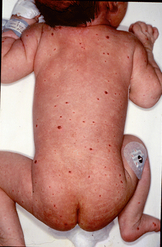

Fig. 7.8
Multifocal hemangiomas in a patient with visceral involvement
Hemangiomas, whether small and innocuos or large and deforming but are united by a predictable biological behavior characterized by three distinct stages: a rapidly proliferating, high-flow stage (8–12 months), followed by a prolonged involuting stage (1–12 years), and finally by a variably prominent end-stage with fibrofatty residuum. [3, 5, 9–11].
However others leave only residual telangiectasias (Fig. 7.9).
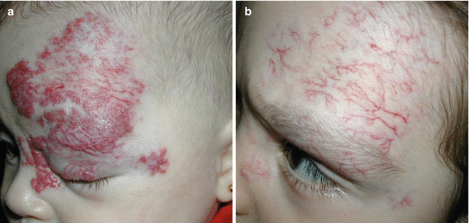

Fig. 7.9
Hemangioma in proliferative phase (a), post-involution telangiectasias (b)
Most hemangioma growth occurs in the first 5–9 months, at which point 80 % of the final size has often been reached [4].
The deep and mixed IH with segmental or indeterminate distribution differ in their growth pattern [12]. These IH sometimes appear later and continue to grow for a longer period than superficial hemangiomas (sometimes the prolonged growth has been observed until 2 years of age). Cervicofacial IH and those involving the parotid gland are a typical example of IH with this growth pattern. This group is known as hemangiomas with prolonged growth phase [12, 13] (Fig. 7.10a, b).
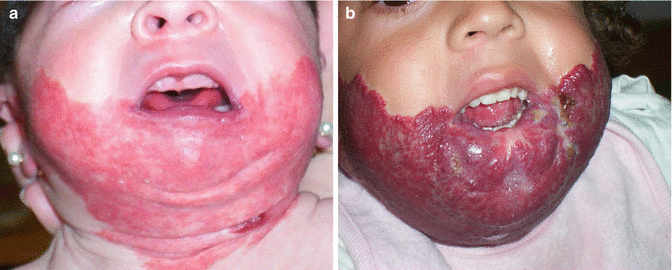

Fig. 7.10
Segmental hemangioma with “beard distribution” in proliferative phase at 2 weeks of age with associated underlying airway hemangioma (a) and at 22 months of age with continued growth (b)
Another subgroup of IH exhibits a pattern of minimal growth, and they presented a reticular or telangiectatic appearance with a proliferative component predominating in the periphery consisting of papules a few millimeters in diameter. They electively are located in the trunk and limbs and lumbosacral area [14–16] (Fig. 7.2). The beginning of involution is even more difficult to predict but usually manifested by changes in color from bright red to a paler red or gray, with a whitish discoloration that occurs always in the center of the hemangioma and extends centrifugally. Deep lesions become less blue and less warm. IH involutes over different periods of time, some in a few years, while others can take several years.
IH during the proliferative phase is composed of well-defined masses of capillaries lined by plump endothelial cells. Normally configured mitotic figures may be numerous. GLUT-1 (the erythrocyte-type glucose transporter protein) is strongly expressed by endothelial cells of infantile hemangiomas at all stages of their evolution and is not expressed by other benign vascular anomalies and reactive proliferations. This appears to be an exclusive marker for IH and is an invaluable tool used to distinguish hemangiomas from other vascular lesions [17].
The diagnosis of a hemangioma is best made by clinical history and physical exam. In cases of unclear diagnosis, the best radiographic modalities to use are either a Doppler ultrasound or MRI. Biopsy should only be considered when history, physical examination, and imaging studies fail to clearly define the pathology.
Complications
The majority of infantile hemangiomas are innocuous (80 %), but a significant minority during the rapid growth phase are associated with important complications such as ulceration, bleeding, risk for permanent disfigurement, compromised organ function, vision and airway obstruction, or high-output cardiac failure. In these cases, quick active and aggressive therapeutic intervention is mandatory [7, 8, 18] (Table 7.1).
Table 7.1
Hemangioma high-risk locations
Location | Complication |
|---|---|
Eyelid | Functional impairment amblyopia |
Ear | Functional impairment |
Beard area | Functional impairment/airway involvement |
Forehead and nasal tip | Disfigurement |
Upper and lower lip | Disfigurement/ulceration |
Neck/anogenital area | Ulceration |
Large segmental facial and scalp area | PHACE syndrome/visceral hemangiomas |
Large segmental lumbosacral area | Spinal dysraphism, anogenital/urogenital/limb abnormalities |
Ulceration
This occurs during the proliferation phase in 15 % of IH (3–4 months of age); however it could occur in precursor lesions prior to the development of IH.
The most commonly affected areas include the anogenital area, head, and neck, particularly the lips and perioral and intertriginous areas; nevertheless IH on any site could be ulcerated (Fig. 7.11). The ulcer could persist for several weeks or months and result in pain, bleeding, infection, and sometimes permanent scarring [19]. The causes of ulceration are understood; the factors that may play a role are friction, maceration, or both. Ulceration is more frequent in mixed or superficial and larger segmental hemangiomas than in the focal subtype [18–21].
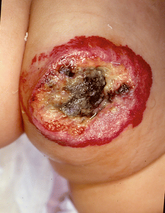

Fig. 7.11
Ulcerated hemangioma in diaper area
Visual Compromise
Periorbital hemangiomas most often involve the upper lid, but the lower lid and retrobulbar space are also common. We can define the anatomic location of the ocular hemangiomas as preseptal, extraconal, and intraconal [22, 23]. All the lesions that affect the visual axis may cause amblyopia, and that is the complication of most concern (Fig. 7.12a, b).
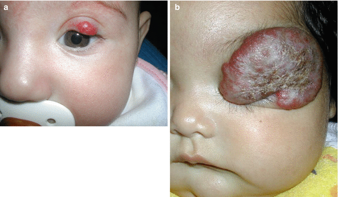

Fig. 7.12
(a) Left upper eyelid focal hemangioma causing astigmatism. (b) Left upper eyelid hemangioma The child presented with complete visual axis obstruction
Amblyopia may result from different mechanisms:
1.
Anisometropia (if there is a refractive difference between the eyes): In the case of hemangiomas, astigmatism is usually secondary to pressure from the tumor on the globe, which causes an irregular corneal curvature.
2.
Strabismic amblyopia can occur if the hemangiomas affect the movement of extraocular muscles.
3.
Deprivation amblyopia arising from partial or complete eyelid closure is particularly devastating if it occurs during the first few months of life, when it can cause profound visual impairment.
Intraconal IH that involves the posterior orbit could result in proptosis, displacement of the globe, or visual axis obstruction [24, 25].
Another relatively common complication of IH is lacrimal duct obstruction because of pressure in the region of the lacrimal duct, resulting in increased tearing and recurrent conjunctivitis.
Airway Hemangiomas
Infantile hemangiomas (IH) involving the “beard area”(which encompasses the preauricular area, mandible, chin, lower lip, and anterior neck) have a high likelihood of concomitant airway hemangiomas, and this association is even more pronounced when the IH involves multiple rather than one component of the “beard area”[26] (Fig. 7.11). Of course, airway hemangiomas can occur in the absence of cutaneous IH, and some cases have been reported with localized cutaneous IH either on the face or in extra-facial locations [27, 28].
Airway hemangiomas can involve any site from the nares to the tracheobronchial tree; however they are most commonly seen in the subglottis, and laryngeal localization represents 1.5 % of congenital lesions of the larynx [29] (Fig. 7.13a, b).
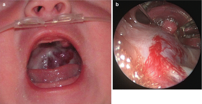

Fig. 7.13
Hemangioma in hard palate without skin involvement (a). Subglottic hemangioma in the same patient (b)
Most of the subglottic hemangiomas become symptomatic during the first year of life. They are located in the posterior or lateral subglottic space, and besides hoarseness, dyspnea, and feeding difficulties, they produce a typical gradually worsening croupous stridor as the main symptom [29].
Stay updated, free articles. Join our Telegram channel

Full access? Get Clinical Tree


