(1)
State University of New York at Buffalo Kaleida Health, Buffalo, NY, USA
The posterior compartment of the arm consists of a single muscle, the triceps. This powerful muscle is the only extensor of the elbow except for the anconeus, which contributes only slightly to this action. The triceps, as its name implies, arises from three heads. The long head, springing from the infraglenoid tuberosity and adjacent capsule of the shoulder joint, descends in front of the teres minor and behind the teres major and the medial head of the muscle.
The lateral head has a linear origin between the insertion of the teres minor and the posterior part of the deltoid tuberosity, with additional fibers from the lateral intermuscular septum. The lateral head, covering the radial nerve and the profunda brachii vessels, inserts into the lateral border of the common tendon.
The medial head of the triceps arises from the back of the shaft of the humerus, distal to the groove for the radial nerve and the whole length of the medial intermuscular septum. It is inserted into the deep aspect of the lower half of the common tendon.
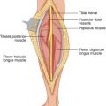
The radial nerve descends between the long and medial heads of the triceps. It then passes the tip of the lateral head in the spiral groove of the humerus, separated from the bone by fibers of the medial head, except for a small area behind the deltoid tuberosity. The nerve pierces the lateral intermuscular septum that lies between the brachialis and brachioradialis. The profunda brachial artery accompanies the radial nerve in the spiral groove.
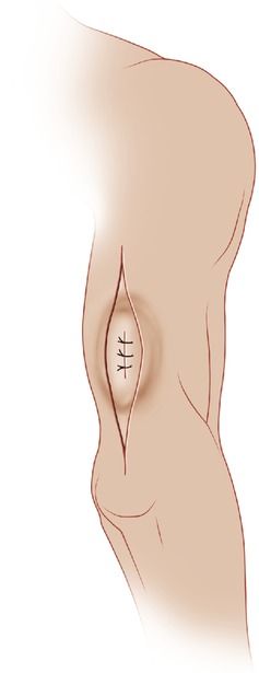
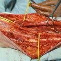
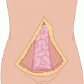
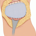
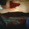
In the example presented of a sarcoma of the triceps, a long elliptical incision is made, including the previous biopsy site (Fig. 8.1). Flaps are raised medially and laterally. The deep fascia is incised (Fig. 8.2). It is clear that the tumor involves the long and lateral heads of the triceps, so both of these heads are divided superiorly well above the site of involvement by the tumor. This exposes the radial nerve coursing in the spiral groove (Fig. 8.3). A portion of the medial head may also be removed if proximity to the tumor makes it necessary. Distally, the insertions for the long and lateral heads are divided at the point at which they join the triceps tendon, preserving the insertion of the medial head in its continuity with the tendon to the olecranon (Fig. 8.4). Sensory branches of the radial nerve, such as the lower lateral cutaneous nerve of the arm, the posterior cutaneous nerve of the forearm, or both may be sacrificed if necessary.
