Fig. 44.1
Infiltrating venous malformation of the middle finger in a girl. (a) Drawing of incisions in the palmar aspect of the middle finger. (b) Bone infiltration evident in plain radiograms, (c) subdermal undermining under the microscope shows the avascular plane and exposition of the involved tissue (skin-sparing technique) and the nerve identified and isolated before tissue excision. (d) Late result showing scars without contracture
3.
Scars of previous surgery influence the choice of new incisions. A flap should not be performed with a previous scar at its base. In extended CVM, incisions should be planned in order to permit extensions, when necessary. Drawing the contour of the CVM mass on the skin before marking the incision lines is helpful in order to better approach the malformation.
Skin-Sparing Technique
Skin sparing is possible by subdermal undermining, which should be carefully done with a scalpel to preserve the subdermal vascular network. This allows minimal blood loss and saves skin flaps for coverage (Figs. 44.1c and 44.7b–c). It is best performed under a microscope or with loupe magnification.
Use of Tourniquet
A tourniquet reduces bleeding and is useful for microsurgical techniques, especially in venous malformations. In arteriovenous (AV) malformations, a tourniquet is best applied at the beginning to expose the tissue and removed to permit Doppler examination after preparation of the fistulous area, as fistulae may be difficult to recognize during ischemia [8].
Microscope
CVMs often enclose nerves and a direct approach may be difficult and risky. In these cases microsurgical techniques permit neurovascular pedicles to be isolated with extreme precision, following their course inside the vascular lesion. The resulting dissection is then much more precise and not only permits the integrity of the nerves to be respected but also can be used as a guide during the dissection of CVM (Figs. 44.1c and 44.6b).
This technique lengthens surgery time but allows a precise dissection which is essential in difficult cases [8].
Tissue Involvement: Clinical Pictures and Surgical Treatment
One of the main challenges in this surgery is the involvement of the surrounding structures, such as the bones, nerves, muscles, tendons, ligaments, and skin. CVM inside tissues may cause infiltration or compression [9, 10].
Skin Involvement
In venous CVM, the skin is thinned by compression. A subdermal dissection is possible, preserving skin flaps and reducing bleeding (“skin-sparing technique”) (Figs. 44.1, 44.2, 44.3, 44.4, 44.5, 44.6, and 44.7) [10, 11].
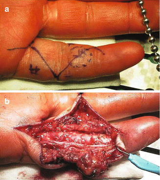
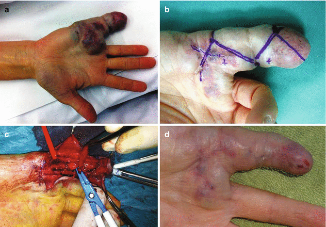
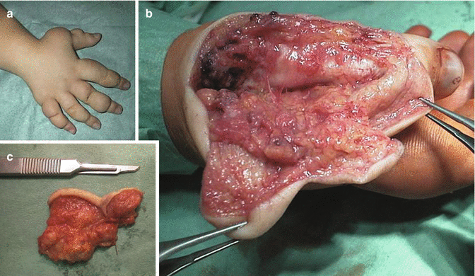
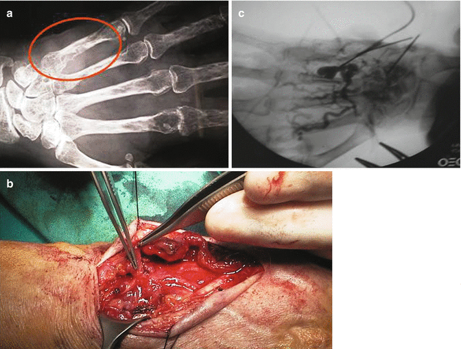
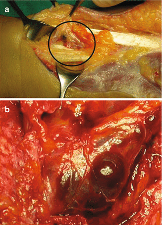
Fig. 44.2
Arteriovenous malformation in the volar aspect of the little finger. (a) Two fistulous areas detected guided by the choice of a Brunner incision. (b) The neurovascular bundles used as a guide for the dissection of the malformed tissue. Tourniquet and microscope control reduced bleeding and resulted in a safe radical excision
Fig. 44.3
Venous malformation in the palm and little finger. (a) Deformity of the involved area with the skin apparently damaged. (b) Skin incision of the second operation (the first was performed in the palm). Note the aspect of the skin after tourniquet inflation. (c) Excision of malformed tissue after isolation of neurovascular bundles. (d) Early result shows good wound healing
Fig. 44.4
En block excision of lymphatic malformation in the thumb of a young girl. (a) Clinical picture. (b) Radical resection of a planned segment of the skin and underlying hypertrophic subcutaneous tissue. (c) Specimen of the removed tissue
Fig. 44.5
Arteriovenous malformation with intraosseous fistulae. (a) Radiogram shows the extent and site of bone involvement. (b) Subcutaneous AV fistulae have been removed and communicating vessels to the bone are clearly seen. (c) Direct alcoholization of bone fistulae reduced the malformed tissue in a few minutes
Fig. 44.6


Arteriovenous malformation involving nerves in the forearm. (a) External compression of the median nerve in the proximal forearm. (b) AV fistulae infiltrating the epineurium of the median nerve near the wrist
Related posts:

Stay updated, free articles. Join our Telegram channel
Full access? Get Clinical Tree
