Topoisomerase II Inhibitors: The Epipodophyllotoxins


Topoisomerase II Inhibitors: The Epipodophyllotoxins


Yves Pommier

François Goldwasser


Introduction and Historical Perspective

DNA topoisomerase II (Top2) inhibitors have been the subject of considerable biochemical, pharmacological, and clinical investigations. In addition to the epipodophyllotoxins that are considered in this chapter, other drugs interact with Top2, including anthracyclines, mitoxantrone, anthrapyrazoles, acridines (amsacrine), ellipticines, and bisdioxopiperazines. However, these other drugs may exhibit other mechanisms of action and are described separately in Chapter 18.

The inhibition of cellular topoisomerase(s) by antitumor agents such as doxorubicin (adriamycin) and ellipticine was first hypothesized by Kohn and coworkers in the late 1970s before eukaryotic topoisomerase II (Top2) had been identified.1 This hypothesis was based on the observations that the DNA breaks induced by doxorubicin and ellipticine had unique characteristics in DNA alkaline elution assays: (a) they were only detectable after full deproteinization and therefore were called protein-associated (or protein-linked) strand breaks, and (b) they were associated with an equal frequency of DNA-protein cross-links. The demonstration that the drug-induced protein-associated strand breaks were mediated through inhibition of Top2 took a few more years. This discovery was facilitated by the cellular pharmacology studies of Zwelling and coworkers reporting the potency of amsacrine as an inducer of protein-linked DNA breaks.2,3 Liu and coworkers, who had isolated mammalian Top2, showed that a number of inducers of protein-linked DNA breaks were acting through Top2.4 Independently, Kohn and coworkers demonstrated that the protein that was linked to DNA upon m-AMSA treatment was Top2.5 The term cleavage (or cleavable) complex is commonly used to define the enzyme-DNA complex because cleavage is only detectable after strong protein denaturation by sodium dodecyl sulfate (SDS). Our knowledge, regarding both the Top2 enzymes and the mechanisms of action of and resistance to Top2 poisons, has considerably increased (for recent review see Refs [6, 7, 8]).

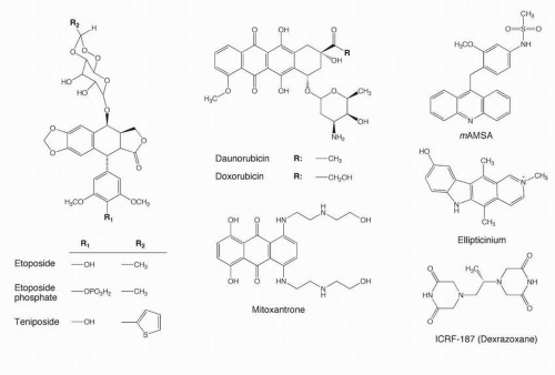
Extracts from the mayapple or mandrake plant have long been used as a source of folk medicine. The active principle in this plant, podophyllotoxin, acts as an antimitotic agent that binds to tubulin at a site distinct from that occupied by the vinca alkaloids. A number of semisynthetic derivatives of podophyllotoxin have been made. Two glycosidic derivatives, VM-26 or teniposide and VP-16 or etoposide (Fig. 19-1), are active against a number of human malignancies. Epipodophyllotoxins, etoposide and teniposide, are characterized as particularly active against rapidly growing tumors. This strong relationship between rapid proliferation and sensitivity to etoposide is well understood: the epipodophyllotoxins—etoposide and teniposide—in contrast to the parent compound podophyllotoxin are inactive against tubulin and are pure inhibitors of DNA topoisomerase II (Top2).

The clinical use of etoposide (VP-16) has evolved through several periods:


  • Identification of solid tumors highly sensitive to etoposide, mainly germ-cell tumors, small cell lung cancers (SCLCs), Ewing’s sarcomas, osteosarcomas, lymphomas, acute leukemias, poorly differentiated adenocarcinomas and poorly differentiated endocrine tumors. Etoposide was approved by the Food and Drug Administration (FDA) for marketing by Bristol Laboratories under the trade name Vepesid in early 1984. Teniposide (VM-26; Vumon) was approved by the FDA in 1992 for refractory childhood leukemia.


  • Evidence of etoposide-induced secondary leukemias, leading to various attempts to reduce the number of cycles of etoposide when given in a curative intent, especially in testicular cancers.


  • Molecular biology and genomic analyses revealed the proximity between the topoisomerase II alpha gene (TOP2a) and the HER2 gene, which raised the question of the potential interest of Top2 inhibitors in HER-2-amplified solid tumors.


  • Molecular pharmacology studies demonstrated that Top2β targeting by etoposide could be responsible for secondary malignancies while Top2α targeting was primarily responsible for the anticancer activity of etoposide. This may justify the search for specific Top2α inhibitors as second-generation etoposide derivatives.


Molecular and Cellular Pharmacology


Enzymology and Functions of DNA Topoisomerase II

The length of eukaryotic DNA and its anchorage to nuclear matrix attachment regions (MARs) limit the free rotation of one strand around the other as the two strands of DNA double helix are separated for DNA transcription, replication, recombination, or repair. DNA topoisomerases catalyze the unlinking of the DNA strands by making transient DNA strand breaks and allowing the DNA to rotate around or traverse through these breaks.9 Three types of topoisomerases known in humans are topoisomerase I (Top1), topoisomerase II (Top2), and topoisomerase III (Top3)9 (Table 19-1). DNA gyrase and topoisomerase IV (Top4) are the
bacterial equivalents of eukaryotic Top2. Quinolones (nalidixic acid, ciprofloxacin, norfloxacin, and derivatives), which are widely used antibiotics, act by inhibiting DNA gyrase and topo IV with little if any, effect on the host human Top2.10,11


FIGURE 19-1 Chemical structures of the epipodophyllotoxins (VP-16, etoposide phosphate and VM-26), anthracyclines (doxorubicin and daunorubicin), mitoxantrone, acridines (m-AMSA), ellipticines (ellipticinium), and bisdioxopiperazine (ICRF-187).

A general feature of the topoisomerase-mediated DNA breaks is the transesterification reaction, wherein a DNA phosphoester bond is transferred to a specific enzyme tyrosine residue, while the enzyme generates a break in the DNA phosphodiester backbone. In the case of Top1, the enzyme becomes linked to the 3′-terminus of the cleaved DNA, whereas in the case of Top3, the enzyme is linked to the 5′-DNA terminus of the DNA single-strand break. In the case of Top2, each enzyme molecule of a homodimer becomes linked to the 5′-terminus of each of the cleaved DNA strands (Table 19-1; Figs. 19-2 and 19-4). Type 1 enzymes (Top1 and Top3) make DNA single-strand breaks, whereas the type 2 enzymes (Top2α and β) make DNA double-strand breaks.


TABLE 19.1 Mammalian DNA topoisomerases


Topoisomerases I


Topoisomerase IIα


Topoisomerase IIβ


Topoisomerases III


Size of monomer


  • 100 kDa (Top1 nuclear)


  • 72 kDa (Top1 mt)


  • Acting as monomer


  • 170 kDa


  • Acting as dimer


  • 180 kDa


  • Acting as dimer


110 kDa


Size of mRNA


  • 4.1kb (Top1 nuclear)


  • 1.8kb (Top1 mt)


6.2 kb


6.5 kb


3.8 kb (main transcript)


Gene location


  • 20q12-13.2 (Top1 nuclear)


  • 8q24.3 (Top1 mt)


17q21-22


3p24


  • 17p11.2-12 (Top3α)


  • 22q11-12 (Top3β)


Catalytic intermediate


  • DNA SSB


  • Covalent linkage to 3′ DNA terminus


  • DNA DSB


  • Covalent linkage to 5′ DNA termini


  • DNA DSB


  • Covalent linkage to 5′DNA termini


  • DNA DSB


  • Covalent linkage to 5′ DNA termini


ATP dependence


No


Yes


Yes


No


Cell cycle expression


Throughout


G2/M


Throughout


Specific inhibitors


Camptothecins, Indenoisoquinolines, Indolocarbazoles (reviewed in Refs [205,206])


Top2 poisons and catalytic inhibitors, intercalators (see Table 19-2)


Same as topoisomerase IIα(preference for amsacrine, mitoxantrone207


Unknown


SSB, single-strand breaks; DSB, double-strand breaks (see Fig. 19-4).
Top1mt: mitochondrial Top1208 is the most recently discovered human Top1 gene.


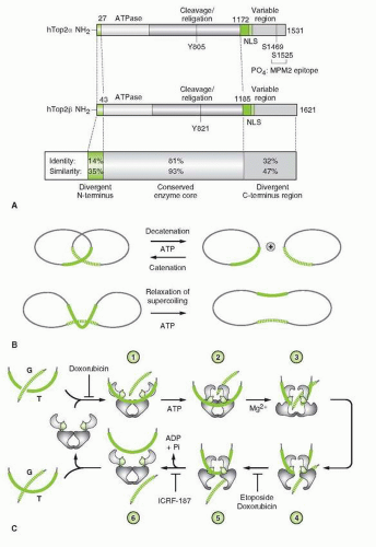
FIGURE 19-2 A. Domain structure of Top2α and β. The three major domains are illustrated, as well as the site of ATP binding (ATPase), the active-site tyrosine (Y), the nuclear localization sequence(s) (NLS), and the sites of phosphorylation (PO4). The N-terminal domain (homologous to the gyrase B subunit) extends from amino acid 1 to about 660. The catalytic core domain (homologous to A subunit of gyrase) extends from about residue 660 to 1,200, and the C-terminal domain (no corresponding homology with gyrase) extends from about residue 1,200 to the C-terminus of the enzyme. Top2 α and β are most divergent in their N- and C-terminus region. B. Schematic representations of two types of topological reactions catalyzed by Top2: catenation-decatenation and relaxation of DNA. C. Model for the catalytic cycle of type II topoisomerases [according to Refs (7,8,12)]. The unliganded enzyme binds to duplex DNA (labeled G) across the TOPRIM domains (step 1). A second duplex DNA strand (labeled T) and ATP bind to the enzyme (step 2). Nucleotide binding promotes dimerization of the ATPase domains and closure of the clamp (step 2). Cleavage of the G-strand (step 3) allows the passage of the T-strand through the cleaved G-strand (step 4). Following G-strand religation (step 5), the T-strand is released through the dimer interface in the C-terminus region (step 5). ATP hydrolysis reopens the ATPase domain and completes the enzyme catalytic cycle. Sites of drug action are indicated: DNA intercalators (doxorubicin at relatively high concentrations) prevent Top2 from binding DNA; Etoposide and lower concentrations of doxorubicin prevent religation of the G-strand (steps 4 and 5), and ICRF-187 traps the G-strand inside the enzyme (steps 5 and 6).

Both Top1 and Top2 can remove DNA supercoiling by catalyzing DNA relaxation (Fig. 19-2B). They can complement each other in this function at least in yeast, where the absence of Top1 can be compensated for by the presence of the other topoisomerase for DNA relaxation. However, yeast strains deficient in Top2 are not viable and die at mitosis because Top2 is essential for chromosome condensation and structure and the proper segregation of mitotic and meiotic chromosomes.9 The reason is that in addition to its DNA relaxing activity, Top2 can separate two linked circles of duplex DNA (decatenation) as well as catalyze the reverse reaction (catenation) by allowing one duplex to go through a double-stranded gap created in the other duplex (strand passage reaction) (see next section and Fig. 19-2B). Decatenation is essential at the end of DNA replication for the separation of daughter DNA molecules and segregation of newly replicated chromosomes. The accumulation of Top2 at the end of S phase and during G2 and its concentration in the chromosome scaffold are consistent with the enzyme’s role in separating chromatin loops and condensing DNA at mitosis (see Table 19-1).


Mammalian cells have two Top2 isoenzymes, termed Top2α and Top2β (Table 19-1), which differ in molecular mass, enzymatic properties, chromosome localization, sequence, cell cycle regulation, and cellular and tissue distribution. While Top2β concentrations are relatively constant throughout the cell cycle, Top2α levels are tightly linked to the proliferative state of the cell. The concentration of the α isoform increases two- to threefold during G2/M and is orders of magnitude higher in rapidly proliferating cells than it is in quiescent populations.


DNA Topoisomerase II Catalytic Cycle

A description of the Top2 catalytic cycle is essential for understanding how Top2 poisons stabilize the “cleavage complex” of the DNA strand passage reaction (see Fig. 19-2) (for more details, see Refs [6, 7, 8, 9, 12, and 13]). In contrast to Top1, Top2 functions as a homodimeric molecule. Its catalytic activity requires the presence of magnesium as well as ATP as an exogenous energy source. Hence, Top2 acts as a DNA-dependent ATPase. Recent structural and mechanistic studies reveal a remarkable dynamic behavior of the enzyme during its catalytic cycle.12,13 Top2 catalyzes DNA strand passage according to the two gate model shown in Figure 19-2C.12,13 The enzyme forms a dimer and initiates its catalytic cycle by binding to its DNA substrate with a preference for DNA crossover regions. Hence, Top2 interactions with DNA are determined by both DNA superstructure (DNA crossovers, bends, etc.) and local DNA sequence. Although Top2 interacts with preferred sequences, its specificity is far less stringent than that of restriction endonucleases. This lack of stringency probably allows the enzyme to act at multiple sites of the genome in order to perform its vital functions. Top2 assumes two alternative conformations: open or closed clamp forms in the absence or presence of ATP, respectively.12,13 The enzyme binds two segments of duplex DNA, referred to as the G and T segments. The G (for Gate) segment is the one cleaved by the enzyme in order to pass the T (for Transported) segment through the enzyme-DNA complex (Fig. 19-2C). Upon ATP binding, Top2 undergoes a conformational change from an open to a closed clamp form (step 3). In the presence of a divalent cation (under physiological conditions Mg2+), the tyrosine active residue of each Top2 monomer (tyrosine 804 for human Top2α or tyrosine 821 for Top2β [Fig. 19-2A]) attacks a DNA phosphodiester bond four bases apart on the G duplex and becomes covalently linked to the 5′ ends of the broken DNA while the 3′-ends are 3′-hydroxyls (see Fig. 19-4). The T segment can then pass through the gap produced in the G segment (steps 3 and 4). Cleavage of the G duplex is reversible in nature, and under normal conditions, the cleavage complex is a short-lived intermediate. After strand passage, the T segment is released from the clamp and the broken ends of the G segment are religated by Top2 (steps 5 and 6). Upon hydrolysis of ATP by the intrinsic ATPase activity of the enzyme, the Top2-DNA complex is converted back to the open clamp form with release of the G segment (step 6). Thus, closing and opening of the Top2 clamp are coupled with ATP binding and hydrolysis, respectively. Through its ability to open both strands of a DNA duplex and to catalyze strand passage in concerted reactions, Top2 can perform a variety of DNA topoisomerization reactions. Whereas DNA relaxation is common to Top1, conversion of circular DNA to knotted forms and removal of preexisting knots are specific to Top2 (Fig. 19-2B only shows catenation-decatenation and relaxation reactions; knotting-unknotting is not shown). These biochemical reactions are commonly used to assay topoisomerase activities in vitro: relaxation of supercoiled plasmid DNA in the absence of ATP and Mg2+ in the case of Top1, decatenation of kinetoplast DNA (kDNA) and unknotting of P4 DNA in the case of Top2.14


DNA Topoisomerase II Poisons


Mechanisms of Top2 Inhibition by Anticancer Drugs

The antitumor Top2 inhibitors presently used in the clinic poison the enzyme by stabilizing the DNA cleavage complexes (steps 3 and 4 in Fig. 19-2C) rather than preventing enzyme catalytic activity.15 The production of DNA cleavage complexes is due to an inhibition of DNA religation in the case of VP-16, VM-26, and the DNA intercalators doxorubicin, daunorubicin, m-AMSA and ellipticines.15, 16, 17 On the other hand, compounds such as quinolones act by inducing the formation of cleavage complexes rather than by inhibiting relegation.18 The cleavage complexes can be detected in cells as protein-linked DNA breaks by alkaline elution or by SDS-KCI precipitation assays (for review, see Ref. [14] and references therein). Cellular topoisomerase-DNA complexes can also be detected using the ICE-bioassay (for Immuno Complex Topo Assay).19 Inhibition of Top2 catalytic activity without trapping of cleavage complexes (Table 19-2; Fig. 19-3) was first demonstrated for strong DNA intercalating agents at drug concentrations that saturate the DNA. It is attributed to DNA structural alterations that prevent the enzyme from binding to DNA (step 1 in Fig. 19-2C) or prevent initiation of the cleavage complex (step 3).20,21 Non-DNA binders such as merbarone and bisdioxopiperazines [ICRF 159, 187 (= dexrazoxane), and 193] produce the “closed clamp” type of inhibition, for example, inhibition of Top2 catalytic activity without trapping of cleavage complexes22,23 (see Table 19-2; Fig. 19-2C). Hence, three types of curves that relate drug concentrations to cleavage complexes can be observed for Top2 inhibitors (Fig. 19-3): (a) monotonal increase of cleavage complexes with drug concentration in the case of non-DNA binders or weak DNA binders (VP-16, VM-26, m-AMSA, quinolones), (b) bell-shaped curve (with initial increase in cleavage complexes with increasing drug concentrations, followed by a decrease in cleavage complexes at higher concentrations) in the case of DNA intercalators (ellipticines, anthracyclines, mitoxantrone, anthrapyrazoles), and (c) monotonal decrease of cleavage complexes in the case of some bulky intercalators (ethidium bromide, ditercalinium, aclarubicin) or non-DNA binders (bisdioxopiperazines) (see Table 19-2; Fig. 19-3) that inhibit catalytic activity without trapping cleavage complexes.

At the biochemical level, Top2 inhibitors exhibit different effects (Table 19-3). The kinetics of cleavage complex formation and reversal in drug-treated cells vary from slow in the case of doxorubicin2 and ellipticine24 to very rapid in the case of VP-16, m-AMSA, and ellipticinium.2,25 The higher cytotoxicity of doxorubicin versus VP-16 may be explained by the importance of persistent cleavage complexes for cytotoxicity. Most drugs induce not only Top2-mediated DNA double-strand breaks but also Top2-mediated single-strand breaks, the ratio of which varies widely among drugs. Ellipticines produce almost exclusively DNA double-strand breaks, while VP-16 and amsacrine produce 10 to 20 single-strand breaks per double-strand break.2,15,25 Anthracyclines produce a mixture of
single-strand and double-strand breaks.2 Hence, the higher cytotoxicity of anthracyclines compared with amsacrine or VP-16 may be due to the higher frequency of DNA double-strand breaks that may be more cytotoxic than single-strand breaks.15 Finally, the DNA sequence and genomic localization of Top2 cleavage complexes vary among drugs.26 Drugs, which are chemically and structurally related, frequently produce closely related patterns of Top2 cleavage, while compounds structurally and electronically unrelated produce different patterns both in purified DNA and in drug-treated cells.15,26,27


TABLE 19.2 Topoisomerase II (Top2) inhibitors


Poisons


References


Suppressors


References


Intercalators and
DNA binders


Doxorubicin


image


See (15,16,30)


Doxorubicin (high conc.)


image


See (15,16,209)


Daunorubicin


Daunorubicin (high conc.)


Epirubicin


Epirubicin (high conc.)


Idarubicin


Idarubicin (high conc.)


Amsacrine


Amsacrine (high conc.)


Mitoxantrone


Mitoxantrone (high conc.)


Elliptinium


Elliptinium (high conc.)


Actinomycin Da


Actinomycin Da (high conc.)


Anthrapyrazoles


(210)


Anthrapyrazoles(high conc.)


(210)


Menogaril


(211)


Menogaril (high conc.)


(211)


Intoplicinea


(212,213)


Intoplicinea (high conc.)


(212,213)


Saintopina


(214)


Saintopina (high conc.)


(214)


Amonafide


(215)


Amonafide (high conc.)


(215)


Streptonigrin


(216, 217, 218)


Bulgarein


(219)


Makaluvamines


(220)


Ethidium bromide


(221)


Alkylating anthraquinones


(222)


Ditercalinium


(223,224)


Olivacines


(225)


Bisantrenes


(226)


Non-Intercalators


VP-16, VM-26


(227, 228, 229)


Distamycin, Hoechst 33258


(230)


(231)


Aza IQDa


(232)


Merbarone


(233)


Flavones-flavonones


(234)


Bisdioxopiperazines


(23)


Isoflavones (Genistein)


(235)


Suramin


(236)


Nitroimidazole (Ro 15-0216)


(237)


Novobiocin


(238)


Terpenoids


(239)


Chloroquine


(240)


Naphthoquinones


(241)


Fostriecin


(242)


Whithangulatin


(243)


Aclarubicin (Aklavin, Oxaunomycin, β-Rhodomycinone)


(244)


Polyaromatic quinones


(245)


Quinobenoxazines


(246)


Quinolones (CP-115,953)


(247)


Azatoxins


(248)


a Dual Top1 and Top2 inhibitor.

See Figure 19-1 and text for definition of poisons and suppressors.


Base-Sequence Preference of Top2 Inhibitors and Drug Binding Model

DNA sequencing of drug-induced cleavage sites shows that each class of inhibitor tends to act at Top2 cleavage sites with different base sequence preferences at the 3′- and/or 5′-terminus of the Top2-mediated DNA double-strand break28, 29, 30, 31, 32, 33, 34 (Fig. 19-4). These drug-specific preferences for certain bases immediately flanking the cleavage sites suggest that the drugs interact directly with these bases. Since all Top2 inhibitors, whether intercalator or not, have a planar aromatic portion that, in some cases, mimics a base pair (see Fig. 19-1), the simplest explanation is that the drugs stack inside the cleavage sites at the enzyme-DNA interface. Depending on the drug structure, preferential base stacking would take place with the base pairs either at the 3′- or the 5′-terminus. This hypothesis implies that topoisomerases first cleave the DNA at many sites and that the drugs bind specifically to some sites and prevent DNA relegation.35, 36, 37

The base sequence analysis data suggest that stacking at one cleavage site is sufficient for the creation of a DNA double-strand break,
a theory consistent with the concerted action of both enzyme monomers during catalysis.28,38 This type of inhibition, which we refer to as “interfacial inhibition” is one of natures’s paradigm for noncompetitive protein inhibition.39 Top2-mediated DNA cleavage tends to be more pronounced at the DNA site of drug binding,40,41 and reactions triggered by cooperative effects with the other monomer appear less stable than those at sites of preferred binding.38 Recent studies show the DNA interface to include not only each of the DNA breaks generated by Top2 but also intercalation sites within the 4 base pair stagger between the two concerted Top1 cleavage sites.41 The drug-binding site on Top2 is not well defined. Analyses of drug-resistant mutant enzymes suggest that both the A′ and B′ regions of the enzyme reversibly bind to the drugs. Recent studies35,38 indicate that the CAP homology domain determines DNA sequence recognition on the G segment of DNA and drug effects, which is consistent with drug binding at or near the DNA cleavage site and formation of a ternary complex: drug-enzyme-DNA.
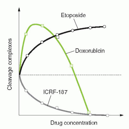


FIGURE 19-3 Different modes of drug inhibition of Top2. Top2 poisons such as the epipodophyllotoxins (VP-16 or VM-26) only trap the Top2 cleavage complexes with increasing efficiency as their concentration increases. Top2 suppressors such as the bis-dioxopiperazines (open circles) are pure catalytic inhibitors that only inhibit the formation of cleavage complexes. Gray squares correspond to biphasic inhibitors such as DNA intercalators (anthracyclines, ellipticines, acridines; see Table 19-2), which enhance Top2 cleavage complexes at low concentrations and suppress cleavage complexes at higher concentrations.


TABLE 19.3 Differences among Topoisomerase II Inhibitors


Other targets besides Top2


  • Free radicals: anthracyclines, mitoxantrone


  • DNA intercalation: e.g., anthracyclines, mitoxantrone, ellipticines


Different effects on Top2


  • Base sequence preferences and location of DNA cleavage sites (see Fig. 19-4)


  • Ratio of DNA double-strand/single-strand breaks: ellipticine > anthracyclines > amsacrine/epipodophyllotoxins


  • Kinetics of trapping Top2 cleavage complexes (slow for anthracyclines; fast for epipodophyllotoxins/amsacrine)


  • Inhibition of cleavage complexes at higher concentration (intercalators)


  • Pure Top2 poisons (epipodophyllotoxins)


Mechanisms of resistance


  • Substrates for transmembrane transporters: anthracyclines and epipodophyllotoxins more than mitoxantrone, amsacrine, and ellipticines


  • Specific Top2 mutations affect drug binding differentially249, 250, 251, 252, 253, 254, 255, 256, 257


See text for references.


Determinants of Sensitivity and Resistance to Top2 Inhibitors

Figure 19-5 summarizes the multiple factors that determine the cytotoxicity of Top2 inhibitors. Before topoisomerase inhibitors reach their nuclear target, they have to be taken up by the cells and transported to the nucleus. Reduced drug accumulation or altered intracellular drug distribution is a dominant feature of many drug-resistant cell lines. Most clinical antitumor Top2 inhibitors are substrates for the 170-kDa transmembrane glycoprotein, Pgp, which is a product of the MDR1 gene and responsible for the classical multidrug resistance (MDR) phenotype.42 MDR sensitive drugs include doxorubicin and analogues, mitoxantrone, anthrapyrazoles, ellipticines, VP-16, and to a lesser extent m-AMSA analogues. Hence, cells overexpressing Pgp are generally resistant to Top2 inhibitors because the drugs are actively extruded from the cells. The amino group on the daunosamine sugar of anthracyclines is probably involved in the drug recognition by Pgp. This is probably why the deamino derivative hydroxyrubicin is less subject to drug resistance while retaining Top2 inhibitory activity.43

Only gold members can continue reading. Log In or Register to continue

Stay updated, free articles. Join our Telegram channel

May 27, 2016 | Posted by in ONCOLOGY | Comments Off on Topoisomerase II Inhibitors: The Epipodophyllotoxins

Full access? Get Clinical Tree
Get Clinical Tree app for offline access