Molecular Targeted Drugs


Molecular Targeted Drugs


Jeffrey W. Clark


This chapter presents the rationale for molecularly targeted drugs and approaches to the selection of and validation of targets and consider the variety of molecules under development in this new category of antitumor drugs. We discuss the basic and clinical pharmacology of BCR-ABL kinase inhibitors as the first and most successful examples of targeted small molecules. Elsewhere in this book the detailed pharmacology of monoclonal antibodies (mAbs), growth factor receptor inhibitors, and epigenetic modifiers is considered.

Until the past decade, cancer therapy depended on drugs lacking selectivity for tumor cells. These were predominantly cytotoxins with narrow therapeutic indices. Most were discovered through cytotoxic screens rather than efforts to exploit targets specific for malignant cells. These drugs have improved treatment of many solid tumors and have cured some hematologic malignancies and selected solid tumors, particularly in the adjuvant setting, but with significant normal tissue toxicity. However, they have cured few patients with metastatic solid tumors. New approaches for treating most cancers are needed.

Rapid growth in knowledge of cancer biology has led to a more rational approach to therapeutic discovery through identifying pathways and proteins that are essential for survival of cancer cells.1, 2, 3, 4, 5, 6, 7 Identification of mutations unique to neoplastic cells has led to development of agents targeting those mutations, providing a means of specifically killing malignant cells while sparing normal tissues. As examples, drugs specific for BCR-ABL or BRAF mutant at the V600E locus have shown remarkable clinical activity, with limited toxicity. Small molecules, mAbs, peptidomimetics, siRNAs, antisense oligonucleotides, expressed genes, and other molecularly targeted therapies are being evaluated for clinical use (Table 29-1). An increasing number of these have sufficient clinical activity to be important components of current therapy (Table 29-2). Figure 29-1 illustrates signaling through growth factor receptors and indicates the site of inhibition of some currently approved targeted anticancer agents.


Druggability of Molecular Targets

Validation of the target in experimental systems is perhaps the first and most important step in developing a successful therapy, and this process is considered in detail in Chapter 2. The targeted protein in the neoplastic cell must be critical for survival of malignant but not normal cells. Targeting a single protein may have limited effects on growth of neoplastic cells unless that protein is vital for proliferation or survival (e.g., “oncogene addiction” such as BCR-ABL in chronic myelogenous leukemia [CML]). Inhibition of any specific gene, even if it is important for neoplastic cell growth, may only be cytostatic. Although these targets can be clinically useful in combination therapy, it is also important to choose as targets molecules critical to the proliferation and survival of the cell. Ultimately, agents must be capable of eliminating (or causing differentiation or prolonged growth suppression of) tumor cells, either by themselves or in combination with other agents, if they are to be useful in long-term control of the cancer. A number of genes could be targeted simultaneously or sequentially, and combinations of approaches inhibiting certain genes (e.g., oncogenes) or their protein products and enhancing expression of others (e.g., tumor suppressor genes) may ultimately be used. A further step is their evaluation in combination with other agents. A recent review considers combination therapies in detail.5

In view of the multiplicity of potential targets, it is important to determine the “druggability” of the target at an early step. It is vastly easier to develop an inhibitor of protein function than it is to replace an inactive or deleted function. Thus, although mutations in tumor suppressor genes, such as those affecting the p53 or retinoblastoma (RB) proteins, play a prominent role in tumorigenesis, it is difficult to
restore function of these proteins without restoring a fully functional gene to each cancer cell, a significant technical challenge.8, 9 However, there are other approaches that can potentially exploit altered tumor suppressor gene function. The synthetic lethal strategy (identifying a gene that becomes essential for tumor cell survival when a suppressor gene is deleted) has led to the development of polyadenosine diphosphate-ribose polymerase (PARP) inhibitors for BRCA1- and BRCA2-mutated ovarian and breast cancers. In cases where function of a tumor suppressor protein is inhibited (such as by binding of MDM2 to a normal p53 protein) as opposed to being absent due to mutation or deletion, blocking the inhibitor (or unblocking the suppressor) might be able to restore function.10


TABLE 29.1 Types of molecularly targeted compounds


Agent


Potential target(s)


Antisense oligonucleotides


RNA, DNA, and proteins


SiRNA


RNA


Gene therapy


Neoplastic cells, immune mediator cells, and normal cells


Ribozymes and DNAzymes


RNA and DNA in tumor cells


mAbs


Circulating proteins (e.g., growth factors), growth factor receptors, cell surface antigens, and other cellular proteins


Modified peptides


Circulating proteins (e.g., growth factors), growth factor receptors, cell surface antigens, and extracellular and intracellular proteins (e.g., enzymes and signal transduction molecules)


Small molecules


All of the above targets


TABLE 29.2 Approved molecularly targeted agents


Type of compound


Agent


Target


Indication


Uncoupled mAbs


Alemtuzumab


CD52


B-CLL


Bevacizumab


VEGF


Colorectal, Lung, Glioblastoma, RCC


Cetuximab


EGFR


Colorectal, Head and Neck


Trastuzumab (Herceptin)


HER2


Breast


Ofatumumab


CD20


B-CLL


Rituximab


CD20


B-cell NHL, B-CLL


mAbs coupled to radioactive or cytotoxic agents


Ibritumomab tiuxetan Y-90


CD20


B-cell NHL


Tositumomab I-131


CD20


B-cell NHL


Small molecules


(Anti-EGFR)


Gefitinib


EGFR


NSCLC


(Anti-EGFR)


Erlotinib


EGFR


NSCLC, Pancreatic


(Anti-HER2)


Lapatinib


HER2, EGFR


Breast


(Anti-BCR ABL)


Imatinib


BCR-ABL, KIT, PDGFR-α


CML/Ph+ALL, GIST, CMML, DFP, SM, HES


(Anti-BCR ABL)


Dasatinib


BCR-ABL, SRC, Eph


CML, Ph+ALL


(Anti-BCR ABL)


Nilotinib


BCR-ABL, PDGFR-α, KIT


CML, Ph+ALL


(Anti-mTOR)


Everolimus


mTOR


RCC


(Anti-mTOR)


Temsirolimus


mTOR


RCC, MCL (in Europe)


Multi-targeted (including anti-VEGFR)


Sunitinib


VEGFRs, KIT, other kinases


RCC, GIST


Multi-targeted (including anti-VEGFR)


Sorafenib


VEGFRs, other kinases


HCC, RCC


Multi-targeted (including anti-VEGFR)


Pazopanib


VEGFRs, other kinases


RCC


Proteosome


Bortezomib


Proteosome


MM


Epigenetic (histone deacetylase)


Vorinostat


Histone Deacetylase


CTCL


Epigenetic (histone deacetylase)


Romidepsin


Histone Deacetylase


CTCL


Immunomodulatory, antiangiogenic, anti-inflammatory


Lenalidomide


Uncertain


MDS


Retinoic acid


Tretinoin


Promyelocytic leukemicretinoic receptor alpha


APML


At the present time, the primary choices for targeting are proteins that are either activated by a mutation (BCR-ABL, Eml4-Alk, and epidermal growth factor receptor [EGFR] mutant forms) or are overexpressed in specific malignancies (such as Her2/neu) and are critical for survival or proliferation of malignant cells.1, 2, 3, 4, 5, 6, 7 Because of the central role that kinases play in pathways impacting cell growth, survival, migration, metabolism, and differentiation, and the ability of small molecules to inhibit the ATP binding sites of different kinases with great specificity, these have been the most successfully targeted proteins to date.1, 2, 3, 4, 5,7


Identifying and Synthesizing Modulators for Molecular Targets

Diverse approaches to developing inhibitors or modulators for identified targets in cancers are now possible. Although small molecules, antibodies, and modified peptides are the agents with clearly established clinical value at this time, this is certain to evolve as approaches such as utilizing siRNA and gene delivery via vectors are evaluated.11,12

A common strategy for developing targeted agents is empirically screening a large number of compounds for activity and subsequently designing better ones based on the structure of the most active compound or compounds (this is the approach by which most anticancer agents have been developed). High-throughput screens allowing rapid evaluation of a large number of compounds have enhanced the utility of this approach. Alternatively, compounds can be designed based on structure of the specific region being targeted, using known sequence data and other information available about the protein (e.g., x-ray crystallography, nuclear magnetic resonance analysis, and computer molecular modeling). Clearly, some combination of these two approaches might be most useful—for example, lead identification by high-throughput screening followed by lead
optimization through structural studies of the inhibitor and target. This allows more rational design of antineoplastic molecules and, at the same time, efficient screening of a wide range of structures.


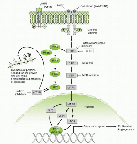
FIGURE 29-1 Two signaling pathways, stimulated by IGF-1 and EGF ligands, which activate downstream signals through PI3 kinase and Ras proteins. Inhibitors of these pathways at the receptor, its intracellular kinase domains, and downstream components are indicated. Cross talk between the pathways occurs at PI3 kinase. Signals in the nucleus stimulate proliferation and angiogenesis, while cytoplasmic signals promote cell growth and survival.

As with all forms of systemic cancer therapy, the ultimate usefulness of these treatment approaches depends on a number of factors, including effective delivery of the agent to tumor cells; adequate binding to neoplastic cells (in the case of mAbs or other compounds used to target proteins on the cell surface or activate the immune system or deliver radioactive compounds) or uptake by neoplastic cells; presence of the agent on or within cells for a sufficient time to lead to meaningful biological effect (inhibition of proliferation, death or differentiation of those cells); selective action of the compound against malignant versus normal cells; rate of elimination of the agent from normal and malignant cells as well as from the body; and unanticipated toxicity of the agent for normal tissues, both acutely and chronically.13, 14, 15 Heterogeneity of neoplastic cells within tumors limits effectiveness of single molecularly targeted therapy. Therefore, approaches that target heterogeneous cell populations, including resistant cells, need to be considered in designing new agents.16, 17, 18

Certain classes of targeted agents, such as mAbs, antisense oligonucleotides, and modified peptides, may additionally be compromised by negative pharmacologic properties of these molecules.13, 14, 15 These negative features include relatively large size, making delivery more difficult; complex structure, contributing to decreased absorption and permeation into tissues (including the central nervous system) as well as susceptibility to rapid clearance or metabolism; and, the presence of alternative binding proteins and receptors for these agents in the blood or expressed on normal cells. Additional problematic features may be immunogenicity: high molecular weight compounds such as biological or polymeric materials often are immunogenic or induce host cytokine release.


Potential Molecular Targets

Discoveries of cancer biology continue to provide a number of potential targets. These include


  • Mutation, amplification, or overexpression of growth factors or their receptors involved in proliferation and survival of various cancers, such as the EGFR family, including HER2/neu;


  • Mutation or enhanced activity of intracellular signaling pathways that promote growth, impede cell death, or enhance metastasis, such as activation of the Ras-RAF-MEK or PI3K pathways;


  • Antiapoptotic mechanisms that antagonize cell death, such as overexpression of bcl-2 or decreased bax expression;


  • Pathways, such as chaperoning, ubiquination, and the proteosome, that protect mutant or oncogenic proteins or affect their turnover rates;


  • Epigenetic factors such as methylation or acetylation of histones, or methylation of DNA, which regulate gene expression and differentiation;


  • MicroRNAs (miRNAs) that promote malignant growth;


  • Tumor promoting factors in the environment, such as angiogenic pathways (e.g., vascular endothelial growth factor [VEGF] or platelet-derived growth factor [PDGF]);


  • Pathways involved in suppressing immune response to cancer.1, 2, 3, 4, 5, 6, 7,19

Somatic mutations provide particularly important and unique targets in cancer (c-KIT mutations in gastrointestinal stromal tumor [GIST], EGFR mutations and eml4-alk translocations in cases of non-small cell lung cancer (NSCLC), and BCR-ABL translocation in CML).20, 21, 22, 23, 24, 25 Important brakes on proliferation can be lost, such as by mutations that inhibit function of p53, RB, or the phosphate and tensin homolog (PTEN), which down-regulates the PI3 kinase pathway,8, 9, 10,26 but replacing these missing functions has not been feasible. An alternative strategy, searching for a synthetic lethal target, has been demonstrated in the experimental setting, as discussed below.

A number of theoretical and practical questions must be answered in order to validate the target, before a large-scale investment is justified. Among the most relevant questions are the following:


  • Are subject gene and its protein found in human tumors, and is there selective expression in tumors versus normal tissues?


  • Is the subject protein’s function essential to the survival and proliferation, and, indeed, the transformed behavior of the malignant cells?


  • Does inhibition of the gene product change the phenotype of these cells and lead to the desired result (such as death or growth arrest of malignant cells or a decrease in metastasis) in an appropriate animal model? It is critical to establish that the gene and associated protein product are important for the biology of human tumors and not just an animal model. Short interfering RNAs (SiRNAs) have become invaluable tools for inhibiting the expression of target genes within cells and determining the role of the target.8,27,28 SiRNAs also have potential as therapeutic agents and early evaluation in clinical trials has begun.


  • Is the protein also expressed in key proliferating normal tissues, such as intestinal epithelium and/or bone marrow progenitors, or even nonproliferating tissues, such as heart, kidney, or brain, and does targeting the protein therefore carry risks for significant toxicity?


  • The profile of gene expression in normal tissues may provide helpful clues about potential toxicity of an agent directed against that gene, as in the case of trastuzumab, an inhibitor of Her2/neu, a growth factor expressed in myocardium. In this case, gene knockout in animal models had early and fatal consequences for the developing embryo, indicating that inhibition of that gene or its protein product might lead to significant toxicity.


  • Are there closely related proteins and what is their physiological function? Are they essential for normal tissue function and survival of the host and might be cross-targeted by the agent producing unwanted toxicities?


  • Are there appropriate biomarkers that can identify likely “responders” and can these be developed as a guides for drug development in early trials?

Identification and optimization of biomarkers that:


  • Aid in selecting appropriate patients likely to respond to a specific agent (e.g., HER2 positivity for trastuzumab, EGFR mutations for erlotinib, and BRAF mutations for BRAF inhibitors);


  • Help validate that the target is being modulated in vivo (e.g., decreased pERK phosphorylation with BRAF inhibitor);


  • Aid in optimizing dose and schedule;


  • Serve as surrogates for response (e.g., HER2 positivity in breast cancer patients treated with trastuzumab and early decline in PET avidity in GIST patients treated with imatinib).

These considerations are essential in determining choice of a target and probability of success. Obviously, even a well-validated target may not be amenable to a drug discovery strategy for a number of reasons. Unanticipated toxicities or pharmacokinetic or pharmacodynamic problems in drug uptake, distribution, or elimination may defeat the most rational strategy.

The reader is referred to excellent reviews of high-priority molecular targets for cancer therapy and the role of biomarkers in the development of targeted agents.1, 2, 3, 4, 5, 6, 7 Angiogenesis inhibitors, mAbs, targeting the EGFR family (EGFRs including Her2) and the PI3K pathway (including mTOR inhibitors), and targeted endocrine therapies are covered elsewhere in this book. The following is a brief review of several of the growth factor receptors and downstream signaling pathways that have yielded substantial new leads for cancer treatment.


Examples of Specific Targets: Growth Factor Receptors

Growth factor receptors or their ligands are among the most attractive molecular targets for cancer therapy1, 2, 3, 4, 5, 6, 7,14,20, 21, 22,29,30 (Fig. 29-1). Their presence on the cell surface makes them more readily accessible to antibodies. They are overexpressed in many malignancies and are mutationally activated in others. Their important role in cellular processes essential for angiogenesis, proliferation, and survival of cells has been demonstrated. Growth factors and their receptors (such as VEGF ligands, VEGFR family, EGF ligands, and EGFR family) promote metastatic potential.29,30

Members of the EGFR family of receptors, including HER2, are especially attractive targets.30, 31, 32 Through either mutation or overexpression, EGFR family members drive cell survival and proliferation as well as the metastatic process in many epithelial tumors.
Inhibition of EGFR in certain preclinical models leads to death of malignant cells and significant antitumor response. HER2/neu is expressed in a subset of patients with breast and gastric cancers and has been successfully targeted.30, 31, 32, 33 Anti-EGFR mAbs or small molecules have successfully treated lung, colorectal, and head and neck cancers.34,35 Finally, as discussed above, mutant proteins provide the most specific target in cancer. EGFRs are mutated in a subset of lung adenocarcinomas, primarily in nonsmokers or light smokers, and these are very sensitive to inhibition by small molecular inhibitors of the tyrosine kinase activity of the EGFR.36

Other growth factor receptors that have been successfully targeted clinically include c-KIT, platelet-derived growth factor receptor (PDGFR), and VEGFR.1,2,4,23,29 Mutations in c-KIT are found frequently in GIST tumors, with mutant PDGFR found in a smaller subset, and targeting of both of these by imatinib or sunitinib has been very successful clinically.23 The VEGFR family (critically involved in tumor angiogenesis) has been successfully targeted in a number of malignancies, including colorectal, lung, renal cell cancer (RCC), hepatocellular cancer (HCC), breast cancers, and glioblastoma.1,2,4,29


Signaling Pathways Downstream of Growth Factor Receptors

Signals from growth factors travel through multiple switches (Ras, PI3 kinase, MAP kinase, SMAD, and others; Fig. 29-1) to reach nuclear and cytoplasmic effectors. Studies continue to evaluate the potential for inhibiting these intracellular pathways either alone or in combination with each other or with anti-growth factor receptor agents as a means of treating cancer. The Ras and PI3 kinase pathways have been the two most extensively evaluated as potential targets to date. The RB pathway is essential in negatively controlling proliferation and various approaches are being pursued to modulating this function in malignant cells.


Ras Pathways

One of the first oncogenes to be recognized in human tumors was mutated Ras genes and the resultant constitutively activated Ras proteins37, 38, 39 (Fig. 29-1). Ras proteins play central roles in transmitting signals important for a variety of critical processes in cells, including proliferation and differentiation. One of the key roles played by Ras proteins (products of the KRAS, NRAS, and HRAS genes) is in transmitting signals from growth factor receptors (such as the EGFR) to downstream signaling molecules. Normally, Ras proteins are activated by binding guanosine triphosphate (GTP), and they subsequently activate downstream targets in signaling cascades, including Raf kinases.37, 38, 39 In the process, GTP is hydrolyzed to guanosine diphosphate and Ras is inactivated. Mutations at codons 12, 13, or 61 of the Ras genes activate Ras proteins by locking them in the GTP-bound state. This leads to constitutive signal transduction in the absence of growth factor stimulation. Ras mutations occur relatively frequently in a number of malignancies. For example, KRAS mutations are found in approximately 35% to 40% of colon cancers, 70% to 90% of pancreatic cancers, and 30% of adenocarcinomas of the lung.40, 41, 42 NRAS is mutated in approximately 25% of acute nonlymphocytic leukemias and KRAS in another 15%.42 HRAS is mutated in a minority of bladder and head and neck cancers. Thus, as a molecular target, Ras proteins have an essential role in maintaining the malignant state.

Attempts to inhibit the activated GTP binding site in mutant Ras have not been tractable to date so a number of other aproaches have been pursued. The unprocessed native protein is inactive and requires sequential extensive posttranslational modification to allow insertion in the plasma membrane, a step required for its active signaling function.43 It must first be farnesylated (attachment of a 15-carbon, lipophilic group) by soluble prenylation enzymes. The carbon terminal (C-terminal) CAAX motif of Ras then directs the prenylated protein to the endoplasmic reticulum and Golgi, in which the C-terminal AAX residues are cleaved by a specific protease. The terminal prenylcysteine is then methylated by a prenyl cysteine methyl transferase found in the endomembrane system. The final product is exported to its active site in the plasma membrane. In the case of N- and H-RAS, this occurs after further lipid attachment (palmitic acid) to another cysteine or cysteines. KRAS, which possesses a polybasic region upstream from the C-terminal peptide, does not require palmitoylation to localize in the plasma membrane.37,38,43 Initial attempts to develop compounds blocking Ras function focused attention on the farnesylation reaction,43 although there has also been interest in exploring inhibition of prenyl cysteine methyl transferase and Ras proteases. The farnesyltransferase inhibitors lacked significant activity in the clinic, leading to new strategies that have examined inhibitors of downstream signaling (e.g., BRAF or the proteins in the MEK/ERK/MAP kinase pathway).44,45 Plx4032, an inhibitor of BRAF kinase (another downstream target) has encouraging activity against melanomas harboring V600E BRAF mutations but only inhibits the mutant BRAF protein.45 Engelman et al.46 showed that the joint blockade of the MAP kinase and PI-3 kinase pathways kills Ras-mutant lung cancer cells, an approach that has entered clinical trials. Barbie et al.47 found that KRAS mutation leads to synthetically lethal sensitivity to inhibition of BKT-1, a component of the NF-κB response to stress, providing another potential target for treating malignancies harboring KRAS mutations.

Ras and BRAF mutations also serve as important biomarkers for determining potential resistance to targeted therapies. Constitutive activation of Ras or Raf proteins in tumors negates the effect of inhibitors of upstream receptor proteins such as EGFR. For example, colorectal cancers that have mutations in KRAS are resistant to antibodies directed against the EGFR.48 Although the data is not as mature, this is probably true for tumors harboring BRAF mutations as well.49


PI3K Pathway

The PI3K pathway also mediates signaling from growth factor receptors and is frequently activated in malignancies.50,51 As mentioned above, many growth factor receptors signal through both MEK/MAP kinase and PI3K pathways and inhibiting both pathways may be necessary to optimally inhibit growth of certain malignant cells.46 As mentioned above, combinations of MEK and PI3K pathway inhibitors are underway. PI3K inhibitors, including inhibitors of downstream mTOR signaling (temsirolimus and everolemis), are reviewed in Chapter 30.


Retinoblastoma Pathway

As opposed to the proliferative and cell survival signals mediated by the above two pathways, the RB pathway serves a critical role in negatively modulating proliferation.9 The RB protein in its underphosphorylated state inhibits E2F, a transcription factor that promotes synthesis
of messenger RNAs (mRNAs) for a number of proteins involved in DNA synthesis. RB’s function is, in turn, tightly controlled by a complex sequence of protein interactions that regulate its phosphorylation state. For example, two of the responsible kinases, cdk4 and cdk6, are activated by cyclin D and inhibited by p16 and p21. Multiple sites of mutation or other alterations in this pathway can be involved in the neoplastic process; essentially any mutation or modification that eliminates or inactivates RB function (including by phosphorylation) will activate E2F and allow cell-cycle progression. Examples of these alterations in human malignancies include loss of RB itself in patients with retinoblastoma; activation of cdk4 in melanoma; overexpression of cyclin D in many human tumors; and loss of p16 function (such as by mutation), which can occur in a number of malignancies.51 Most human tumors display an alteration of at least one component of this pathway, most frequently p16 deletion or cyclin D overexpression.9 Experimental models of RB loss or inactivation have confirmed the tumorigenic effect of mutations in this pathway.

A number of agents have been developed targeting regulators of RB function. For example, flavopiridol (an inhibitor of a number of CDK proteins but especially CDK9) has been in clinical trials.52 Thus far it has had modest antitumor activity against solid tumors, although it has had sufficient activity against B-cell chronic lymphocytic leukemia (B-CLL) to continue to be evaluated for this indication.52


Mutations in Malignant Cells


BCR-ABL Kinase

The 9:22 chromosomal translocation in CML has proven to be a particularly attractive target.24 This translocation places the Abl tyrosine kinase activity on chromosome 9 in juxtaposition to the breakpoint cluster region of chromosome 22. The resulting fusion protein has a complex variety of functions. It is a constitutively active tyrosine kinase that affects a number of signaling pathways within the cell. It is capable of cell transformation and oncogene addiction.24 Antisense to the BCR-ABL gene reverses the malignant phenotype and induces apoptosis in CML cells in vitro. Thus, a large body of preclinical data argues that BCR-ABL acts as a driver mutation and that inhibiting it should produce significant anti-CML effect. A number of potent inhibitors of the BCR-ABL tyrosine kinase activity are highly effective against CML, as discussed below.


Inhibitors of BCR-ABL and Other Tyrosine Kinases: Imatinib


Mechanism of Action

Imatinib mesylate (Fig. 29-2A) is a member of the diphenylaminopyrimidine class of tyrosine kinase inhibitors and potently inhibits certain protein tyrosine kinases, including BCR-ABL, which is constitutively active in CML; c-KIT, which is frequently mutated in GIST; and PDGFR-α, which is overexpressed in a number of malignancies and mutated in a subset of GIST.22, 23, 24 PDGFR mutations also are found in dermatofibrosarcoma protuberans (DFPs), chronic myelomonocytic leukemia (CMML), and hypereosinophilic syndrome (HES), all of which respond to imatinib. Similar to the majority of other approved tyrosine kinase inhibitors, imatinib targets the ATP binding site of these receptors. It binds to and fixes the enzyme in its inactive conformation (Fig. 29-3).7


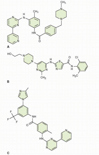
FIGURE 29-2 Structure of the small molecule tyrosine kinase inhibitors of BCR-ABL. A. imatinib; B. dasatinib; and C. nilotinib.


Clinical Pharmacology and Metabolism

Imatinib is orally bioavailable as is true of the other currently approved small molecular weight tyrosine kinase inhibitors (Table 29-3). It is primarily metabolized by the CYP3A4 member of the cytochrome P450 family of enzymes in the liver involved in metabolism of many drugs with minor metabolism by other P450 enzymes.53, 54, 55, 56, 57 The elimination half-life is approximately 20 hours for the parent compound and 40 hours for the major active metabolite, the N-demethylated piperazine derivative (N-desmethyl-imatinib), which has approximately the same potency as the parent compound. Elimination of metabolites is primarily (˜85% to 90%) in the feces with approximately 10% to 15% in the urine. The majority of a dose of the drug is excreted within 7 days, only 25% as the parent drug. Clearance rates can vary by up to 40% between patients and are decreased in patients with renal dysfunction lower CYP3A4 activity. Thus, it
is important to monitor for toxicity in individual patients and to consider the possibility of underdosing in nonresponsive patients (see below).


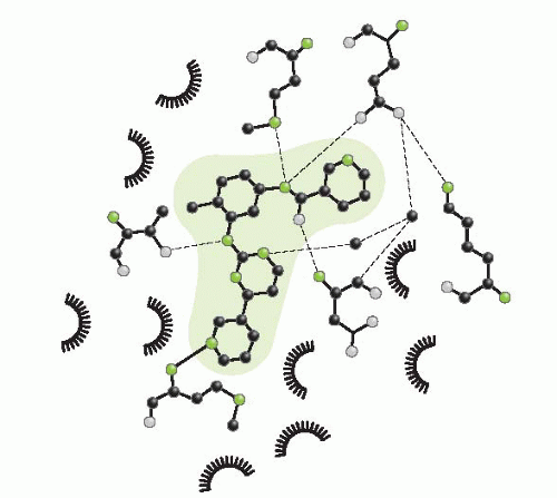
FIGURE 29-3 Detailed diagram of the intermolecular interactions between the drug (shaded in blue) and amino acids in BCR-ABL. Hydrogen bonds are indicated by dashed lines, while van der Waals interactions (indicated by halos around the amino acid name and its position in the protein sequence) are shown for nine amino acids with hydrophobic side chains.

Many other compounds are substrates for CYP3A4, and it is important to carefully evaluate these and minimize their use whenever possible when prescribing imatinib. Inducers of CYP3A4, such as phenytoin, significantly decrease imatinib exposure. There is a significant increase in imatinib exposure (with increases in both maximum concentration [Cmax] and area under the curve [AUC]) when imatinib is given with CYP3A4 inhibitors or competitive substrates such as ketoconazole or simvastatin.

After oral administration, the drug is relatively quickly absorbed (peak plasma concentrations are seen within 2 to 4 hours) and exhibits high bioavailability (98%). It is 95% bound to plasma proteins, primarily albumin and alpha-1 acid glycoprotein. The AUC increases proportionally with increasing dose. Because of its long t1/2, it slowly accumulates to a steady state when the dose is given daily. Its steady state trough concentration in plasma when 400 mg is given once daily averages about 1 mg/mL but with significant variability.55 This variability is important to keep in mind when evaluating toxicity and response. Imatinib poorly penetrates into the cerebrospinal fluid (CSF).


TABLE 29.3 Features shared by most small molecule tyrosine kinase inhibitors approved to date


Binding to the ATP site (active or inactive conformation or both)


Usually inhibit more than one kinase, although the degree of selectivity varies significantly


Oral therapy; half-lives that allow either daily dosing or twice daily dosing (depending on the specific agent)


Many are cytochrome P450 (most commonly CYP3A4) substrates, so it is important to determine if the patient is on inhibitors or inducers of these enzymes.


Acceptable toxicity at clinically effective doses


Significant component of side effects seen are due to predominant protein inhibited (e.g., skin toxicity with EGFR inhibitors, hypertension with VEGF or VEGFR inhibitors). However, other side effects vary somewhat even within inhibitors of same predominant protein.


Imatinib Toxicity

Major imatinib toxicities include


  • Neutropenia, thrombocytopenia, and anemia


  • Hepatotoxicity, with elevated liver enzymes. Toxicity can be decreased by holding or adjusting the dose, but occasionally it can be severe, especially if given with acetaminophen; thus, acetaminophen should be used with caution in patients taking imatinib.


  • Fluid retention or edema (often periorbital edema, which is usually manageable though occasionally serious, with a rare incidence of ascites, pleural effusions, brain edema, and congestive heart failure)


  • Musculoskeletal pains and cramps


  • Abdominal pain


  • Rash and other skin conditions, including blistering


  • Nausea and vomiting


  • GI irritation (imatinib should be taken with food and a large glass of water)


  • GI bleeding or intratumoral bleeding (especially in patients with GIST), which can be significant


  • GI perforation

Doses should be held in the face of evidence of hepatic dysfunction and should be modified for myelosuppression or when strong inhibitors or inducers of CYP3A4 are used concurrently. Overall, imatinib is well tolerated, as only 2% to 4% of patients discontinue the drug because of intolerance.

Imatinib may inhibit metabolism of simvastatin, cyclosporine, pimozide, certain HMG-CoA reductase inhibitors, and warfarin (Coumadin). Whenever possible, alternate drugs that do not interact with CYP3A4 should be utilized, such as low molecular weight heparin instead of warfarin. Grapefruit juice inhibits CYP3A4 and should be avoided. In addition, a number of compounds used as alternative or complementary therapies might influence CYP3A4 function and should be avoided.

Resistance to imatinib arises primarily by point mutations in the kinase domain of the target enzyme (Fig. 29-4). Most of these mutations remain sensitive to dasatinib and nilotinib, but not the enzyme
with T315 I substitution. Resistant forms of the enzyme have been isolated from patients prior to therapy, indicating that mutations arise spontaneously, but are selected by the presence of drug.133 The most common mutations conferring resistance occur at Glu 255 and Thr 315, both creating high-level resistance to imatinib and nilotinib. Dasatinib remains effective against Glu 255. Mutations at Met 351 and Glu 355 create low-level resistance to imatinib, but not to the other drugs. Monitoring of peripheral blood BCR-ABL transcripts by polymerase chain reaction (PCR) aids the assessment of levels of response, detects early evidence of the emergence of resistance, and aids in the selection of secondary therapies.134

Uncommonly, other mechanisms, besides mutations, may be associated with resistance to imatinib and include amplification of the BCR-ABL, especially in Ph-positive acute lymphoblastic leukemia, decreased uptake or increased efflux of imatinib from malignant cells, clonal evolution with development of new chromosomal abnormalities, and activation of other signaling pathways (e.g., overexpression of SRC family kinases).132 Again, it is important to identify those patients without initial response that may be due to altered pharmacokinetics with low serum drug levels that might benefit from a trial of dose escalation as opposed to those whose disease is resistant to one of the above mechanisms.


Clinical Effectiveness


Chronic Myelogenous Leukemia

Imatinib is approved for treatment of patients with CML.24 It produces hematologic responses in the vast majority (>95%) of CML patients in chronic phase, either previously untreated or refractory to interferon, at doses that have acceptable toxicity (400 mg/d with possible escalation to 600 or 800 mg/d depending on response and tolerance).24,55,57 About one quarter of patients who have incomplete cytogenetic responses to imatinib will show an improvement in response to the higher dose. Cytogenetic complete remissions are seen in the majority of patients. The 8-year survival is approximately 85%, and approximately 60% continue to take imatinib at standard doses for 6 years. Median time to cytogenetic response is rapid (˜1 month), but the quality of remissions improves over time. At higher dose (600 mg/d), it also has activity against CML in accelerated phase, with approximately 28% to 37% short-lived complete hematologic response and 20% major cytogenetic response. Although less active in patients with blast crisis when used at 600 mg/d, it still produces a small percentage of complete hematological responses (˜5%) and major cytogenetic responses (˜15%). It has less clinical activity in patients with acute lymphoblastic leukemia who have the 9:22 chromosomal translocation, possibly because of a greater role for SRC family kinases, although the actual mechanism of decreased responsiveness compared to CML remains to be fully elucidated.


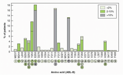
FIGURE 29-4 The amino acid substitutions, found in BCR-ABL kinase domain in 219 patients with CML and in 26 patients with Ph-positive acute lymphoblastic leukemia. Substitutions are shown below each amino acid position. Their color coded (as shown in the box) ties to their frequency, as depicted above each amino acid position. The T315I substitution creates resistance to all approved kinase inhibitors. (Figure from Hughes T, Deininger M, Hochhaus A, et al. Monitoring CML patients responding to treatment with tyrosine kinase inhibitors: review and recommendations for harmonizing current methodology for detecting BCR-ABL transcripts and kinase domain mutations and for expressing results. Blood 2006;108:28-37, with permission.)


Gastrointestinal Stromal Tumor

GISTs are the most common mesenchymal tumors arising from the GI tract although they are uncommon tumors overall.22,23 They originate from the interstitial cell of Cajal, an intestinal pacemaker cell. Although they can arise from anywhere in the GI tract or even the omentum, mesentery, or retroperitoneum, they most commonly occur in the stomach, followed by the intestine. Primary therapy for GIST is surgical resection. However, a fairly high rate of recurrence or metastatic disease is noted. These tumors
have been largely unresponsive to standard chemotherapeutic agents.

Imatinib has significant inhibitory effects on both c-KIT and the PDGFR tyrosine kinase activity.22,23 Immunohistochemical staining for c-KIT is usually positive in GIST tumors and the c-KIT receptor is mutated in a high proportion (˜85%) of patients with GIST. A variety of mutations in the c-KIT receptor in GIST tumors have been identified (Table 29-4). The clinical antitumor activity of imatinib against GIST correlates with specific mutations in c-KIT, as shown in Table 29-4. The most common mutation is in exon 11, occurring in approximately two thirds of patients. Highest response rates are seen in patients with exon 11 mutations. The second most common mutation is in exon 9, which occurs in approximately 17% of cases. Mutations also occur occasionally in exons 13 or 17. In those GIST tumors with wild-type C-KIT, the PDGFR-α is mutated in approximately 40% of such cases and these patients also respond to imatinib.22,23

The approved dose of imatinib for metastatic GIST is 400 to 600 mg/d. In a randomized trial, 400 mg/d gave equivalent results to 400 mg po BID.58 However, there was an approximately 33% response rate in patients initially treated with lower dose upon dose escalation, so it is reasonable to try this approach in appropriate patients before considering switching to alternative therapy. Imatinib is also approved at 400 mg/d for adjuvant therapy of patients with resected c-KIT positive tumors (>3 cm) who are at high risk of recurrence.56


Other Malignancies

Imatinib is also active against a number of other uncommon diseases, including myelodysplastic/myeloproliferative diseases with PDGFR-α gene rearrangements (e.g., CMML with EVT6-PDGFR translocation), unresectable DFPs (constitutive production of ligand), aggressive systemic mastocytosis (without the D816V c-KIT mutation), HES (FIP11-PDGFR), or chronic hypereosinophilic leukemia.


TABLE 29.4 c-KIT mutations and response to imatinib


Mutation


Response to imatinib therapy (PR) (%)


c-KIT exon 11


84


c-KIT exon 9


48


c-KIT exon 13


100a


c-KIT exon 17


50a


PDGFR-a sensitive


67a


PDGFR-a resistant


0a


No c-KIT or PDGFR-α mutations


0


a Based on very small numbers, with large margin for error. Adapted from Heinrich MC, Corless CL, Demetri GD, et al. Kinase mutations and imatinib response in patients with metastatic gastrointestinal stromal tumors. J Clin Oncol 2003;21:4342-4349; Table 1 with permission.


Dasatinib


Mechanism of Action

Dasatinib (Fig. 29-2B) is also a potent inhibitor of the BCR-ABL tyrosine kinase with in vitro activity approximately 100 to 300 times that of imatinib. Its IC50 varies depending on the exact mutation but is generally in the range of 0.5 to 5 nM.57,59, 60, 61 In contrast to imatinib or nilotinib, it binds to both the active and inactive conformations of the BCR-ABL kinase.7 It inhibits most, but not all, mutations that confer resistance to imatinib (e.g., it does not inhibit the T315I mutation).7,57,59, 60, 61 It inhibits a number of other tyrosine kinases including c-KIT, EPHA 2, PDGFR-β, and SRC family members.

May 27, 2016 | Posted by in ONCOLOGY | Comments Off on Molecular Targeted Drugs

Full access? Get Clinical Tree

Get Clinical Tree app for offline access