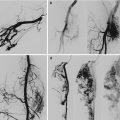
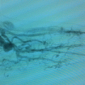
Fig. 45.1
Venous malformation of the thorax wall involving the pleura
Surgery is the elective choice in small and superficial VMs, located on skin and subcutaneous tissue [4]. The excision should be done by microinvasive techniques, using if possible a spiral purse-string suture. When the malformation is very large, a double-stage approach is preferable, performing the implant of a skin expander in the first step and the removal of the malformation followed by plastic reconstruction in the second step. The best positioning of skin expanders depends on the malformation site. In many cases, it is useful to implant the skin expander on the anterior abdominal or lumbar area, to perform a rotational flap after the exeresis of the thorax wall malformation.
Sclerotherapy is useful in very extensive VMs with muscle or bone involvement, when a total surgical removal is impossible or dangerous [5]. It is recommendable to perform sclerotherapy under radioscopic guidance because these malformations often show a direct drainage in the azygos, hemiazygos, brachiocephalic, or superior cava veins. A preoperative direct puncture phlebography allows to confirm the needle position within the lesion and to evaluate the outflow veins. At this time, the scleroembolization is carried out injecting the sclerosing agent mixed with a contrast medium to control its spread within the vessels (Fig. 45.2).
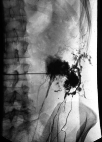

Fig. 45.2
Percutaneous sclerotherapy of a thoracic venous malformation
Various sclerosing agents are commonly used. The choice will depend on the morphologic patterns, anatomical site, and extent of the malformation. For small-caliber VMs, it is possible to use 2–3 % polydocanol or 0.2–3 % sodium-tetradecyl-sulfate solution. For large-caliber VMs, it is preferable to use 95 % ethanol because a more powerful sclerosant is necessary. The dosage of the sclerosing agent will be established in proportion to the size of the malformation. The volume of contrast material required to fill the space is then used to estimate the quantity of sclerosant needed. Anyway, ethanol injected must not exceed a maximum dose of 2 ml/kg body weight. It is preferable to inject the sclerosant dose by multiple direct punctures of the malformation in different sites on the chest wall (Fig. 45.3). A low molecular weight heparin and steroid treatment is recommendable after sclerotherapy of thorax VMs in order to prevent thrombophlebitis, skin necrosis, or nerve damage.
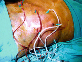

Fig. 45.3
Sclerotherapy of a chest wall venous malformation by multiple direct punctures
A combined treatment is the preferred option in the majority of cases. The combination of percutaneous and surgical treatment offers the best clinical, morphological, and functional results. In cases of extensive thorax VMs, it is necessary to perform multiple sequential surgical or endovascular procedures to obtain a complete regression.
Lymphatic Malformations
Lymphatic malformations (LMs) of the chest wall are usually macrocystic extratruncular forms (cystic hygromas) arising from the subclavian or axillary regions. These malformations induce severe clinical symptoms because they are often very large and tend to gradually grow over the years, especially in concomitance with hormonal, traumatic, or infective events. Early treatment is recommendable to avoid the risk of a pleuric or mediastinal involvement with compression of vital structures such as trachea or central veins.
Surgical removal is considered the best cure for thorax LMs [6]. When the cystic sac is very large and deep with infiltration of muscle and peripheral nerves, it is very difficult to achieve a complete surgical excision. In these cases, it is possible to perform a multistaged surgical removal, so reducing the risk of hemorrhage or nerve injury.
Percutaneous sclerotherapy is accepted as safe and satisfactory treatment for LMs of the thorax wall, particularly in pediatric patients [7]. Intralesional injection of sclerosants is a low-invasive procedure which gives good clinical results with marked or complete shrinkage of the cystic mass and minimum recurrence (Fig. 45.4).
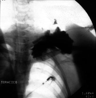

Fig. 45.4
Sclerotherapy of a macrocystic lymphatic malformation of the subclavian region
The size of the malformation dictates the choice of the sclerosing agent. Sodium-tetradecyl-sulfate solution is preferred for circumscribed forms. Ethanol is more effective in larger LMs of the thorax but induces a considerable risk of pleuric reactions or brachial plexus injuries. OK-432, bleomycin, doxycycline, and pingyangmycin are good alternative scleroagents in these malformations, because they allow to achieve the mass shrinkage with a moderate inflammatory reaction.
Stay updated, free articles. Join our Telegram channel

Full access? Get Clinical Tree


