The Intensive Care Unit, Part B: Antibiotic Resistance and Prevention of CVC-BSIs, Catheter-Associated Urinary Tract Infections, and C. difficile


The Intensive Care Unit, Part B: Antibiotic Resistance and Prevention of CVC-BSIs, Catheter-Associated Urinary Tract Infections, and C. difficile


Nasia Safdar

Dennis G. Maki


INTRODUCTION

Intensive care units (ICUs) have revolutionized the care of critically ill patients with trauma, shock states, and other life-threatening conditions, leading to greatly improved outcomes (1,2). However, healthcare-associated infection (HAI) (ICU-acquired) remains a major challenge in the ICU patient; the rates of infection in the ICU are three to five times higher than the rates in other hospital wards (3,4). Although patients in the ICU represent only 10% of all hospital admissions, they account for nearly 50% of all HAIs in U.S. hospitals. Major advances in our understanding of the epidemiology and pathogenesis of ICU-acquired infections have occurred over the past two decades, leading to the development of measures to greatly reduce or prevent these HAIs.


EPIDEMIOLOGY

Currently, HAIs affect >2 million patients in U.S. hospitals annually, and are associated with approximately 90,000 deaths each year (5).

Surveillance of HAIs, especially in high-risk hospital settings, such as the ICU, has become an integral feature of infection control and quality assurance in all U.S. hospitals. The Centers for Disease Control and Prevention (CDC) Study of the Efficacy of Nosocomial Infection Control (SENIC) Project showed that surveillance can help prevent HAIs (6).


TABLE 25.1 Rates of Device-Associated Infections per 1,000 Device-Days by Type of ICU in CDC NHSN Hospitals, January to December 2010


Type of ICU


Infection


Medical Rate, Mean (25%, 75%)


Medical-Surgical Rate, Mean (25%, 75%)


Surgical Rate, Mean (25%, 75%)


Coronary Rate, Mean (25%, 75%)


Catheter-associated urinary tract infection


2.4 (0.9, 3.7)


2.2 (0.6, 3.4)


3.0 (1.0, 4.4)


1.9 (0.3, 3.1)


Central line-associated bloodstream infection


1.8 (0.8, 2.3)


1.4 (0.0, 2.1)


1.4 (0.4, 1.9)


1.3 (0.0, 1.8)


Ventilator-associated pneumonia


1.4 (0.1, 2.2)


1.8 (0.0, 2.5)


3.5 (0.4, 4.8)


1.3 (0.0, 2.1)


Adapted from Dudeck MA, Horan TC, Peterson KD, et al. National Healthcare Safety Network (NHSN) Report, data summary for 2010, device-associated module. Am J Infect Control. 2011;39:798-816.


The National Nosocomial Infections Surveillance (NNIS) system was established in the early 1970s to measure the impact of HAIs, better understand their associated risk factors, and develop effective strategies for their control (7). NNIS is now called the National Healthcare Safety Network (NHSN) and includes approximately 5,000 hospitals. Surveillance of HAIs has been standardized by the NNIS/NHSN system by providing definitions, especially for device-associated infections (DAIs) (8). These definitions have recently been updated to increase specificity and accuracy of surveillance for DAIs, especially ventilator-associated pneumonia (VAP). Targeted surveillance and calculation of DAI rates per 1,000 device-days allows benchmarking with similar hospitals and detection of unique institutional problems that need redress and a mechanism for assessing institutional trends and even HAI outbreaks.

Since the length of stay heavily impacts the HAI risk, infection rates should be expressed per 1,000 patient-days. Device utilization affects DAI rates, and the CDC recommends surveillance of DAIs and calculation of rates of DAI per 1,000 device-days. The rates of HAI vary among the different types of ICUs, and are highest in neonatal, surgical, and burn units followed by medical ICUs. Patients in coronary care units have a very low risk of infection (Table 25.1) (9,10,11).


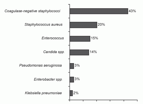
Figure 25.1. Microbiology of infections in the ICU. (Adapted from Richards M, Thursky K, Buising K. Epidemiology, prevalence, and sites of infections in intensive care units. Semin Respir Crit Care Med. 2003;24:3-22.)

The epidemiology of ICU-acquired infection in developing countries has recently been characterized by a new, large multinational surveillance system in developing countries in South and Central America, Asia, Africa, and the Middle East using NNIS/NHSN definitions of HAI. In a recent report, the overall rates of DAI in 55 ICUs of the consortium were 22.5 infections per 1,000 ICU-days; 41% of infections were VAP, followed by central line-associated bloodstream infection (CLA-BSI; 12.5 episodes per 1,000 catheter-days) and catheter-associated urinary tract infections (CA-UTIs; 8.9 episodes per 1,000 catheter-days) (12). These rates are 2- to 3-fold higher than reported in North American ICUs, and highlight the extraordinary vulnerability to HAIs in ICUs around the world.

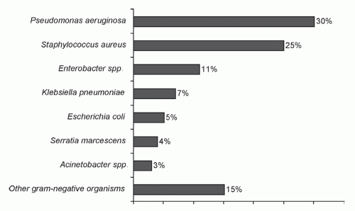
Aerobic gram-negative bacilli, especially Pseudomonas aeruginosa, account for 50% of ICU infections; gram-positive cocci (20%) and Candida spp. (10%) make up the remainder (3,4). Figures 25.1, 25.2, 25.3 and 25.4 show the microbiology of ICU infection overall and with VAP, CLA-BSIs, and CA-UTIs (4).


GENERAL ASPECTS OF INFECTION CONTROL

The U.S. Joint Commission (TJC), formerly the Joint Commission Accreditation of Healthcare Organizations (JCAHO), and similar regulatory agencies in many other countries mandate that every hospital have an active program for surveillance, prevention, and control of HAIs (13). Surveillance is the cornerstone of an effective control program. In most institutions, surveillance focuses on infections caused by antibiotic-resistant bacteria and infections that greatly increase morbidity and mortality (e.g., surgical site infections [SSIs], BSIs, and VAP).


Figure 25.2. Microbiology of bloodstream infections in the ICU. (Adapted from Richards M, Thursky K, Buising K. Epidemiology, prevalence, and sites of infections in intensive care units. Semin Respir Crit Care Med. 2003;24:3-22.)


Figure 25.3. Microbiology of healthcare-associated pneumonia in the ICU. (Adapted from Richards M, Thursky K, Buising K. Epidemiology, prevalence, and sites of infections in intensive care units. Semin Respir Crit Care Med. 2003;24:3-22.)

Although it is unclear whether environmental contamination with resistant bacteria translates into greater infections in patients, the inanimate environment is a reservoir of resistant HAI pathogens. Several studies have shown that methicillinresistant Staphylococcus aureus (MRSA), vancomycin-resistant enterococcus (VRE), Clostridium difficile, and gram-negative bacteria can be recovered from a variety of hospital surfaces. Although the ICU environment cannot be made microbe-free, certain architectural and environmental issues warrant attention. ICUs should be located in areas that limit traffic flow to essential ICU personnel. An adequate number of sinks and dispensers of waterless alcohol-based hand rub or antimicrobial soap must be available for all entering personnel who will have contact with the patient and the immediate environment. Separate areas and sinks should be used for cleaning, for storage, and for reprocessing contaminated equipment. All ICUs
should have airborne infection isolation rooms for patients with tuberculosis or other airborne infections. For ICUs involved in the care of bone marrow transplant patients or hematologic malignancy, positive pressure isolation rooms using high-efficiency particulate air (HEPA) filtration should be available. All surfaces contiguous to the ICU patient should be wiped down with the general hospital disinfectant at least daily, and urine-measuring devices, a frequent reservoir of gram-negative bacilli, should be rinsed with a disinfectant after each use. Each ICU patient should have a dedicated stethoscope and sphygmomanometer.


Figure 25.4. Microbiology of catheter-associated urinary tract infections in the ICU. (Adapted from Richards M, Thursky K, Buising K. Epidemiology, prevalence, and sites of infections in intensive care units. Semin Respir Crit Care Med. 2003;24:3-22.)


HAND HYGIENE

Infected or colonized patients represent the main reservoir of HAIs in the ICU, and the major mechanism of spread of HAI pathogens in the ICU is by carriage on the hands, apparel, or equipment of healthcare workers (HCWs). This has been most clearly shown in outbreak settings and for gram-positive pathogens in studies predating the advent of novel agents, such as waterless alcohol-based hand rubs, for hand hygiene; the role played by HCWs in horizontal transmission of gram-negative bacilli in the ICU in the presence of waterless alcohol-containing hand rubs remains to be elucidated. In a recent well-conducted cohort study, Waters et al. sought to determine whether or not hand carriage of gram-negative bacilli by neonatal nurses was associated with endemic HAIs caused by gram-negative bacilli in neonates cared for by those nurses (14). The investigators found that 192/2,935 neonates enrolled acquired an infection caused by gram-negative bacilli; 70% of the isolates were available for molecular typing, and 9% (11/119) of strains causing infection were recovered from the hands of neonatal ICU nurses. An additional 33% (39/119) of strains were shared among infants, providing indirect evidence of hand carriage by HCWs. In this study, sampling of nurses’ hands was performed quarterly immediately after hand hygiene using waterless alcohol-containing hand rubs, and because carriage is typically transient, it is possible that more frequent culturing would have yielded a larger number of shared strains. It is important to note that the role of the environment as a reservoir of HAI gram-negative pathogens was not assessed in this study.

Given the importance of hands as a major vehicle of horizontal transmission, hand hygiene remains the fundamental measure advocated to prevent HAIs (15,16,17,18,19,20). Despite the universal acknowledgment of handwashing/hygiene as a cornerstone of HAI control programs, compliance rates of >50% have been difficult to achieve, and handwashing/hygiene rates have ranged from 9% to 50% in HCW studies (21,22).

Recent investigations have strived to better understand the reasons for poor compliance in the face of the compelling evidence of the importance of handwashing/hygiene for HAI prevention (21), identifying cutaneous irritation, inconvenient sink location, time constraints, high workload, or understaffing. Of concern, risk factors for noncompliance with hand hygiene include being a physician (rather than a nurse), working in an ICU, and, paradoxically, engaging in patient-care activities with a high risk of cross-transmission (21). Interventions to redress these deficiencies have included targeted education; feedback; convenient location of sinks and hand hygiene agents; use of alternative, less irritating hand hygiene agents; and patient education (17). Table 25.2 summarizes the strategies to enhance compliance with hand hygiene (23).


TABLE 25.2 Strategies to Improve Hand Hygiene Compliance


  • Healthcare worker education


  • Routine observation and feedback


  • Engineering controls


  • Easy, convenient availability of alcohol-based hand rub


  • Patient education


  • Reminders in workplace


  • Administrative sanctions or rewards


  • Improved skin care for healthcare workers


  • Active participation at individual and institutional levels


Adapted from Pittet D. Improving adherence to hand hygiene practice: a multidisciplinary approach. Emerg Infect Dis. 2001;7:234-240.


Hygienic hand care with antiseptics is clearly more effective than conventional handwashing with soap and water; the advantage is most pronounced when contamination is heavy (24,25). Conventional handwashing with plain soap and water results in minimal reduction or even, paradoxically, an increase in bacterial counts compared to the baseline count before the handwashing (Figure 25.5) (18,26). The increase is probably caused by promotion of bacterial release and dispersal through shedding of colonized skin squames (27,28). In addition to superior antimicrobial activity, some antiseptics, such as chlorhexidine, bind to the stratum corneum, producing longterm anti-infective activity on the skin surface (29).

Antiseptics commercially available in the United States in a variety of formulations include chlorhexidine, iodophors, triclosan,
parachlorometaxylenol, and alcohol-based products (17). A number of before-after studies using time-series analysis and HAIs as the primary outcome in ICUs (15,16,30,31,32,33,34) have shown that alcohol-containing waterless hand rubs were associated with significant HAI reductions. Three large, well-conducted, randomized trials assessing the efficacy of chlorhexidine-containing hand hygiene products showed a 27% to 47% relative reduction in HAIs (30,33,34). The CDC recommendations for hand hygiene have been published (35), emphasizing hand antisepsis with an antiseptic-containing soap or detergent, or an alcohol-based hand rub: (a) before and after direct contact with patients or the environment and equipment in the immediate vicinity of the patient and (b) before performing invasive procedures, such as insertion of an intravascular device or urinary catheter. Alcohol-based waterless hand rubs are now widely used in hospitals for hand hygiene because of their convenience and broad-spectrum activity (17). However, all have limited efficacy with gross soilage so that visibly soiled hands should always be washed with antiseptic soap and water (36).


Figure 25.5. Immediate bacterial removal with 3 handwashing agents. Each agent was studied in 10 individuals with 1 week between the tests. Cultures were obtained immediately before (B) and after (A) handwashing with the agent. The bacterial count increased after hand washing with soap alone. (Adapted from Maki DG. The use of antiseptics for handwashing by medical personnel. J Chemother. 1989;1(suppl 1):3-11.)

The major factor limiting the acceptance of alcohol products for hand antisepsis in the past was desiccation and irritation of skin. This is now obviated by incorporating emollients into alcohol-based hand rubs, which has enhanced HCW acceptance, and may augment antibacterial activity by slowing the evaporation of alcohol (37). A recent randomized clinical trial in 50 ICU HCWs compared a conventional 2% chlorhexidine gluconate wash with water to a waterless alcohol-based hand rub (61% ethanol with emollients) and showed that the use of the waterless alcohol-based product produced significantly less skin scaling and irritation (38); unfortunately, degerming was not assessed.


TABLE 25.3 Handwashing and Hand Antisepsis Recommendations from the CDC/HICPAC Guideline on Hand Hygiene


Strength of Recommendationa


When hands are visibly dirty or contaminated with proteinaceous material or are visibly soiled with blood or other body fluids, wash hands with either a nonantimicrobial soap and water or an antimicrobial soap and water.


IA


If hands are not visibly soiled, use an alcohol-based hand rub or wash hands with an antimicrobial soap and water for the following situations:


IB


Before direct contact with patients


Before putting on sterile gloves when inserting a central vascular catheter


Before inserting a urinary catheter, peripheral vascular catheter, or other invasive procedure not requiring surgery


After contact with patient’s intact skin


After contact with body fluids, mucous membranes, and wound dressings if hands are not visibly soiled


Moving from a contaminated body site to a clean body site during patient care


After contact with inanimate objects in the immediate vicinity of the patient


After removing gloves


Before eating and after using a restroom, wash hands with a nonantimicrobial soap and water or with an antimicrobial soap and water.


IB


Antimicrobial-impregnated wipes are not a substitute for using an alcohol-based hand rub or antimicrobial soap.


IB


If exposure to Bacillus anthracis, wash hands with nonantimicrobial soap and water or antimicrobial soap and water.


II


IA: strongly supported for implementation and strongly supported by well-designed experimental, clinical or, epidemiologic studies.


IB: strongly recommended for implementation and supported by certain clinical or epidemiologic studies and by strong theoretical rationale.


II: suggested for implementation and supported by suggestive clinical or epidemiologic studies or by strong theoretical rationale.


a Categorization of recommendations.


Adapted from Boyce JM, Pittet D. Guideline for hand hygiene in health-care settings. Recommendations of the Healthcare Infection Control Practices Advisory Committee and the HICPAC/SHEA/APIC/IDSA Hand Hygiene Task Force. MMWR Recomm Rep. 2002;51:1-45.


The recent CDC guidelines have been endorsed by the American Medical Association (39) and the American Society for Microbiology (40), both of which have played an active role in emphasizing hand hygiene in all areas of healthcare. Institutional commitment is essential to improve compliance with recommended hand hygiene practices. The CDC guideline recommends that institutions (a) monitor and record adherence to hand hygiene by ward or service, (b) provide feedback to HCWs about their performance, and (c) monitor the volume of alcohol hand rub used per 1,000 patient-days.

Monitoring compliance with hand hygiene is most often undertaken using direct observations by trained observers. Although this remains the gold standard, there are limitations to this method including interobserver variability as well as being time- and labor-intensive. More recently, a number of electronic monitoring systems for capturing hand hygiene compliance have become available; however, these systems need to be validated and tested for efficacy before coming into widespread use. Table 25.3 summarizes the recommendations for hand hygiene in the 2002 CDC Guideline (35).


ANTIMICROBIAL RESISTANCE IN THE ICU

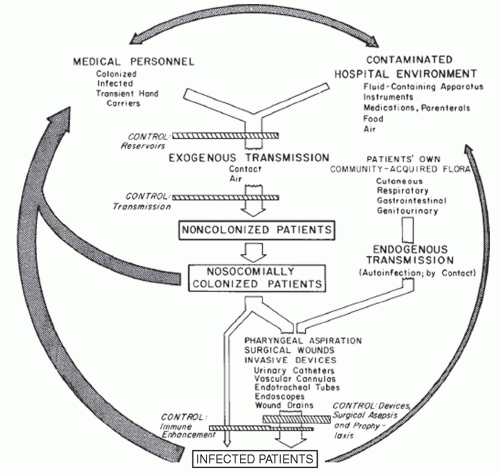
The global crisis in antimicrobial resistance has had a huge impact in the ICU where antibiotic pressure, critically ill patients, invasive devices, and procedures all contribute to increased spread of multidrug-resistant pathogens (Figures 25.6 and 25.7) (41,42,43). Stemming the tide of antimicrobial resistance
mandates a multifaceted approach, encompassing antimicrobial stewardship, hand hygiene, and barrier precautions for HCWs in contact with high-risk patients. The CDC’s Campaign to Prevent Antimicrobial Resistance aims to prevent antimicrobial resistance in healthcare settings (44). The campaign centers on four main strategies: prevent infection, diagnose and treat infection, use antimicrobials wisely, and prevent transmission.


Figure 25.6. The epidemiology of nosocomial infection. Transmission occurs mainly by contact spread to a much lesser extent by the airborne route. Aspiration, surgical wounds, exposure to invasive devices, and antimicrobial use amplify transmission, colonization, and susceptibility to infection. (Adapted from Maki DG. Control of colonization and transmission of pathogenic bacteria in the hospital. Ann Intern Med. 1978;89:777-780, with permission.)


CONTROL OF ANTIMICROBIAL RESISTANCE: OPTIMIZING ANTIMICROBIAL USAGE

Antimicrobial use drives antimicrobial resistance (45,46). Studies have shown that inappropriate antimicrobial use is common in healthcare institutions (47,48). Antimicrobial stewardship is essential to limit unnecessary antimicrobial use, optimize patient outcomes, and reduce the problem of antimicrobial resistance (49). Various strategies have been proposed to improve antimicrobial use and limit the emergence of resistance (42). These include the use of protocols or guidelines, formulary restriction of key drugs, infectious disease consultation, computerized physician order entry, and increased use of diagnostics to confirm the presence of an infection (Table 25.4).


PREVENTION OF HEALTHCARE-ASSOCIATED SPREAD OF RESISTANT ORGANISMS

Isolation of infected or colonized patients is widely regarded as the most important measure to prevent the spread of resistant pathogens through the healthcare institution (50). The most recent CDC guideline categorizes isolation precautions into (a) standard precautions and (b) transmission-based precautions (51). Standard precautions specify the use of gloves for any anticipated contact with blood, any body fluid, secretions or excretions (except sweat), nonintact skin, and mucous membranes. Gowns are recommended if patient-care activities are likely to generate splashes of blood, body fluids, and secretions. Hand hygiene is expected after removing gloves and between patients. Standard precautions apply to all patients without regard to clinical diagnosis.

Transmission-based precautions include contact, droplet, and airborne precautions, each based on the mode of transmission of the infectious agent within the healthcare setting. Acknowledging that multidrug-resistant HAI pathogens, particularly MRSA or VRE, are spread primarily by direct (and indirect) contact with HCWs, the guideline specifies that patients known to be colonized or infected by resistant bacteria are to
be placed in contact isolation, which requires a private room for the patient (or cohorting the patient in a semiprivate room with another patient who is also colonized or infected by the same organism). HCWs are expected to wear gloves on entry to the room and gowns if substantial contact with the patient or the environment is anticipated. Gloves and gowns should be removed and hands treated with a medicated hand hygiene product while still in the isolation room. Noncritical patient-care items should be dedicated; if reused, they must be disinfected between patients.


Figure 25.7. Major antimicrobial-resistant pathogens associated with nosocomial infections in ICUs in 1989, 1993, 1997, 2002 and 2004. (Adapted from National Nosocomial Infections Surveillance. (NNIS) System Report, data summary from January 1992 through June. 2004, issued October 2004 and adapted from Richards M, Thursky K, Buising K. Epidemiology, prevalence, and sites of infections in intensive care units. Semin Respir Crit Care Med. 2003;24:3-22.)


TABLE 25.4 Antimicrobial Usage Strategies for Reducing the Emergence of Antimicrobial Resistance in the Intensive Care Unit


Recommendation


Strength of Recommendationa


Limit unnecessary antibiotic administration


Develop hospital-based guidelines for antibiotic use


II


Create an antibiotic use-quality-improvement team


II


Provide professional education and detailing on antibiotic use for physicians


II


Restrict hospital formulary


I


Use quantitative cultures for nosocomial pneumonia


Optimize antimicrobial effectiveness


Avoid inadequate treatment by using automated guidelines


II


Use combination antimicrobial treatment


II


Consult with infectious diseases staff


II


Cycling antibiotics


II


Automatic stop orders for surgical prophylaxis


I


Avoid routine selective digestive decontamination


I


Computer-assisted provider order entry


a Level I, supported by randomized controlled trials; Level II, supported by nonrandomized trials and observational studies. Adapted from Kollef MH, Fraser VJ. Antibiotic resistance in the intensive care unit. Ann Intern Med. 2001;134:298-314.


Unfortunately, the existent paradigm for preventing the spread of resistant organisms in the hospital—waiting until colonization or infection by MRSA, VRE, or some other resistant organism is serendipitously identified by the clinical laboratory, following which the patient is placed in isolation, usually in a single room, requiring the use of gloves, with or without a gown, for all contacts with the patient—is failing dismally, viewing the inexorable growth in antimicrobial resistance (52).

A recent guideline from the Society for Healthcare Epidemiology of America (53) recommends that surveillance cultures/testing to detect silent VRE or MRSA carriage be performed in roommates of VRE- or MRSA-colonized or infected patients and other high-risk patients at the discretion of infection control staff; patients found to be colonized must also be placed in contact isolation (53). If these measures fail to contain the spread, efforts should be intensified in the highest risk areas, such as the ICU. Cohorting of staff and screening of staff for carriage, if epidemiologic data point to a link, is recommended. Verification that environmental cleaning/disinfection procedures are effective by environmental surveillance cultures before and after cleaning areas containing VRE- or MRSA-colonized or infected patients also is recommended.

We believe that a simpler strategy for preventing the spread of all types of multidrug-resistant bacteria is the preemptive use of barrier isolation precautions (gowns and gloves) and dedicated patient-care items (e.g., stethoscopes and sphygmomanometers) for all high-risk patients from the time of admission to prevent HCWs from acquiring hand contamination by multidrug-resistant organisms (MDROs) when they have contact with patients with unrecognized colonization or infection and block transmission to other as yet uncolonized patients. Numerous studies have shown that the preemptive use of barrier precautions, can effectively prevent the spread of MDROs, such as MRSA or VRE, in an epidemic setting (54), and other studies have shown the effectiveness of preemptive isolation in high-risk populations, such as patients in an ICU, for prevention of endemic HAIs, including by MDROs (55,56,57,58). Three prospective randomized trials have been conducted to assess the efficacy of preemptive barrier precautions
(55,56,59); two showed benefit with a reduction in all HAIs in ICU patients (relative risk reduction, 52% to 81%) (55,56).


SPECIFIC INFECTIONS


Intravascular Device-Related Bloodstream Infections

The use of intravascular devices (IVDs) has become an essential component of delivering care to many patients, particularly those with cancer. Unfortunately, vascular access is associated with substantial and generally underappreciated potential for producing iatrogenic disease, particularly BSIs originating from infection of the percutaneous device used for vascular access. Nearly 40% of all healthcare-associated BSIs derive from vascular access in some form (60), and can be associated with excess mortality approaching 35% (61), increased length of hospitalization, and excess healthcare costs (62,63).

Individual types of IVDs pose different risks of infection. In a recent systematic review of 200 prospective studies, we showed that point incidence rates of IVD-related BSI (IVDR-BSI) were lowest with peripheral intravenous (0.1%, 0.5 per 1,000 IVD-days) or midline catheters (0.4%, 0.2 per 1,000 catheter-days). Far higher rates were seen with short-term noncuffed and nonmedicated central venous catheters (CVCs) (4.4%, 2.7 per 1,000 catheter-days). Arterial catheters used for hemodynamic monitoring (0.8%, 1.7 per 1,000 catheter-days) and peripherally inserted central catheters (PICCs) used in hospitalized patients (2.4%, 2.1 per 1,000 catheter-days) posed risks approaching those seen with short-term conventional CVCs used in the ICU. Surgically implanted long-term CVCs—cuffed and tunneled catheters (22.5%, 1.6 per 1,000 IVD-days) and central venous ports (3.6%, 0.1 per 1,000 IVD-days)—appear to have high rates of infection when risk is expressed as BSIs per 100 IVDs, but actually pose much lower risk when rates are expressed per 1,000 IVD-days (64).

Figure 25.3 summarizes the microbial profile of IVD-related BSIs (IVDR-BSIs) (4). As might be expected from knowledge of the pathogenesis of these infections, skin microorganisms account for the largest proportion of IVDR-BSIs.

Recent evidence-based guidelines provide the best current information on the evaluation of the ICU patient with fever or other signs of sepsis (65). Before any decision regarding initiation of antimicrobial therapy or removal of an IVD is taken, the patient must be thoroughly examined to identify all plausible sites of infection, including VAP, CA-UTI, SSI, antibiotic-associated colitis, or line sepsis.

Despite the challenge of identifying the source of a patient’s signs of sepsis (65), several clinical, epidemiologic, and micro-biologic findings point strongly toward an IVD as the source of a septic episode. Patients with abrupt onset of signs and symptoms of sepsis without any other identifiable source should prompt suspicion of infection of an IVD. The presence of inflammation or purulence at the catheter insertion site is now uncommon in patients with IVDR-BSI (66). However, if purulence is seen in combination with signs and symptoms of sepsis, it is highly likely that the patient has IVDR-BSI and should prompt removal of the IVD. Finally, the recovery of certain microorganisms in multiple blood cultures (e.g., staphylococci, Corynebacterium or Bacillus spp., or Candida or Malassezia spp.) strongly suggests infection of the IVD.

It is indefensible to start anti-infective drugs for suspected or presumed infection in the critically ill patient without first obtaining blood cultures from two separate sites, at least one of which is drawn from a peripheral vein by percutaneous venipuncture. In adults, if at least 30 mL of blood is cultured, 99% of detectable bacteremias should be identified (67,68,69). Similar operating characteristics are achieved in the pediatric population using a weight-based graduated volume approach to blood cultures (70). Standard blood cultures drawn through CVCs provide excellent sensitivity for diagnosis of BSI, but are less specific than cultures obtained from a peripheral vein (71,72). If the patient has a long-term multilumen catheter, a specimen should be obtained from each lumen of the catheter because studies have found a high rate of discordance (approximately 30%) between cultures obtained from different lumens of the same catheter (73).

Short-term IVDs should be removed from the outset in unstable patients with suspected IVDR-BSI (as follows); however, it often is undesirable or difficult to do this in patients with surgically implanted IVDs, such as Hickman and Broviac catheters. Only 15% to 45% of long-term IVDs that are removed for suspected infection are truly colonized or infected at the time of removal (74,75,76). To avoid unnecessary removal of IVDs, methods have been developed to identify infection while allowing the device to stay in place: (a) paired quantitative blood cultures drawn from the IVD and percutaneously from a peripheral vein (77), (b) differential time to positivity (DTP) of paired standard blood cultures, one drawn from the IVD and the second from a peripheral vein (78), and (c) Gram stain (79) or acridine orange staining of blood samples drawn through the IVD (80,81).

Quantitative blood cultures are labor-intensive and cost almost twice as much as standard blood cultures. The DTP of paired blood cultures, one drawn through the IVD and the second concomitantly from a peripheral vein, has been shown to reliably identify IVDR-BSI of both short-term and long-term IVDs if the blood culture drawn from the IVD turns positive >2 hours before the culture drawn peripherally (78).

If a short-term vascular catheter is suspected of being infected because the patient has no obvious other source of infection to explain the fever, there is inflammation at the insertion site, or cryptogenic staphylococcal BSI or candidemia has been documented, blood cultures should be obtained and the catheter should be removed and cultured. Failure to remove an infected catheter puts the patient at risk of developing septic thrombophlebitis with peripheral IV catheters, septic thrombosis of a great central vein with CVCs (82), or even endocarditis. Continued access, if necessary, can be established with a new catheter inserted in a new site. Although small studies have found some utility of guidewire exchange in the management of CVCs suspected of being infected (83,84,85,86), we believe that, in the absence of randomized studies demonstrating its safety, guidewire exchange generally should not be performed if there is suspicion of IVDR-BSI, especially if there are signs of local infection such as purulence or erythema at the insertion site or signs of systemic sepsis without a source. In these instances, the old catheter should be removed and cultured and a new catheter should be inserted in a new site.

Jun 16, 2016 | Posted by in INFECTIOUS DISEASE | Comments Off on The Intensive Care Unit, Part B: Antibiotic Resistance and Prevention of CVC-BSIs, Catheter-Associated Urinary Tract Infections, and C. difficile

Full access? Get Clinical Tree

Get Clinical Tree app for offline access