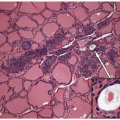
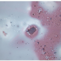
The classification of thyroid pathology includes congenital, inflammatory, and neoplastic disorders. These are listed in Table 3.1. Many of these are diagnosed based on clinical and biochemical features and do not require biopsy. In general, biopsy plays a key role in the diagnosis of nodular disease. However, biopsies of nodules may contain nonnodular thyroid tissue with variable pathologies.
Congenital aplasia and hypoplasia and enzymatic disorders give rise to hypothyroidism that is usually detected by screening early in life. When enzymatic deficiency results in subclinical disease and goiter ensues, the patient develops diffuse hyperplasia, known as dyshormonogenetic goiter; nodules may arise in this setting [1]. The commonest congenital lesion is the thyroglossal duct cyst that presents as a mass; this entity is discussed in Chapter 7.
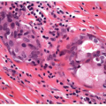
Inflammatory lesions of the thyroid include infectious and autoimmune disorders. Infections give rise to acute and subacute or granulomatous inflammation. Autoimmune disease is usually characterized by a chronic lymphocytic process, but the granulomatous inflammatory process known as de Quervain’s thyroiditis is thought to be of autoimmune etiology. These lesions are reviewed in Chapter 8.
Hyperplasia can be diffuse, as in compensatory goiters of patients with thyroid hormone deficiency due to any cause (congenital, endemic, or chemically induced). In areas of iodine deficiency, endemic goiter is common, but in many parts of the world, this entity is disappearing due to the use of iodized salt. In places where dietary iodine is sufficient, the most common form of diffuse hyperplasia is Graves’ disease, an autoimmune disorder characterized by stimulating antibodies directed at the TSH receptor. Unlike other autoimmune lesions, inflammation is scant, and the disorder is mainly characterized by papillary hyperplasia; this is discussed in Chapter 9.
Sporadic nodular goiter is a common nodular proliferation of thyroid follicular epithelium. The follicular proliferations have been considered to be hyperplastic based on morphologic features, but molecular studies have suggested that there may be a neoplastic component. The distinction between hyperplasia and neoplasia is not clear, and it may be that there is progression from hyperplasia to clonal adenomas. This interesting, common, and diagnostically challenging disorder is discussed in Chapter 10.
TABLE 3.1 Classification of Thyroid Pathology
Congenital Lesions
Aplasia
Hypoplasia
Enzymatic defects resulting in goiter
Thyroglossal duct cyst and other aberrant thyroid nodules
There is only one benign thyroid neoplasm recognized: the follicular adenoma. This lesion has several morphological variants, the majority with follicular patterns that are discussed in Chapter 10. One variant has papillary architecture and is reviewed in Chapter 9.
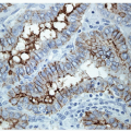
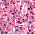
The critical objective of thyroid biopsy is the diagnosis, classification, and management of malignancy. The vast majority of thyroid malignancies derive from thyroid follicular epithelium. These are the most common malignancy of endocrine organs [2, 3, 4]. Age-adjusted global incidence rates vary from 0.5 to 10 cases per 100,000 population [5]. Thyroid cancer incidence rates have steadily increased over the last decades [6, 7, 8, 9] and continue to rise [10]. This represents one of the most challenging biopsy diagnoses [11], and the controversies in this field are clearly important, given the fact that these cancers frequently affect patients 20 to 50 years of age, and the disease is two to four times more frequent in females than in males, giving them high impact to health care programs and society in general as well as the affected individuals. Radiation exposure, iodide intake, lymphocytic thyroiditis [12], hormonal factors, and familial history are putative causative factors for thyroid carcinoma [13], and the recognition of specific mutations implicated in the various subtypes is providing novel diagnostic markers as well as targets for individualized therapies [14].
Only gold members can continue reading. Log In or Register to continue