Importance
Anyone can have a stroke, including babies and children, but the vast majority (90%) of strokes affect people aged over 55. Each year over 150,000 people in the UK have a stroke. Stroke is the third most common cause of death, after heart disease and cancer (11% of all deaths). Stroke is the largest single cause of severe adult disability in developed countries and is very expensive as patients need prolonged rehabilitation and, sometimes, care for life. Half a million people in the UK are living with long-term disability as a result of stroke. Managing stroke in a stroke unit (multidisciplinary, coordinated care) produces marked benefit.
Definitions
A stroke is defined as ‘rapidly developing clinical signs of focal disturbance of cerebral function, with symptoms lasting 24 h or longer or leading to death, with no apparent cause other than of vascular origin’. This includes subarachnoid haemorrhage (SAH) but excludes subdural haematoma and haemorrhage into a tumour. By definition, a transient ischaemic attack (TIA) lasts less than 24 h, so TIAs are classified separately, but the causes of TIA and stroke are very similar. TIAs are a risk factor for stroke. The term ‘stroke’ is now used in preference to cerebrovascular accident (CVA).
Outcome of Stroke
The outcome of a stroke (Table 7.1) depends on the aetiology, volume and part of the brain that is affected, the age and fitness of the patient and co-morbidities. You will see a range of figures for outcome, depending on the population in the study. The case fatality rate has fallen steadily since the 1980s but it remains higher in women and for haemorrhagic stroke.
Table 7.1 Outcome of stroke
| Outcome | Percentage |
| 1-month mortality (UK) | 15–20 |
| • ischaemic | 10 |
| • haemorrhagic | 50 |
| 1-year mortality of survivors | 25 |
| Around 3 months: | |
| Dead | 15 |
| Full or almost full recovery | 10 |
| Minor impairment | 25 |
| Moderate to severe impairment (need significant help) | 40 |
| Long-term high-dependency care | 10 |
| Further stroke in the first year | 15 |
| Return to work if previously working | 35 |
| Unable to walk unaided | 20 |
| Unable to walk outdoors | 40 |
Aetiology and Pathology
A stroke results from interruption to the brain’s blood supply due to an infarct or a haemorrhage. An infarct is an area of ischaemia, usually due to thrombosis in situ or an embolus from the carotids or heart but occasionally due to low BP from any cause or damage to the blood vessel wall with dissection. A primary haemorrhage may be due to an arterial abnormality, such as an aneurysm, but most infarcts and bleeds occur in vessels damaged by hypertension and atheroma. Cerebral amyloid angiopathy (see Chapter 4) is increasingly recognized as a cause of lobar intracerebral haemorrhage (ICH) in elderly patients. T2*-weighted MRI demonstrating chronic cerebral microbleeding at the white/grey matter junction supports this diagnosis. Secondary bleeding may occur in an area of brain damaged by an infarct. The proportions of the types of stroke vary in different countries: in the UK atherothrombotic strokes comprise 85% of cases whereas in Japan haemorrhagic strokes are much more common. The biggest risk factor for stroke is increasing age. AfroCaribbean and SE Asian ethnicity and apolipoprotein E ε2 or ε4 carriage (for ICH) also increase risk, but it is more useful to consider risk factors that can be modified.
Risk factors for stroke are similar to those for other vascular diseases such as coronary artery disease (CAD) and peripheral vascular disease (Table 7.2). However, there are differences in relative risk that are not understood, e.g. smoking is a bigger risk for CAD and hypertension is a bigger risk for stroke. In future, there may be increased understanding of the role of inflammation: a high CRP on a sensitive assay is emerging as a risk factor for vascular disease. Primary and secondary risk factors are similar. Risk factors tend to be multiplicative. As there is more chance of another vascular event once one has occurred, the risk–benefit ratio for treatments changes. (This is why healthy 40 year olds are advised not to take aspirin as their chance of a bleed outweighs the likely benefits.)
Table 7.2 Management and goals for risk factors for stroke and primary and secondary prevention
| Risk factor | Management | Goal |
| BP high | Promote healthy lifestyle; diet (see below), reduce alcohol intake and increase exercise. Treat BP if still high with drugs individualized to patient (but ACE inhibitor/diuretic combinations may have a particular role) | < 130/90 mm/Hg; < 130/85 mm/Hg if renal insufficiency or heart failure; < 130/80 mm/Hg if diabetic |
| Heart disease | As appropriate for the condition, also aiming to reduce platelet stickiness, minimize LVH and maintain sinus rhythm | Reduce chances of embolization |
| Atrial fibrillation | Verify AF on ECG or paroxysmal AF (PAF) on 24-h tape. For patients in chronic or intermittent AF, use warfarin aiming for INR 2.0–3.0. Aspirin is used if there are contraindications to oral anticoagulation or the patient is low-risk (CHADS2 score) | Antiplatelet drugs if ‘low risk’ but anticoagulation for AF or PAF |
| Sticky platelets | Secondary prevention. Aspirin (75–300 mg) except in intolerance and brain haemorrhage, caution with asthma and history of GI haemorrhage, with modified release dipyridamole for 2 years after vascular event. Clopidogrel if aspirin intolerant. However, a recent review suggests clopidogrel should be used first-line in stroke or multivascular disease (NHS HTA 2011) | Concordance with long-term antiplatelet therapy |
| Carotid stenosis | Antiplatelet therapy, document degree of stenosis and offer endarterectomy if CT confirms stroke in ipsilateral hemisphere, function worth preserving and stenosis > 70% or > 50% in men with cortical symptoms | Ensure eligible patients with anterior circulation strokes are screened: the fit elderly have most to gain |
| Smoking | Advise quitting and refer for support, e.g. clinic/pharmacological help (nicotine or buproprion, varenicline) | Quit and avoid passive smoking |
| Unhealthy diet | Advocate low-fat, low-salt, high fruit and vegetable diet with weight loss if needed. Discourage excess consumption of any food, however ‘healthy’, to avoid health scares, e.g. heavy metals in oily fish | Varied healthy diet, evidence for ‘Mediterranean diet’ |
| Obesity | Calorie restriction and increased caloric expenditure | Achieve and maintain desirable weight (BMI 20–25 kg/m2). Higher BMI is less of a risk if central obesity is not present |
| Excess alcohol | Low/moderate alcohol intake protects from atherothrombotic stroke (amount probably depends on individual as increase in BP and obesity may be adverse) but high or binge intake is a risk for haemorrhage | Avoid binge or excessive drinking |
| Adverse lipid profile | Advice about diet, weight and exercise. For 2° prevention start simvastatin 40 mg regardless of cholesterol. Refer to specialist if triglycerides/total cholesterol remain high | It is likely that the lower the total cholesterol, the better |
| Diabetes and impaired glucose tolerance | After diet and exercise, use oral hypoglycaemic drugs, particularly metformin, unless contraindicated; insulin if adequate control not achieved | Normal fasting plasma glucose (< 7 mmol/L) and near normal HbA1c (< 7%) |
| Lack of exercise | Medical check before initiating vigorous exercise programme: start slowly if older or unfit. Moderate-intensity activities (40–60% of maximum capacity) are equivalent to a brisk walk. Additional benefit from vigorous (> 60% of maximum capacity) exercise for 20–40 min on 3–5 days/week | At least 30 min of moderate-intensity physical activity on most days of the week |
| Previous TIA | Treat all risk factors |
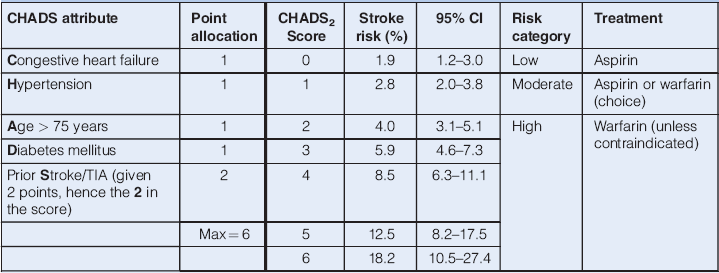
The CHADS2 score (see Table 7.3) is a clinical prediction rule for estimating the risk of stroke in patients with non-rheumatic atrial fibrillation, based on age and major clinical risk factors.
Table 7.3 CHADS2 score for predicting risk of stroke in 1 year in non-valvular atrial fibrillation

The inclusion of additional ‘stroke risk modifiers’ has been proposed, giving the following CHA2DS2-Vasc score:
- Age: 65–74 scores 1, 75+ scores 2
- Sex: female gender scores 1
- Vascular disease present scores 1
A total score of 0/9 is so low risk that even aspirin is not indicated, a score of 1 indicates aspirin would be beneficial, and a score of 2 and above indicates oral anticoagulation should be used.
Older people should be considered for warfarin, though contraindications include recurrent falls and dementia. In the BAFTA (Birmingham Atrial Fibrillation Treatment of the Aged) study (mean age 81.5 years) warfarin was much more effective than aspirin and actually had a trend towards causing fewer haemorrhages. A risk score for bleeding had been devised (HAS-BLED). See atrial fibrillation (AF) in Chapter 9.
Because of the complexity of dosing with warfarin (vitamin K antagonist) and the need for regular blood tests (INRs) huge effort has been put into developing easier to use anticoagulants. Dabigatran [a direct thrombin (IIa) inhibitor] is the newly licensed front runner and factor Xa inhibitors (e.g. rivaroxaban and apixaban) are near to market and may offer more consistent anticoagulation and decreased risk of ICH, albeit at a high cost.
Presentation
Typically, the onset is abrupt. Occasionally a hemiparesis develops over 12 or more hours. If symptoms progress over days/weeks, suspect an alternative diagnosis such as tumour or subdural haematoma. Other common stroke mimics include hypoglycaemia, partial seizures, Todd’s paresis after a partial seizure, hemiplegic migraine and metabolic disturbances.
Reduced conscious level at presentation usually indicates non-stroke pathology but a stroke may present with coma, in which case neurological examination requiring cooperation is impossible. Stroke presents with coma in three situations:
In a brain stem stroke, signs are often bilateral and the pupils may be small. In a major cortical stroke, the cheek on the paralysed side may flap in and out with respiration and the limbs on that side are likely to have completely lost all tone. Reflexes may be unhelpful at this stage. An almost pathognomonic sign is conjugate deviation of gaze towards the side of the lesion, due to the unopposed effect of the contralateral frontal eye field (remember, the patient ‘looks away’ from a tumour but ‘towards’ a stroke). Loss of consciousness points towards a severe stroke, but there are no reliable clinical predictors to distinguish haemorrhage from infarct.
The conscious or slightly drowsy patient usually presents little diagnostic difficulty. The peak time of onset for stroke is in the early hours of the morning and the patient will describe waking up and trying to get out of bed, only to find themselves unable to walk. An eyewitness may relate that the patient dropped their cup and developed facial asymmetry and difficulty with speech and then became unable to stand or perhaps even sit properly. Examination will then usually reveal characteristic deficits: in the early stages, the paralysed limbs are more often flaccid than spastic and in some cases they stay that way. Establish the exact time of onset (unfortunately impossible if the patient woke from sleep, around 25% cases). If it is less than 4.5 h, act fast as thrombolysis may be possible (see subsequent text).
Educational programmes are encouraging the public to treat stroke with as much urgency as a heart attack and the ambulance service uses a validated triage such as FAST (the Face Arm Speech Time score) to identify stroke patients for urgent transfer to a specialist stroke service with accurate documentation of the time of onset. Any one of facial asymmetry, arm or leg weakness or speech disturbance suggests a stroke with a sensitivity of 82% and specificity for stroke diagnosis of 37%.
Transient Ischaemic Attacks
These are isolated or recurrent focal neurological symptoms (usually negative) which resolve within 24 h. They are usually due to platelet emboli from an atheromatous plaque or ulcer in the aorta, the common carotid artery or, most often, the carotid bifurcation or red cell emboli from the heart. Occasionally the pathology is thrombosis or low flow. Monocular loss of vision (amaurosis fugax) hemi- or monoparesis, dysphasia and unilateral sensory disturbance are examples within the internal carotid territory. Most TIAs last less than a couple of hours, usually just a few minutes. The distinction between a TIA and a very small infarct is an anachronism and many older people with no clinical history will have a stroke on a scan.
TIAs also occur within the territory of the vertebrobasilar circulation, although true vertebrobasilar ischaemia or insufficiency is something of a diagnostic dustbin and is very rare. The main features are true vertigo (a sensation of rotary movement of either patient or surroundings), true drop attacks (sudden falls due to total loss of tone without disturbance of consciousness and with rapid and complete recovery) and diplopia, although cortical blindness, tetraparesis, ataxia and dysphagia may also occur. Vertebrobasilar TIAs carry a similar risk of stroke to anterior circulation TIAs and should be treated just as aggressively. TIAs are not a cause of loss of consciousness.
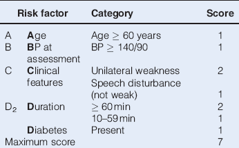
The risk of stroke after a hemispheric TIA is up to 15% in the first month and highest in the first 72 h, so after a probable TIA give aspirin 300 mg (unless on warfarin or contraindicated) and refer to a rapid access TIA clinic according to the degree of risk as assessed by the ABCD2 score (Table 7.4). If confident about the diagnosis some start a statin and antihypertensives at once.
Table 7.4 ABCD2 score

Patients with a high risk of subsequent stroke:
- ABCD2 score of 4 or above,
- two or more TIAs in a week regardless of score (crescendo TIA),
- any patient with neurological symptoms on warfarin,
should have specialist assessment and investigation within 24 h of onset of symptoms.
Patients with a lower risk of subsequent stroke:
- ABCD2 score of 3 or below,
- who present after a week,
should have specialist assessment within 1 week.
The role of a TIA clinic is to:
- Confirm clinical diagnosis.
- Arrange investigations including imaging of the brain, carotids and heart.
- Initiate aggressive secondary prevention.
Patients thought to have had a TIA usually have a brain scan. It is especially important to scan those with isolated aphasia, atypical history or limb jerking as the incidence of space occupying lesions is higher with these presentations.
Management of Carotid Disease
Doppler duplex imaging or magnetic resonance (MR) angiography is mandatory following anterior circulation TIA or acute non-disabling stroke if the patient would be suitable for carotid endarterectomy (depending on fitness and life expectancy). The standards (with which many stroke services struggle) are that if there is carotid stenosis on the relevant side of between 50 and 99% (according to American criteria) or between 70 and 90% (according to the more conservative European criteria), the patients should be assessed and referred for carotid endarterectomy within 1 week and have surgery if indicated within 2 weeks of onset. Carotid stenting gives worse outcomes than endarterectomy at 70+ years (due to higher procedural risk) (NICE 2008, 2011).
Data are accumulating favouring endarterectomy for severe asymptomatic stenosis for those under 75 if life expectancy is > 10 years. If intracerebral arterial stenosis is found management should be medical as angiographic stenting has been demonstrated to carry a higher risk of harm in Western populations.
The Established Stroke
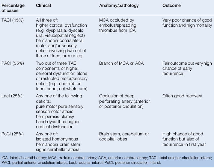
Detailed descriptions of the enormous variety of syndromes explicable in terms of the precise anatomy of the damage sustained are beyond the scope of this book. The identification of the major deficits is more important. The classification devised by Bamford (Bamford et al. 1990; Table 7.5) is simple and provides useful prognostic information.
Table 7.5 Brain infarction (Bamford et al. 1990)

Clinical Problems Following Stroke
- Dysphagia: poor swallow is common initially (see Management of hydration and nutrition p. 101).
- Delirium occurs in over 10% of patients in the first week, predicted by pre-existing cognitive impairment, right hemisphere stroke, large lesion, post-stroke infection; marker of poor prognosis.
- Dysphasia: disorder of language affecting some right-handed patients with left-hemisphere lesions:
 Anterior dysphasia – non-fluent, impaired naming.
Stay updated, free articles. Join our Telegram channel

Full access? Get Clinical Tree


