Age Changes
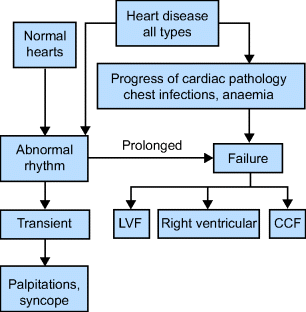
Manifestations (Figure 9.1)
Coronary Artery Disease (CAD)
CAD is the most common cause of death in the UK and the USA. Over 80% of all cardiac deaths in the UK occur in those aged over 65. The sex incidence is equal, unlike in younger age groups. The risk factors are the same as for cerebrovascular disease; see Table 7, Chapter 7.
Managing Cardiovascular Disease in Older People
There is a huge body of research and guidelines on managing cardiovascular disease. Some of the guidance regarding the management of older people is contradictory. This is because older people are hugely under-represented in trials and registries of heart disease. Many studies looking at older people count over 75 year olds as being representative of the ‘very’ old! In addition, these 75 year olds have been screened to exclude many co-morbidities. Thus much of the advice is extrapolated to older people without considering other problems, functional and cognitive abilities, not to mention pharmacological considerations. The cardiovascular community is finally beginning to recognize this, so future guidance will be more age-appropriate.
Presentations
- Angina.
- Acute coronary syndromes (ACS): unstable angina and myocardial infarction (MI).
- Sudden death.
- Heart failure.
- Arrhythmias.
Chronic Stable Angina
Angina is pain secondary to ischaemia caused by reduced blood flow to the heart muscle secondary to atherosclerotic plaques. Stable angina suggests stable plaque disease.
- The classical presentation is retrosternal pain radiating to the left arm and throat on exertion.
- The pain is reproducible by increased oxygen demand, e.g. on exertion and stress.
- The pain is not affected by inspiration, coughing or changing position.
- It is relieved by rest or glyceryl trinitrate (GTN) spray, usually in less than 5 min.
- Patients may deny pain but describe a heaviness or feeling of pressure across the chest.
- They may describe breathlessness on exertion, fatigue or even faintness.
- If the chest pain becomes more frequent, lasts longer or is more severe, this is suggestive of plaque rupture, i.e. an ACS.
- In older people, atherosclerosis is often diffuse throughout the coronary arteries and co-existent left ventricular failure (LVF) is more likely.
Investigations
- ECG: may be normal at this point or show signs of LV hypertrophy or features of ischaemia, e.g. Q waves, ST and T wave flattening or inversion or left bundle branch block (LBBB) from previous ischaemic events.
- CXR: likely to be normal, but may show cardiomegaly or signs of LVF.
Management
- Patient education: stop smoking, increase exercise.
- Aspirin, 75 mg daily; clopidogrel, 75 mg if allergic or intolerant.
- Advise GTN prior to exercise as it is fast acting and lasts for 30–45 min.
- Commence anti-anginal treatment, for example:
 Long-acting nitrates (ensure nitrate-free period by giving isosorbide mononitrate modified release once daily or remind patient to take the patch off at night).
 Beta-blockers, e.g. bisoprolol, slow the heart rate and give more time for coronary perfusion.
 Calcium antagonists, e.g. diltiazem, amlodipine (but watch out for ankle oedema).
 Nicorandil, a potassium channel opener, is a good choice for patients with low BP and symptoms of orthostatic hypotension secondary to other anti-anginal therapy. Avoid doses greater than 30 mg bd in older people because of the risk of large, painful mouth ulcers and occasionally anal ulcers which may bleed.
 Ranolazine is an add-on therapy for refractory angina. It inhibits the sodium current which occurs late in systole in ischaemic myocytes. It does not reduce the heart rate or BP during exercise. It does prolong the QT interval, but does not seem to be pro-arrhythmogenic. It may also improve HbA1c in diabetics.
 Ivabradine is a selective and specific blocker of the If channel of the sino-atrial node cells. Thus heart rate is reduced without lowering BP. It is most beneficial where patients cannot tolerate enough beta-blocker to slow the heart; use reduces hospital admissions.
- In fit older people, consider lowering cholesterol, usually with simvastatin 40 mg.
- Address all risk factors (optimize diabetic control; treat anaemia, hypertension and aortic valve disease).
- Stratification of fit older people for angiography and revascularization can be difficult. Exercise testing has a lower specificity and sensitivity in women and is not useful in LBBB. Older people are likely to have background calcification in CT calcium scoring. Therefore a MIBI scan (technetium 99 2-methoxy isobutyl isonitrile) is likely to be most useful.
- Angiography is appropriate for patients with severe symptoms or being considered for vascular surgery including aortic repair, femoral bypass and carotid surgery.
- Angioplasty, bare metal stents and drug eluting stents are currently being evaluated in older people.
Acute Coronary Syndromes (ACS)
Definition
ACS can be divided into two broad groups: ST elevation myocardial infarcts (STEMI) and unstable angina/non-ST elevation myocardial infarcts (NSTEMI). This is because they are managed differently and they have a different prognosis.
ST-Elevation Myocardial Infarction
Increasing evidence shows that older people with acute STEMI respond just as well to primary percutaneous coronary angiography and intervention (PCI) and thrombolysis as younger patients.
Examination
- The patient is usually severely unwell, clammy and peripherally shut down.
- Monitor the pulse rate and rhythm and blood pressure frequently.
Investigations
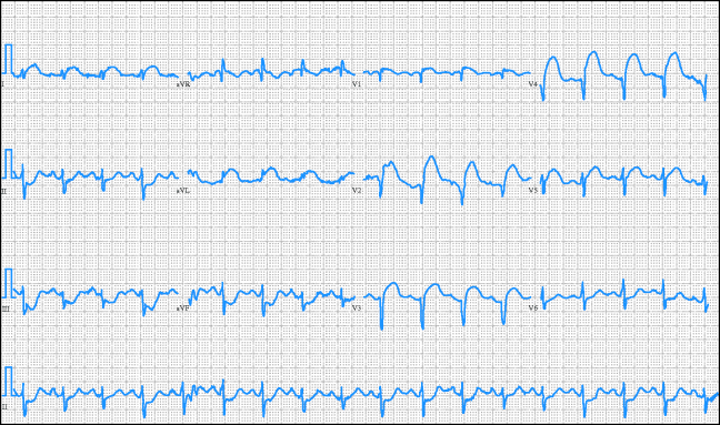
- ECG changes: by definition, ST elevation (see Figure 9.2), new left bundle branch block.
- Serial troponin I measurements to demonstrate myocyte damage.
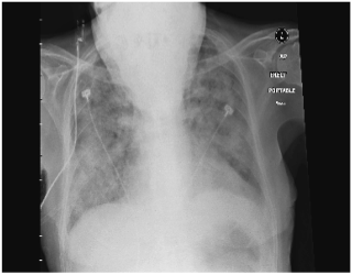
- The CXR may show cardiomegaly or signs of pulmonary oedema (see Figure 9.3).
Figure 9.2 ECG showing acute STEMI. Note hyperacute ‘tombstone’ elevation of the anterior leads and tachycardia. From Wikimedia Commons (public domain).

Figure 9.3 Chest radiograph demonstrating acute pulmonary oedema. Note: (1) portable film with head obscuring part of chest suggesting the patient is sick; (2) cardiomegaly even allowing for portable film; and (3) extensive pulmonary fluid.

Management
Pre-hospital:
- Give oxygen 2–4 litres via nasal prongs.
- Treat pain with intravenous (IV) opiates to reduce sympathomimetic drive on heart.
- Stat dose of 300 mg aspirin. Continue 75 mg on daily basis if no contraindication.
- Best practice is to admit to specialist centre offering PCI. Angiography will demonstrate the nature of the lesion causing the ECG changes and will lead to appropriate intervention including angioplasty or stenting. Evidence suggests that older people do better with primary PCI because of the lower risk of cerebral haemorrhage compared with thrombolysis.
- If PCI is not locally available and there no contraindications thrombolyse with IV streptokinase. Tissue plasminogen activator (tPA) is associated with increased risk of cerebral haemorrhage in older people.
- Give low-molecular-weight heparin (LMWH), e.g. enoxaparin.
- Treatment with oral beta-blockers, again to be continued if no contraindications; this has been proven to be of benefit to older people.
- ACE inhibitors in patients with evidence of LV failure (to reduce LV remodelling).
- Consider cholesterol-lowering treatment.
- Older people should have equal access to cardiac rehabilitation facilities post MI.
Unstable Angina and NSTEMI
This is chest pain secondary to enlargement or rupture of atherosclerotic plaque. The unstable plaque is profoundly thrombogenic so treatment is aimed at reducing this. Features include:
Investigations
- Serial troponin I measurements to assess myocyte damage.
- The ECG may show signs of ischaemic heart disease: LVH, Q waves from previous MI, ST depression.
- The CXR may show cardiomegaly or signs of pulmonary oedema (see Figure 9.3).
Management
- Admit to a monitored bed if possible.
- Bed rest.
- Aspirin 300 mg stat.
- P2Y12 inhibitors including clopidogrel. Of the newer agents, ticagrelor may have a role in older people with chronic kidney disease (CKD), but prasugrel has been associated with a higher risk of bleeding.
- GTN spray/tablet.
- IV GTN or buccal nitrate.
- Low-molecular-weight heparin (LMWH).
- Beta-blockers (metoprolol is short acting and can be stopped if side-effects develop), unless contraindicated.
- If the pain does not improve, add calcium antagonist (diltiazem).
- The use of IV glycoprotein IIa/IIIb blockers (e.g. tirofiban and abciximab, which prevent platelets from cross-linking and therefore prevent platelet aggregation) has not been fully established in older people. There seems to be increased risk of intracranial haemorrhage.
- Caution – watch for bradycardia and hypotension.
- Following successful management, reduce all possible risk factors and establish an oral medication regime.
- If the patient is biologically fit, they should be considered for revascularization, either angioplasty or coronary bypass grafting. If no intervention is planned/appropriate, then a 12-month course of clopidogrel is associated with a reduced risk of further events.
Heart Failure
Heart failure is an extremely common cause of hospital admissions, readmissions, reduced function and institutionalization. Although usually biventricular, it is conventionally divided into acute LV failure and congestive cardiac failure.
Acute Left Ventricular Failure
Clinical Features
- Severe breathlessness, orthopnoea, paroxysmal nocturnal dyspnoea.
- Frothy pink sputum.
- On examination, there may be tachycardia, a third heart sound, crackles at the lung bases and, sometimes, pleural effusions.
Investigations
- ECG may show evidence of an acute infarction, LV hypertrophy, tachycardia or LBBB. Also look for AF and other arrhythmias.
- Blood tests may show a troponin rise.
- In early failure, the CXR may show upper lobe blood diversion and fluid in the horizontal fissure or septal lines. With increasing pulmonary oedema, there may be perihilar shadowing and bilateral pleural effusions. If the effusion is unilateral, it is more likely to be on the right. See Figure 9.3.
Management
- The patient is likely to be sitting bolt upright already, but be sure to check as frail older patients slide down modern hospital beds!
- Give oxygen via a face mask.
- Urinary catheter to monitor urine output.
- GTN infusion to offload right heart and reduce angina if present. Aim to keep systolic BP above 90 mmHg.
- IV loop diuretics, i.e. furosemide.
- Opiates (with an anti-emetic) are very useful in this situation, as they not only reduce anxiety and pain therefore reducing oxygen demand, but also reduce pre-load.
- Treat precipitant, e.g. fast AF.
- If there is evidence of an acute MI, treat as above.
- Treat co-existing pneumonia with IV antibiotics.
Chronic Heart Failure
- In the UK 900,000 patients have heart failure.
- Average age at diagnosis is 76.
- The prevalence will rise with the ageing population and owing to better management of CAD.
- Prognosis of heart failure is poor: 30–40% die within 1 year of diagnosis.
- The annual mortality after that ranges from 10–50% depending on severity.
- Frequent features include fatigue, functional decline, confusion, falls and cachexia.
Note: Ankle oedema is often non-cardiac: e.g. chair-bound immobility increases venous pressure, due to gravity, loss of muscle-pump activity and pressure on thigh veins by the chair.
Causes include:
- Coronary artery disease (most common).
- Hypertension.
- Valvular heart disease.
- Cor pulmonale including secondary to pulmonary emboli.
- Thyrotoxicosis.
- Severe anaemia.
- Arrhythmias, especially fast AF.
- Drugs, including non-steroidal anti-inflammatory drugs (NSAIDs) leading to fluid overload.
- Cardiomyopthies.
Investigations
NICE guidelines on heart failure recommend:
- Early transthoracic echo.
- Measure brain natriuretic peptide (BNP) or N terminal pro-B-type natriuretic peptide (NTpro BNP).
- High BNPs confer poor prognosis. (NB: Also high in other conditions including PE and may be low in obese patients and those on beta blockers.)
- Baseline urea, electrolytes, eGFR, TSH, LFTs, fasting lipids and glucose, FBC.
- CXR.
- Peak expiratory flow rate (PEFR) to exclude COPD.
Treatment
Diastolic Heart Failure
- This is thought to account for up to 50% of cases with symptoms and signs of heart failure with a normal LV ejection fraction on echo.
- Estimated annual mortality rate of 7%.
- Risk factors include:
 Aged over 75.
 Female.
 Overweight.
 History of hypertension (leads to stiff arteries).
 Diabetes.
 CAD.
CAD.
 AF.
AF.
- The cause is thought to be impaired LV relaxation (especially during exercise) owing to changes in cytoskeletal proteins of the myocardium and increased LV diastolic stiffness secondary to increased myocardial fibrosis. This leads to incomplete LV filling in diastole, which in turn increases diastolic pressures causing pulmonary congestion and poor ability to increase cardiac output.
- It can cause flash pulmonary oedema precipitated by AF or fluid overload.
- The ECG may show LVH secondary to hypertension or a large left atrium (p mitrale).
- Echocardiography is essential for diagnosis: the LV ejection fraction should be above 50%, the LV volumes should be normal with no valvular lesions.
- Management involves lifestyle changes such as losing weight and reducing salt intake.
- Treat contributing disease such as respiratory problems and anaemia.
- Treat fluid overload with furosemide or spironolactone. Aldosterone antagonists reduce fibrosis. Their role in diastolic heart failure is being investigated by the TOPCAT study (ongoing).
- Treat hypertension with an ACEi.
- Drug treatment has been shown to improve symptoms of diastolic heart failure, but not improve mortality.
Hypertension
Definition
The definition of hypertension is constantly being updated. Current NICE/British Hypertension Guidelines suggest a clinic BP of <140/90 under 80y and <150/90 over 80y. This is in line with the European guidelines. Isolated systolic hypertension implies that only systolic pressure is elevated.
The American College of Cardiology and American Heart Association are now advocating a systolic measurement of 140–145 and a diastolic of less than 90 for older people.
Prevalence
Among people aged 65–74, prevalence ranges from 45% to 64% according to different surveys.
Effects of Hypertension
Hypertension is a major risk factor for atherosclerosis and thus stroke, coronary artery disease, diastolic heart failure and peripheral vascular disease.
Effects of Treatment
Up to age 80, treatment produces considerable benefit (more than in younger subjects) in total mortality and cardiovascular morbidity and mortality, but sometimes at the expense of making the patient feel worse. The Hypertension in the Very Elderly Trial (HYVET) now shows definite advantages of treating relatively fit elderly patients. However, remember to consider risk of falls and symptoms of dizziness in each individual patient before treating.
Treatment
Stay updated, free articles. Join our Telegram channel

Full access? Get Clinical Tree


