Age Changes
The following facts are given because they are interesting, but they are of more theoretical than practical importance.
Red Blood Cells
There is evidence of reduced deformability in health and this is even more marked in those with cerebrovascular disease.
White Blood Cells
Plasma Proteins
Conflicting reports on age changes relate to the difficulty in distinguishing those changes due solely to age from those due to other factors, such as malnutrition or disease. The main points to emerge are as follows:
Anaemia
- Estimates of the prevalence of anaemia vary dramatically.
- Prevalence in over 70s in the community is 15%; 24% in nursing homes.
- People with anaemia have a higher mortality, greater risk of hospitalization, poorer mobility and functional abilities and a higher risk of falls compared with those who do not.
Haemopoiesis and Ageing
Basic Investigations
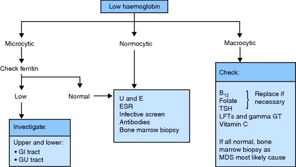
Anaemia is the most common abnormality and the red cell size (mean corpuscular volume, MCV) and morphology often suggest the aetiology (Figure 14.1). Always request a peripheral blood film; it is inexpensive and easy and will often lead you directly to the cause. White cell morphology will also be helpful, especially in myeloproliferative disorders.
Measurement of the acute-phase response is also useful. ESR is the cheapest investigation but it reflects changes slowly (i.e. days). CRP is better for monitoring more rapid changes (i.e. hours). A protein electrophoretic strip is needed to diagnose myeloma.
Clinical History and Examination
The symptoms of anaemia, i.e. tiredness, breathlessness and general malaise, are too common and non-specific to be of great value. Anaemia may exacerbate symptoms due to other pathologies, e.g. angina, heart failure, intermittent claudication, increased confusion in dementia, and falls may occur more frequently. The patient’s symptoms may therefore be greater than expected from the haematological abnormality.
The history may suggest the cause. Ask about previous surgery and transfusions, family history (pernicious anaemia), medication and dietary change, weight loss, GI symptoms and blood loss. Physical examination may not be helpful, but look for pallor, jaundice, lymphadenopathy, abdominal organomegaly, rectal masses and neurological abnormalities, as these may direct you to the correct diagnosis.
Two important final points: remember that anaemia is a sign of disease and is not a diagnosis and that there are often multiple causes in each patient.
Types and Causes of Anaemia
The WHO definition of anaemia is haemoglobin of less than 13 g/dL in males or less than 12 g/dL in females. The validity of this definition in older people is often disputed, but it improves the pick-up rate of disease.
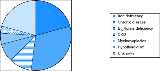
The main categories (see Figure 14.2) are:
- Due to bleeding.
- Due to cell destruction (haemolysis).
- Deficiency states.
- Marrow dysfunction.
- Anaemia of chronic disease.
Iron-Deficiency Anaemia (Microcytic)
Iron-deficiency anaemia, the most common cause of microcytic (low MCV) anaemia, affects 2–5% of the older population.
Aetiology
History
Examination
See also Chapter 11 for full gastrointestinal examination.
- General: pallor, lymphadenopathy or jaundice.
- Tachycardia, hypotension, murmurs if there is volume loss from bleeding.
- Mouth: stomatitis, telangiectasia around the mouth and palate.
- Hands: koilonychia (spoon-shaped nails associated with iron deficiency).
- Abdomen: epigastric tenderness, masses, scars from previous surgery, hepatomegaly or splenomegaly.
- Examine the rectum for masses and exclude melaena.
Investigations
- Peripheral blood film: the red cells are small, hypochromic and vary in size (anisocytosis) and shape (poikilocytosis). There may also be pencil cells and target cells.
- Red cell distribution width (RDW) may increase reflecting greater variability in the size of red blood cells. Remember that if there is also B12 or folate deficiency, the red cells may not be microcytic.
- A low serum ferritin is good evidence of iron deficiency. However, it is an acute-phase protein and may be falsely elevated in the context of inflammation, in which case CRP will also be high.
- A raised reticulocyte count implies bleeding or haemolysis with appropriate increased bone marrow activity.
- As 10–15% of patients have both upper and lower GI causes of anaemia, most patients need both an oesophago gastro duodenoscopy (OGD) with duodenal biopsies and at least a CT abdomen with contrast (see Chapter 11).
Treatment
- Oral iron should be continued for 3 months to replenish the iron stores; it is sufficient to prescribe ferrous sulphate 200 mg twice daily, as more than this may not be absorbed and increases side-effects such as constipation or diarrhoea. Warn the patient that their stools will become black and gritty. Encourage the patient not to chew the tablets or their teeth or dentures may become stained.
- If the response to oral iron treatment is not satisfactory (i.e. 2 g/dL after 2 months), check the patient is taking the tablets and that any bleeding has stopped. Also consider additional diagnoses that may explain the problem, such as co-existent anaemia of chronic disease. Consider giving ascorbic acid to increase absorption.
Blood Transfusion
- Blood transfusion is needed after acute haemorrhage, commonly a GI bleed. If there has been volume loss, there is no need to give furosemide to prevent overload.
- Injuries such as scalp lacerations, large haematomas and hip and pelvic fractures may cause substantial blood loss (especially in those taking warfarin) which may not be obvious. Watch for a drop in haemoglobin and transfuse if symptomatic or the Hb drops below 8.
- Patients with acute anaemia who are not shocked need full investigation but emergency transfusion is not needed – transfuse in the day which is less disruptive and safer.
- Patients with chronic anaemia may benefit from transfusion. The usual cutoff is below 8 g/dL, but the threshold may be higher if the patient has symptoms such as angina. Usually, 2 units are sufficient; oral or IV furosemide is usually needed.
- In conditions like myelodysplastic syndrome, regular transfusions can be done in day care facilities.
Haemolytic Anaemia
- Haemolysis is the premature destruction of red blood cells. When the bone marrow can no longer match the rate of haemolysis, the patient becomes anaemic.
- Haemolytic anaemias are rare in old age and therefore in danger of being overlooked, especially as the peripheral blood film may not show any specific features.
- Autoimmune haemolytic anaemias are more common in older age as part of an autoimmune illness (e.g. SLE), due to antibody production secondary to infection (hot and cold antibodies following a viral infection) or as an iatrogenic disease (e.g. methyldopa treatment).
- Patients with hypersplenism may also have a haemolytic element to their anaemia.
- Most patients with metal heart valves have minor haemolysis.
- Relevant investigations are those indicating increased red cell destruction, i.e. raised bilirubin level, lactate dehydrogenase, raised reticulocyte count (raising the MCV), haptoglobins and direct antibody test (Coombs’ test).
- Supportive management includes stopping culprit drugs, replacing folate and transfusions only for severe cases.
Deficiency States Causing Macrocytosis
The macrocytic (high MCV) anaemias are usually secondary to deficiency of folic acid, vitamin B12 or, more rarely, thyroxine. They are much less common than anaemia due to iron deficiency or chronic disease. The bone marrow precursors are megaloblastic in B12 and folate deficiency and drug toxicity (as DNA replication is affected). In other macrocytic anaemias the marrow is normoblastic. See Table 14.1 showing causes of macrocytosis.
Table 14.1 Causes of macrocytosis
| Folate deficiency |
| B12 deficiency |
| Alcohol excess |
| Hypothyroidism |
| Myelodysplastic syndromes |
| Liver disease |
| All causes of reticulocytosis |
| Drugs: |
| • Folic acid antagonists (methotrexate, phenytoin) |
| • Purine synthesis antagonists (6-mercaptopurine) |
| • Pyrimidine antagonists (cytosine arabinoside) |
| Spuriously raised MCV – cold agglutinins, paraproteins |
Vitamin B12 Deficiency
- Pernicious anaemia (PA) is the cause of B12 deficiency in 80% of cases in the UK.
- Other causes are gastrectomy, bacterial colonization of intestinal strictures or diverticula and disorders of the terminal ileum, especially Crohn’s disease.
- Only strict vegans are likely to suffer from dietary vitamin B12 deficiency.
- B12 stores last for several years, hence the lag between a pathology affecting B12 absorption and clinical manifestations.
Pernicious Anaemia
- PA develops insidiously due to autoimmune gastric atrophy and failure of intrinsic factor secretion.
- The incidence is said to be 1:10,000 with a female preponderance.
- Cancer of the stomach occurs in about 10% of cases.
- Other complications are peripheral neuropathy, subacute combined (corticospinal and dorsal column) degeneration of the spinal cord so with the neuropathy there are UMN and LMN signs. There may be cognitive impairment.
- These features may predate the haematological changes and continue if folate is given when B12 is low.
- There may be a family history of other autoimmune conditions such as Hashimoto’s thyroiditis, vitiligo and Addison’s disease.
Clinical Features
- Symptoms and signs of anaemia, with lemon tinge to skin (jaundice secondary to mild haemolysis).
- Glossitis, anorexia and weight loss.
- In severe cases, hepatosplenomegaly and heart failure.
- Neurological signs.
Diagnosis
Stay updated, free articles. Join our Telegram channel

Full access? Get Clinical Tree


