Postmenopausal androgen hypersecretion may be due to an ovarian tumor, an adrenal tumor, or ovarian hyperthecosis. It is important to assess the patient with a full androgen profile and imaging studies of the ovaries and adrenal glands. When an adrenal tumor is identified in a postmenopausal woman with androgen excess, adrenal and gonadal venous sampling may be needed to localize the source of androgen hypersecretion.
Case Report
This 59-year-old woman was evaluated for weight management and was noted to have hirsutism on physical examination. She had no signs or symptoms of glucocorticoid excess. The patient had a long-standing history of mild facial hirsutism and noticed exacerbation of hirsutism and development of coarse facial features over several years before current presentation. She did not have acne but noticed oily skin. She had to shave daily. Her medical history was significant for rheumatoid arthritis treated with methotrexate and frequent courses of prednisone. She also had chronic depression, treated with duloxetine. Twenty years prior, she had hysterectomy and right oophorectomy (for menorrhagia). She did not have vasomotor symptoms to help estimate the onset of menopause. She was not treated with estrogen.
On physical examination her body mass index was 42.8 kg/m 2 , blood pressure 134/72 mmHg, and heart rate 70 beats per minute. Hirsutism was evident over abdomen, chest, chin, back, and face. Her degree of hirsutism was marked with a modified Ferriman-Gallwey score of 30 (normal, <8; maximum, 36).
INVESTIGATIONS
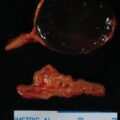
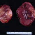
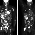
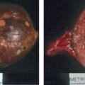
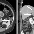
Laboratory test results are shown in Table 89.1 . Testosterone hypersecretion was documented. Transabdominal and endovaginal ultrasound of the pelvis showed a 2.2-cm solid left ovarian mass, also visualized on a pelvic CT ( Fig. 89.1 , right). Adrenal CT showed a 1.9-cm lipid rich left adrenal adenoma (see Fig. 89.1 , left). The question was whether the source of testosterone hypersecretion was adrenal or ovarian. Adrenal and gonadal venous sampling was the next step. Adrenal venous sampling was performed without cosyntropin infusion. Testosterone concentration in the left gonadal vein was 100-fold higher than the testosterone concentration in the inferior vena cava, adrenal vein, and the right gonadal vein ( Box 89.1 ). Testosterone hypersecretion from the left ovarian mass was confirmed.
| Biochemical Test | Result | Reference Range |
| Sodium, mEq/L Potassium, mEq/L Creatinine, mg/dL Total testosterone, ng/dL Androstenedione, ng/dL Corticotropin, pg/mL DHEA-S, mcg/dL 17-hydroxyprogesterone, ng/dL LH, IU/L FSH, IU/L Estradiol, pg/mL Aldosterone, ng/dL Renin plasma activity, ng/mL per hour 1-mg overnight DST cortisol, mcg/dL 24-Hour urine cortisol, mcg | 137 4.2 0.96 510 113 9.4 128 119 18 25 28 9.9 1.3 2.2 40 | 135–145 3.6–5.2 0.6–1.1 8–60 30–200 7.2–63 15–157 Postmenopausal: <51 Postmenopausal: 5.3–65.4 Postmenopausal: 16–157 Postmenopausal: <10 ≤21 0.6–3 <1.8 mcg/dL 3.5–45 |
Related posts:
 45-Year-Old Woman With Corticotropin-Independent Cushing Syndrome and Bilateral Adrenal Adenomas
 Adrenocortical Carcinoma Associated With Lynch Syndrome
 Metastatic Paraganglioma: Role for Systemic Chemotherapy
 Cushing Syndrome Associated With Ectopic Corticotropin and Corticotropin-Releasing Hormone–Secreting Pheochromocytoma
 Bilateral Macronodular Adrenal Hyperplasia in the Setting of Multiple Endocrine Neoplasia Type 1
 Catecholamine-Secreting Paraganglioma in Pregnancy
Stay updated, free articles. Join our Telegram channel

Full access? Get Clinical Tree


