Figure 1.1
Ear wide excision plan
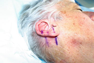
Figure 1.2
Ear interpolation flap plan
The Technique
The mastoid interpolation flap, which is the staged pedicle flap described herein, is very useful for helical ear defects when cartilage needs to be removed. It helps re-create a normal-looking ear. For smaller helical rim defects, a skin graft, cutaneous helical rim advancement flap, primary closure, or wedge resection often provides an excellent reconstructive result. Helical rim area can be a problem in itself with thin skin. However when cartilage needs to be removed, this increases the risk of perichondritis, which can be a surgical nuisance.
In a large series of patients after ear reconstruction, it was shown 2–4 % of cases become infected and may progress to perichondritis, if untreated [9]. Trauma, surgical or otherwise is the most common cause in nearly half the incidences of perichondritis and Pseudomonas aeruginosa the most common micro-organism isolated [10]. The treatment of such perichondritis is primarily antibiotics and surgical debridement when needed, and the antibiotic of choice is Ciprofloxacin [11].
It is important to secure hemostasis during the procedure and place a drain to prevent hematoma formation. If perichondritis develops in spite of antibiotics, which is rare, then it is important to aggressively drain any abscess early. Fortunately, this is very rare after elective surgery for skin cancer.
As a general rule, the initial reconstructive effort is aimed only at repair of the anterior portion of the primary defect. Re-creation of the helical rim and posterior primary defect coverage is done at the second stage – when the pedicle is detached and the ear is reconstructed. Some surgeons cut a template of foil or paper and lay over the mastoid to mark the outlines of the flap. It is important to avoid hair-bearing areas to avoid a hairy ear post-operatively. Rather than cutting out a template, I prefer to press the ear and lay it flat against the mastoid. Given that the excision margins are already marked on the ear, this allows to accurately plan the flap by continuing the markings onto the mastoid skin surface (Fig. 1.2). The combination of posterior ear, post-auricular sulcus, and mastoid skin usually provides an excellent tissue match for the anterior helical soft tissue defect. The flap is elevated and the secondary mastoid scalp and primary anterior helical defect margins are slightly undermined to provide increased flap mobility (Fig. 1.3) [12]. In our case, some thinning of the flap was needed as the mastoid skin was thicker than the excised skin of the anterior helical defect. If the ear has a prominent helical rim curl with or without loss of the rim cartilage, a few double-armed or basting sutures may be placed through the cartilage into the anterior lip of the helical rim to recreate the natural curl [12].
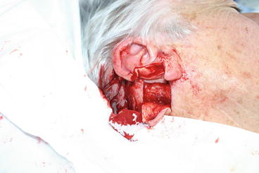
Figure 1.3
Ear mastoid interpolation flap being raised
Even with such sutures in place, it is often necessary to later on thin the flap to achieve perfect contours. I normally wait for 6 months post-operatively before planning any tertiary procedure such as this.
If the defect extends only to the helical rim, the flap can be started at the junction where mastoid skin meets the posterior ear. If the defect extends further medial to the scaphoid fossa or beyond, the flap incision is then ideally started on the posterior ear and extended onto the mastoid area. The flap should be sized slightly larger than the measured width of the defect and be long enough so that excessive tension is not placed on the flap after it is sutured [13].
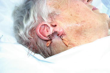
Once the flap is sutured in place, a drain is inserted to avoid any post-operative collection or hematoma formation. I tend to use a Penrose or a ‘glove’ drain fashioned using a sterile surgical glove. Some authors prefer to use nasal packing or gel foam but in my experience, I have found this unnecessary. It is standard to apply a pressure dressing for 48 h, after which I usually remove the drain. I avoid suction drains (and prefer the glove drain) as the former are more bulky. I prefer to use a light dressing post-operatively which is not as noticeable (Fig. 1.4).
 Topical Treatment of Skin Cancers and the Risks of ‘Fighting Fire with Fire’
When a Lipoma Wasn’t a Lipoma: A Discussion About Granular Cell Tumors of Skin
Metastatic Cutaneous Adenocarcinoma
Adenocystic Carcinoma
Islands on the Cheek: Island Flaps on the Cheek and a Modified Oblique-Sigmoid Flap
Rotation Flaps of the Scalp: Study of the Design, Planning and Biomechanics of Single, Double and Triple Pedicle Flaps
Topical Treatment of Skin Cancers and the Risks of ‘Fighting Fire with Fire’
When a Lipoma Wasn’t a Lipoma: A Discussion About Granular Cell Tumors of Skin
Metastatic Cutaneous Adenocarcinoma
Adenocystic Carcinoma
Islands on the Cheek: Island Flaps on the Cheek and a Modified Oblique-Sigmoid Flap
Rotation Flaps of the Scalp: Study of the Design, Planning and Biomechanics of Single, Double and Triple Pedicle Flaps


Related posts:
Topical Treatment of Skin Cancers and the Risks of ‘Fighting Fire with Fire’
When a Lipoma Wasn’t a Lipoma: A Discussion About Granular Cell Tumors of Skin
Metastatic Cutaneous Adenocarcinoma
Adenocystic Carcinoma
Islands on the Cheek: Island Flaps on the Cheek and a Modified Oblique-Sigmoid Flap
Rotation Flaps of the Scalp: Study of the Design, Planning and Biomechanics of Single, Double and Triple Pedicle Flaps
Stay updated, free articles. Join our Telegram channel
Full access? Get Clinical Tree
